System and Method For Statistical Shape Model Based Segmentation of Intravascular Ultrasound and Optical Coherence Tomography Images

a technology of intravascular ultrasound and optical coherence tomography, which is applied in image enhancement, instruments, and recognition of medical/anatomical patterns, etc., can solve the problems of inability to use statistical shape models in previous ivus segmentation algorithms, inability to accurately segment the arterial wall boundaries, and inability to detect the presence of a single artery, etc., to achieve adequate flexibility, enhance the segmentation results, and increase the segmentation quality

Inactive Publication Date: 2008-03-27
SIEMENS MEDICAL SOLUTIONS USA INC
View PDF5 Cites 58 Cited by
  • Summary
  • Abstract
  • Description
  • Claims
  • Application Information

AI Technical Summary

Benefits of technology

[0007] Exemplary embodiments of the invention as described herein generally include methods and systems for a model-based approach to segmentation of the arterial wall from both intravascular ultrasound images and optical coherence tomography images in the rectangular domain. Both the lumen and medial / adventitial contour variations are modeled within a shape space in the resampled rectangular domain. In a properly built shape space using training data, the lumen and media-adventitia contours are constrained to a smooth, closed geometry, which increases the segmentation quality without any tradeoff with a regularizer term, yet with adequate flexibility. In addition, a non-parametric intensity model based on a probability density based image energy is used with global image measurements rather than pointwise measurements used in previous methods. This enhances the segmentation results. Experiments demonstrate that a method according to an embodiment of the invention can be used in plaque analysis, 3D reconstruction of the arterial wall, and for measurements such as lumen size, lumen radius, wall radius, etc.
[0020] According to a further aspect of the invention, the boundary contour is initialized by dividing an image into a plurality of columns, finding a position of a maximal gradient for every column wherein a contour is initialized, and passing a median filter over the initial contour wherein noise is eliminated.

Problems solved by technology

The disruption of an atherosclerotic plaque is considered to be the most frequent cause of heart attack and sudden cardiac death.
Manual segmentation and processing is tedious, time-consuming, and susceptible to intra- and inter-observer variability.
None of the previous IVUS segmentation algorithms used a statistical shape model based approach.
Medical IVUS image segmentation algorithms are almost always hampered by noise, stents, shadowing due to calcium deposits and have weak or missing boundaries of structures.

Method used


Image

  • System and Method For Statistical Shape Model Based Segmentation of Intravascular Ultrasound and Optical Coherence Tomography Images
  • System and Method For Statistical Shape Model Based Segmentation of Intravascular Ultrasound and Optical Coherence Tomography Images
  • System and Method For Statistical Shape Model Based Segmentation of Intravascular Ultrasound and Optical Coherence Tomography Images

Examples


Embodiment Construction

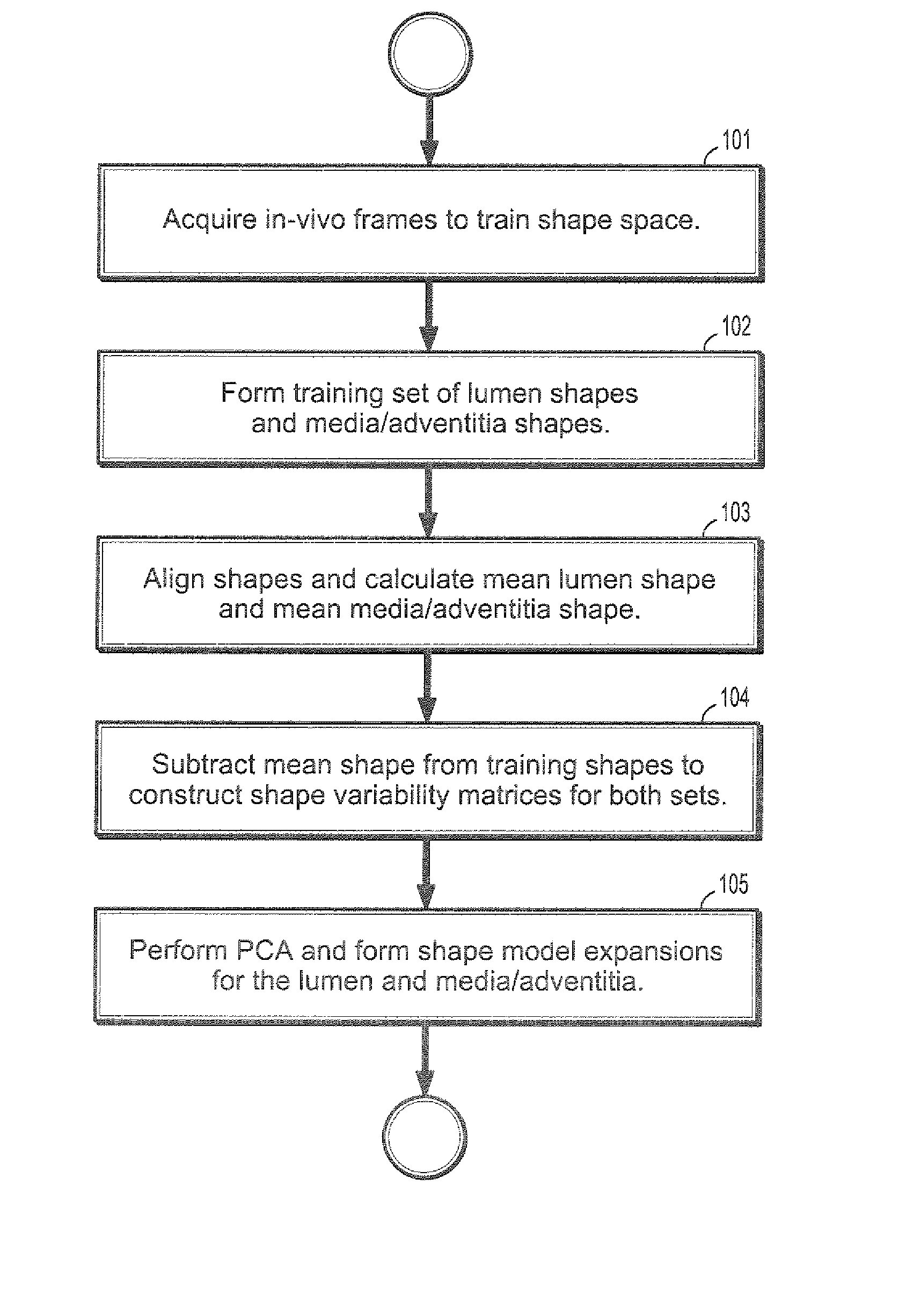
[0040] Exemplary embodiments of the invention as described herein generally include systems and methods for segmentation of arterial wall boundaries from intravascular images. Accordingly, while the invention is susceptible to various modifications and alternative forms, specific embodiments thereof are shown by way of example in the drawings and will herein be described in detail. It should be understood, however, that there is no intent to limit the invention to the particular forms disclosed, but on the contrary, the invention is to cover all modifications, equivalents, and alternatives falling within the spirit and scope of the invention.

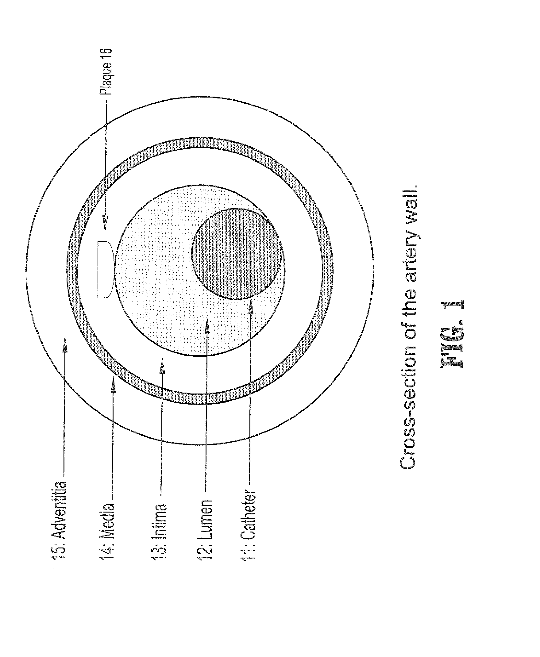
[0041] As used herein, the term “Image” refers to multi-dimensional data composed of discrete image elements (e.g., pixels for 2-D images and voxels for 3-D images). The image may be, for example, a medical image of a subject collected by computer tomography, magnetic resonance imaging, ultrasound, or any other medical imaging system known to o...


PUM


Abstract

A method for segmenting intravascular images includes acquiring a series of digitized images acquired from inside a vessel, each said image comprising a plurality of intensities corresponding to a 2-dimensional grid of pixels, providing a precomputed set of shapes for modeling contours of vessel wall boundaries, wherein a contour can be expressed as a sum of a mean shape and a inner product of shape modes and shape weights, initializing a boundary contour for one of said set of images, initializing said shape weights by projecting a contour into said shape modes, updating said shape weights from differential equations of said shape weights, and computing a contour by summing said mean shape and said inner product of shape modes and updated shape weights.

Description

CROSS REFERENCE TO RELATED UNITED STATES APPLICATIONS [0001] This application claims priority from “Shape-driven Segmentation of Intravascular Ultrasound and Optical Coherence Tomography Images”, U.S. Provisional Application No. 60 / 820,139 of Unal, et al., filed Jul. 24, 2006, the contents of which are herein incorporated by reference.TECHNICAL FIELD [0002] This disclosure is directed to the segmentation of arterial wall boundaries from intravascular images. DISCUSSION OF THE RELATED ART [0003] Intravascular ultrasound (IVUS) and optical coherence tomography (OCT) allows real time tomographic assessment of the arterial wall, which is useful for studying vascular wall architecture for diagnosis and assessment of the progression of the cardiovascular diseases. Atherosclerosis is a disease affecting arterial blood vessels, commonly referred to as a “hardening” of the arteries. It is a chronic inflammatory response in the walls of arteries, in large part due to the formation of multiple...

Claims


Application Information

Patent Timeline
Patent Type & Authority Applications(United States)
IPC IPC(8): G06K9/36
CPCG06K9/621G06K2209/05G06T7/0083G06T2207/30101G06T7/0097G06T2207/10081G06T2207/10132G06T7/0089Y10S128/922G06T7/12G06T7/149G06T7/174G06V10/7557G06V2201/03
Inventor UNAL, GOZDEBUCHER, SUSANNSLABAUGH, GREGORY G.FANG, TONG
Owner SIEMENS MEDICAL SOLUTIONS USA INC
Why Eureka
Try Eureka