Scatter radiation correction in radiography and computed tomography employing flat panel detector

a detector and scatter radiation technology, applied in the field of scatter radiation correction in radiography and computed tomography with flat panel detectors, can solve the problems of contrast loss, quantitative distortion, significant problem of scatter radiation (which contains only very little image information), etc., and achieve the effect of worsening the signal-to-noise ratio

Active Publication Date: 2008-04-03
SIEMENS HEALTHCARE GMBH
View PDF7 Cites 29 Cited by
  • Summary
  • Abstract
  • Description
  • Claims
  • Application Information

AI Technical Summary

Benefits of technology

[0008] An object of the invention is to provide an improved scatter radiation correction method that worsens the signal-to-noise ratio as little as possible, or not at all.
[0037] Under the assumption that the correction error is small relative S and P, in the following the effect of the scatter radiation correction on the differential contrast, the noise and the dCNR is examined. For this the logarithmized projection data are also required: pcor=−ln Pcor=−ln(Ps−Scor)  (#10a) pcor=−ln(P+εcor)  (#10b)
[0038] The differential contrast is now reestablished via scatter radiation correction. The interference calculation applied to equation (#10b) provides Δ⁢ ⁢pcor=Δ⁢ ⁢P+Δ⁢ ⁢ɛcorP+ɛcor≈Δ⁢ ⁢PP⁢11+ɛcor / P=Δ⁢ ⁢p⁢11+ɛcor / P,(#11) wherein it was utilized that εcor runs very smoothly and the derivation can locally be set to approximately equal 0. Under the assumption of a negligible correction error |εcor|<<|P|, it thus results that a precise scatter radiation correction of the original differential contrast can be regained, meaning that the scatter radiation-conditional differential contrast loss is compensated by the factor (1+S / P) by the correction through a differential contrast amplification.
[0080] The described image quality analysis and the inventive solution suggestion have the following advantages with regard to the elimination and adaptive noise filtering of the scatter radiation correction term:
[0082] the specific noise filtering of the scatter radiation correction term considerably reduces the increase of the noise via the scatter radiation correction,

Problems solved by technology

Due to the large irradiated patient volume in each projection image, the scatter radiation (which contains only very little image information) represents a significant problem.
The effects of the scatter radiation are contrast loss, distortions of the quantitative results (such as cupping and artifacts) and increase of the noise.
The use of scattered-ray grids is generally not sufficient.
Although the quality measure of contrast-to-noise remains, an increase of the image noise normally occurs with the corrections, such that the noise increases in equal measure to the contrast.
However, since the scatter radiation within the measurement field is not measured, this method is only very crude.
A problem of these known scatter radiation correction methods lies in that the “signal-to-noise” ratio or the ratio of scatter radiation intensity to primary radiation intensity is worsened by the scatter radiation correction.

Method used


Image

  • Scatter radiation correction in radiography and computed tomography employing flat panel detector
  • Scatter radiation correction in radiography and computed tomography employing flat panel detector
  • Scatter radiation correction in radiography and computed tomography employing flat panel detector

Examples


Embodiment Construction

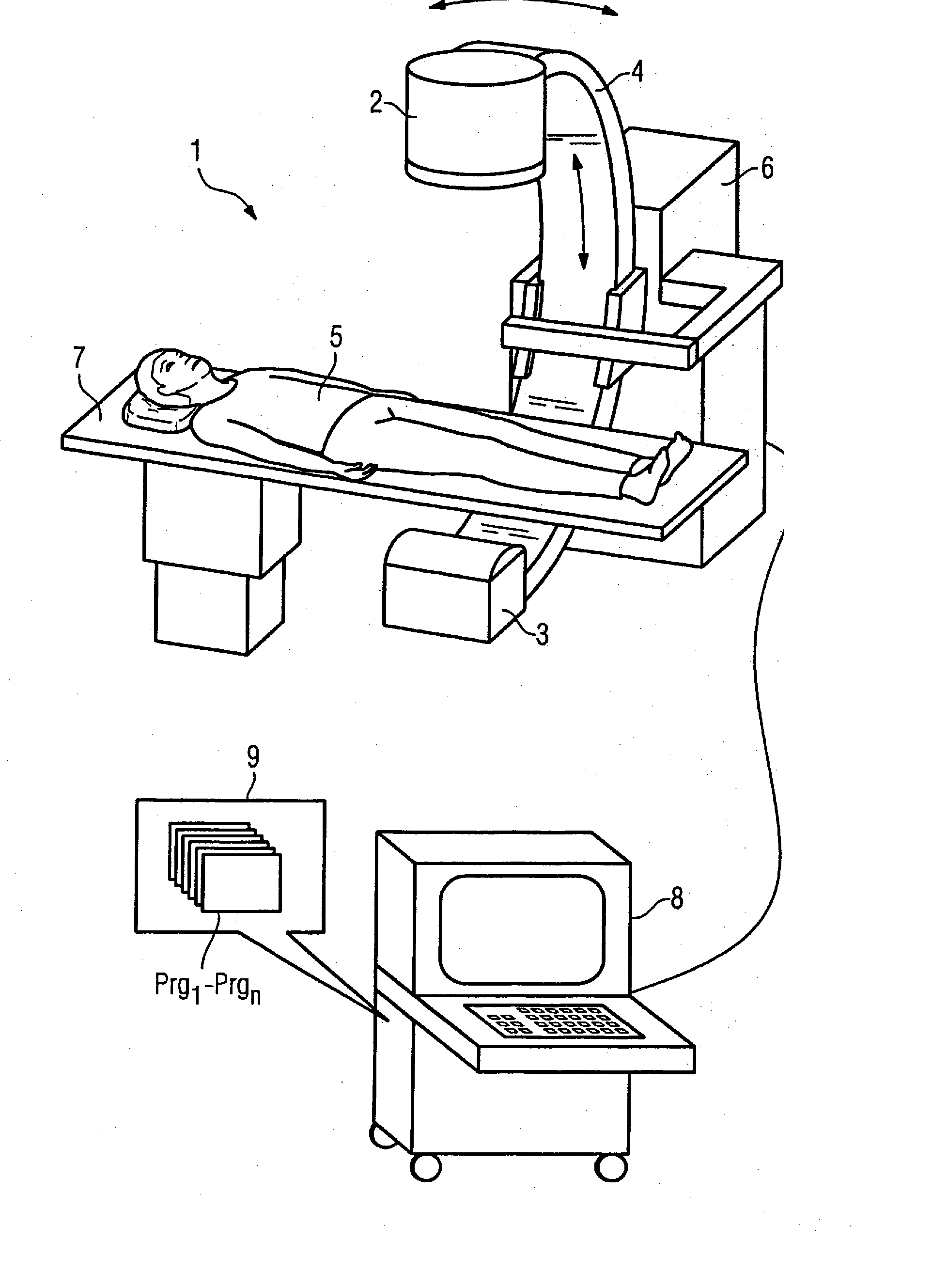
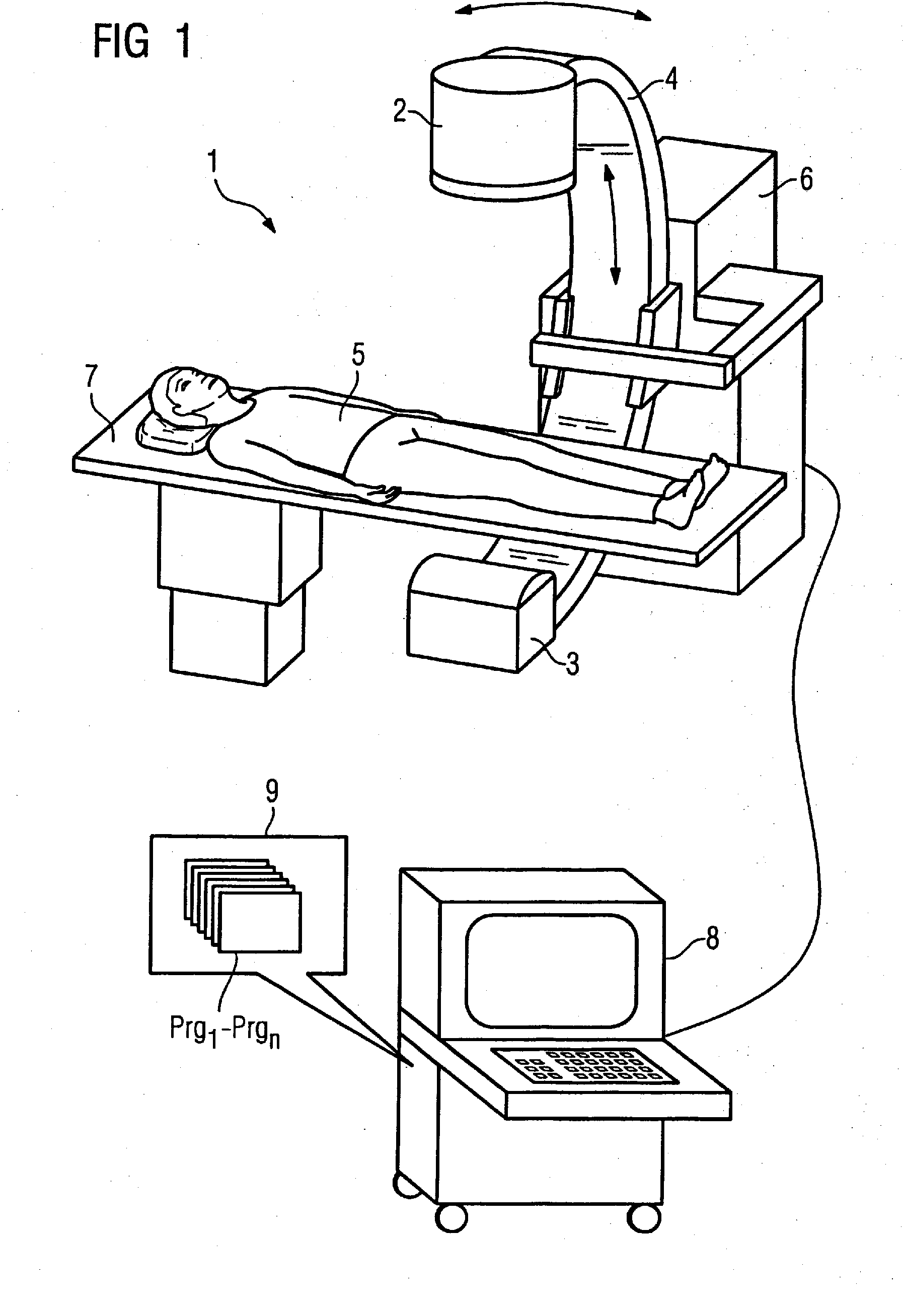
[0145]FIG. 1 illustrates a C-arm system 1 with which projective and tomographical x-ray exposures can be generated. It has a C-arm 4 attached to a housing 6, the C-arm 4 being movable around a patient 5. An x-ray tube 2 is arranged at one end of the C-arm 4 and a flat detector (FPD) 3 is arranged at the other end of the C-arm 4. The patient 5 is located on a movable patient bed 7, such that the patient 5 and a region thereof to be examined can be moved into the measurement field between the x-ray tube 2 and the detector 3 of the C-arm system 1. A control and computation unit 8 with a memory 9 in which corresponding control and image processing programs Prg1-Prgn can run as needed is used for control of the system 1. Program code for implementation of the inventive method is also stored in the memory 9, such that the scatter radiation occurring in the system can be corrected.

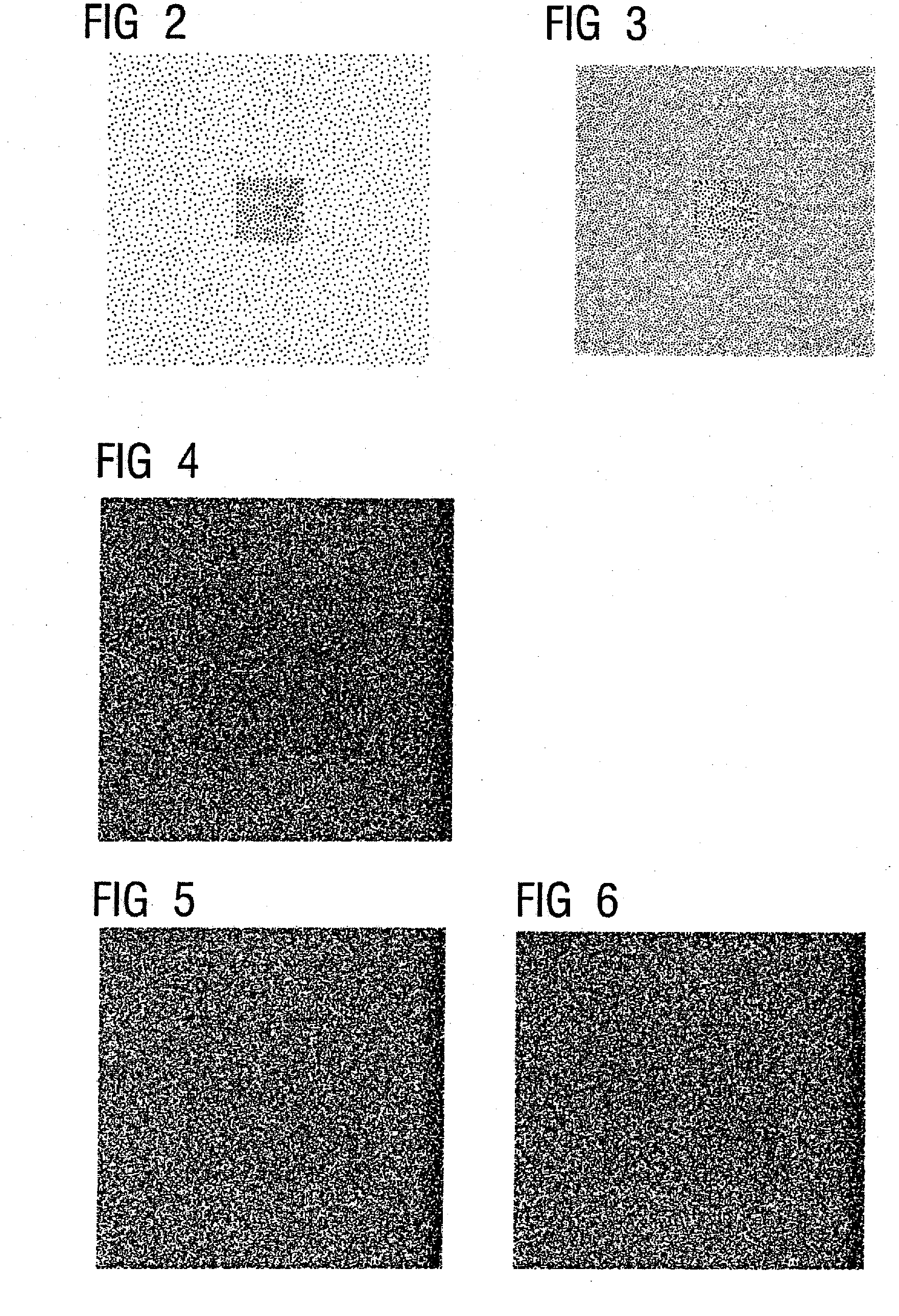
[0146] The following illustrations of FIG. 2-17 are based on simulation calculations, whereby a simulation su...


PUM


Abstract

In a method for correction of scatter radiation errors in radiography and computer tomography, using flat panel detectors, initially an estimation of a scatter radiation distribution Scor(y,z) is undertaken, and a standard correction term δpcor is subsequently calculated. Noise filtering of the standard correction term δpcor with F(δpcor) is implemented and subsequently the noise-filtered standard correction term F(δpcor) is added to the logarithmized measured total projection data ps. The noise filtering can be implemented adaptively dependent on a previously-determined local noise variance. The method can be implemented in a radiography system and / or by a computer for generation and / or processing of projective and / or tomographic exposures, with a memory containing program codes causing the computer in operation to execute the steps of the method.

Description

BACKGROUND OF THE INVENTION [0001] 1. Field of the Invention [0002] The invention concerns a method for correction of scatter radiation fields in radiography and computed tomography with flat panel detectors. [0003] 2. Description of the Prior Art [0004] From the printed document M. Zellerhoff, B. Scholz, E.-P. Rührnschopf, T. Brunner: “Low Contrast 3D Reconstruction from C-Arm Data”, Proceedings of SPIE. Medical Imaging 2005, Vol. 5745, 645-655. It is known that the technology of the flat image detectors (FPD) enables a low-contrast resolution in three-dimensional reconstruction of the tissue density distribution with C-arm computed tomography. Due to the large irradiated patient volume in each projection image, the scatter radiation (which contains only very little image information) represents a significant problem. The effects of the scatter radiation are contrast loss, distortions of the quantitative results (such as cupping and artifacts) and increase of the noise. The use of ...

Claims


Application Information

Patent Timeline
Patent Type & Authority Applications(United States)
IPC IPC(8): A61B6/00
CPCA61B6/027G06T5/002G06T5/003G06T5/20G06T2207/30004G06T2207/10116G06T2207/20012G06T2207/20192G06T2207/10081
Inventor HAERER, WOLFGANGRUEHRNSCHOPF, ERNST-PETER
Owner SIEMENS HEALTHCARE GMBH
Why Eureka
Try Eureka