
Methods To Identify, Prepare, And Use Naive T Cell Recent Thymic Emigrants

a technology of thymic emigrants and t cells, applied in the field of methods to identify, quantify and purify recent thymic emigrants, can solve the problems of difficult sorting and functional characterization of rte, failure to provide convincing data that compared, and limited purity of the isolated population

Inactive Publication Date: 2008-05-15
SEKALY RAFICK PIERRE +4
View PDF0 Cites 7 Cited by
  • Summary
  • Abstract
  • Description
  • Claims
  • Application Information

AI Technical Summary

Benefits of technology

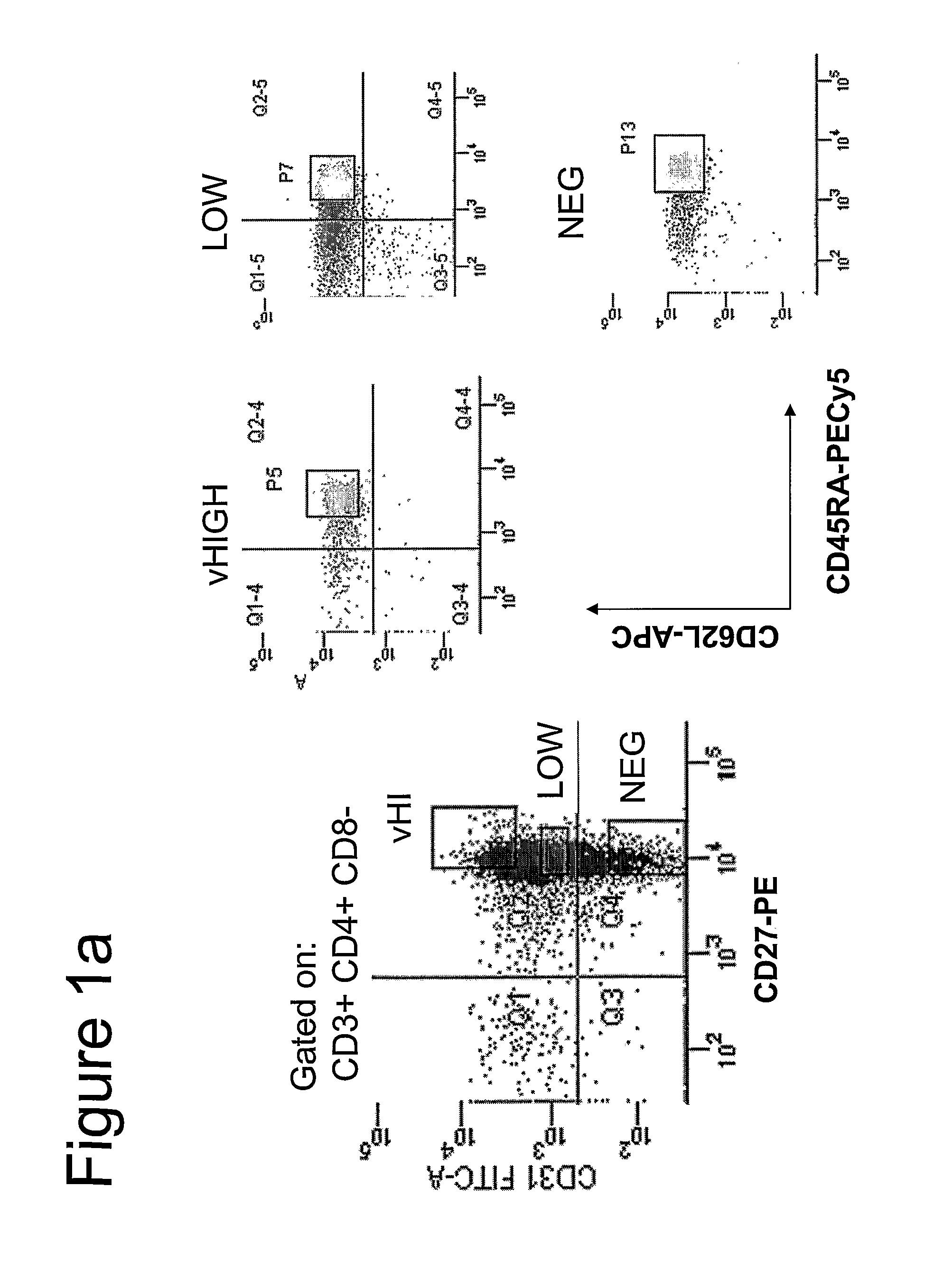
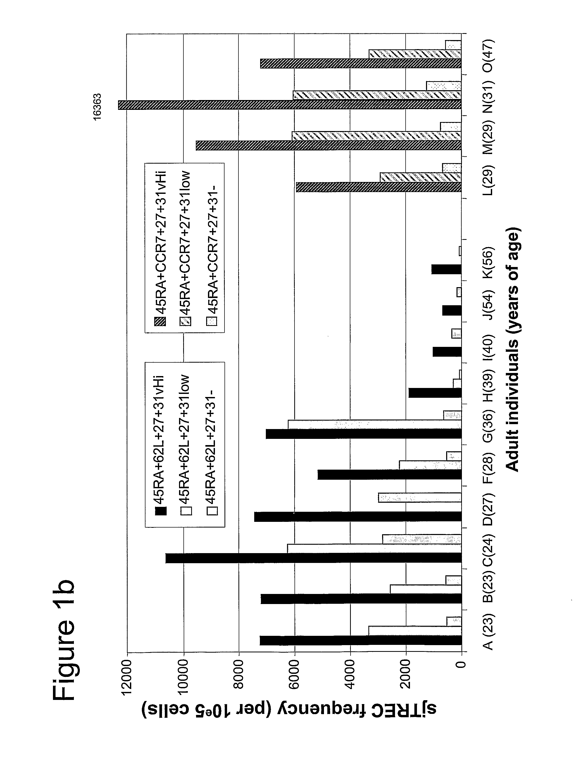
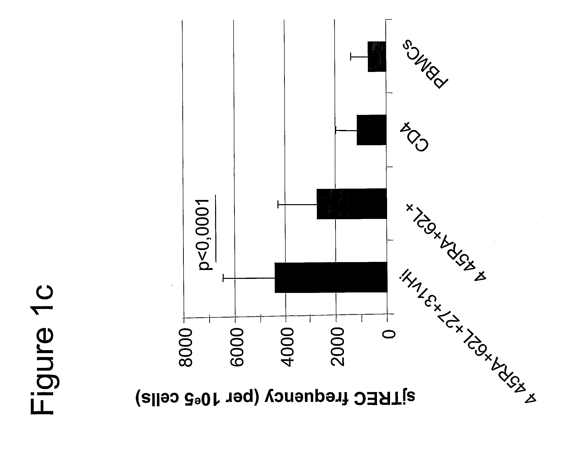
[0032]As used herein the terminology “CD31hi” refers to the phenotype of cells that are at least in the 80% higher percentile of cells in the tested biological sample in terms of CD31+ expression levels at their cell surface (as defined by number of molecules per cell) within “positivity” (as defined below). Preferably these cells are in the 70% higher percentile, more preferably 60%, even more preferably 50%, even more preferably 40%, even more preferably 30%, even more preferably 20% and most preferably 10%. The 80% higher percentile (CD31hi) is the sum of the very high (vHi) and the intermediate expression level of CD31+. The CD31+ expression levels within “positivity” is calculated after having substracted the background. Indeed, in order to determine the level that defines CD31 vHi, intermediate, low and neg, the level of expression is split according to background. Indeed, in the present invention, the cells bear up to 6 fluorochome-coupled monoclonal antibodies. In addition, the array of fluorescence produced may “leak” within the detectors aimed at measuring the level of CD31. This creates a certain background level of fluorescence that is compensated (with standard flow cytometry compensation protocols using the FACSDiVa™ software) but remains present at low levels. With this in mind, a threshold of “positivity” is defined by standard flow cytometry comparison methods (i.e comparison to single stain and “fluorescence-minus-one” controls) as understood by a person of ordinary skill in the art. All cells with a fluorescence level above that threshold are then considered positive for the antigen. Thus, in Examples presented herein, cells in the lowest 50%, namely background levels, are considered negative for CD31 (CD31neg). Within the CD31 positive fraction of cells (all cells with fluorescent levels above background), the level of CD31 expression was sub-defined in three subsets. Firstly, CD31low cells were determined to be within the 20% lowest fluorescent intensity levels of positivity. Secondly, CD31vHi cells were defined to be within the highest 40% in fluorescent intensity level of positivity. The middle 40% is the third intermediate subset. Thus, cells referred to CD31hi naïve CD4 T cells are composed of both CD31vhi and the CD31 intermediate population.
[0033]Other objects, advantages and features of the present invention will become more apparent upon reading of the following non-restrictive description of specific embodiments thereof, given by way of example only with reference to the accompanying drawings.

Problems solved by technology

However, as TRECs are DNA molecules, their PCR-based quantification implies the lysis of cells, rendering sorting and functional characterization of RTE quite difficult.
Indeed, this recent study failed to provide convincing data that compared TREC levels within CD31+ cells to levels found within the late stage thymocytes.
In addition, the many steps used by Kimmig et al. to purify the thymic naive T cells may have introduced biases and limited the purity of the isolated population.

Method used


Image

  • Methods To Identify, Prepare, And Use Naive T Cell Recent Thymic Emigrants
  • Methods To Identify, Prepare, And Use Naive T Cell Recent Thymic Emigrants
  • Methods To Identify, Prepare, And Use Naive T Cell Recent Thymic Emigrants

Examples


example 1

Isolation of PBMCs from Blood Samples

[0044]Blood and leukapheresis samples were obtained from healthy adult donors. Samples were obtained with the informed consent of the subjects and according to the guidelines of the bioethical committee of Centre hospitalier de l'Université de Montreal (CHUM) and McGill University Health Center (MUHC).

[0045]PBMCs were obtained from blood samples using density gradient sedimentation with Ficoll Paque Plus™ (Amersheam Bioscience). Cells were washed twice with PBS 2% FCS before staining with mAbs for flow cytometry.

example 2

Isolation of Thymocytes from Thymus Samples

[0046]Pediatric thymic tissues were sampled from children undergoing elective cardiac surgery from Saint-Justine Hospital as well as CHEO, with the informed consent of the child's parents and according to the guidelines of the bioethical committee of Centre hospitalier de l'Université de Montreal (CHUM) and Research Ethics Board of the Children's Hospital of Eastern Ontario. Following mechanical dissociation of thymic tissue using cell strainers (Falcon), isolated thymocytes were harvested and stained.

example 3

Cell Surface Phenotying and Sorting by Flow Cytometry

[0047]PBMCs. Freshly isolated cells were stained simultaneously with the following mAbs : FITC labeled anti-CD31(BD Pharmingen), PE labeled anti-CD27(BD Pharmingen), PECy5 labeled anti-CD45RA(BD Pharmingen), PECy7 labeled anti-CD3(BD Pharmingen), APC labeled anti-CD62L(BD Pharmingen), APC-Cy7 labeled anti-CD4 (BD Pharmingen) and ECD labeled anti-CD8 (Coulter). In the event of phenotyping, the cells were analysed by 7-color flow cytometry with the FACS LSR II™ (Becton Dickinson) using FACSDiVa™ (Becton Dickinson) software. Previously isolated cells and frozen cells were thawed and stained simultaneously with the following mAbs: PE labeled anti-CD31 (BD Pharmingen), PECy5 labeled anti-CD45RA(BD Pharmingen), PECy7 labeled anti-CCR7 (BD Pharmingen), APC labeled anti-CD3(BD Pharmingen), APC-Cy7 labeled anti-CD27 (BD Pharmingen) Alexa 700 labeled anti-CD4 (BD Pharmingen), and ECD labeled anti-CD8 (Coulter). Seven-color cell sorting was ...


PUM

PropertyMeasurementUnit
timeaaaaaaaaaa
frequencyaaaaaaaaaa
cell adhesionaaaaaaaaaa

Abstract

A method of purifying a subpopulation of naive T cells that have recently emigrated from the thymus, comprising (a) contacting a biological sample susceptible of containing T cells with at least four different ligands, namely ligands directed to the markers CD3, CD4, CD45RA, and CD31; (b) sorting the cells so as to recover T cells having a phenotype comprising at least the following four markers CD3+, CD4+, CD45RA+ and CD31hi, whereby a subpopulation of naive T cells is purified.

Description

CROSS REFERENCE TO RELATED APPLICATIONS[0001]This application claims priority on U.S. provisional application No. 60 / 630,195 filed on Nov. 24, 2004. All documents above are herein incorporated by reference.FIELD OF THE INVENTION[0002]The present invention relates to methods to identify, quantify and purify recent thymic emigrants (RTEs); method to assess thymic function; kits for use in these methods; and purified recent thymic emigrants. More specifically, the present invention is concerned with a more precise phenotype for RTEs.BACKGROUND OF THE INVENTION[0003]The thymus is the main source of T cells replenishing the naive T cell pool with a diverse T cell repertoire. These newly produced cells are exported to the periphery as recent thymic emigrants (RTEs), a sub-compartment of antigen-naive T cells. The exact nature of RTEs remains unclear and determining their phenotype is key in understanding their physiology and overall behavior in pathological settings, such as HIV. In such ...

Claims


Application Information

Patent Timeline
Patent Type & Authority Applications(United States)
IPC IPC(8): A61K35/55G01N33/567C12N5/08A61K35/17C12N5/0783
CPCA61K35/17C12N5/0636G01N2333/70589G01N2333/7051G01N2333/70514G01N33/56972
Inventor SEKALY, RAFICK-PIERREFILION, LIONEL G.DION, MARIE-LISECHEYNIER, REMITHIEBOT, HUGUES
Owner SEKALY RAFICK PIERRE
Learn More