
Multi-aperture single photon emission computed tomography (SPECT) imaging apparatus

a computed tomography and single-photon emission technology, applied in the field of medical imaging, can solve the problems of increasing the design complexity of the nuclear medicine imaging device, the inability to correct geometric misalignment, and the use of relatively more complicated iterative 3d image reconstruction techniques, etc., to achieve the effect of reducing image noise and high detection efficiency

Inactive Publication Date: 2008-05-22
GAMMA MEDICA IDEAS
View PDF5 Cites 23 Cited by
  • Summary
  • Abstract
  • Description
  • Claims
  • Application Information

AI Technical Summary

Benefits of technology

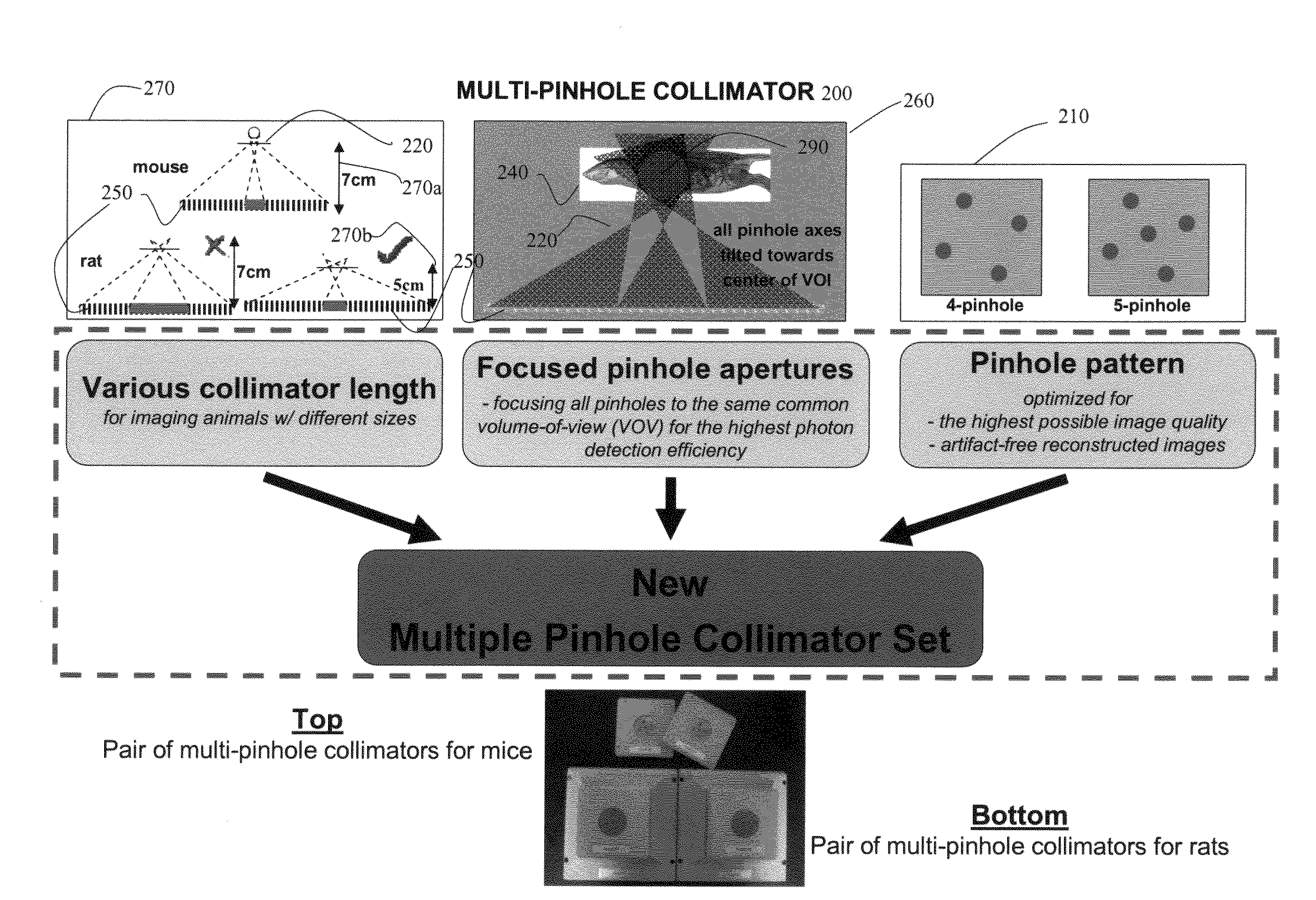
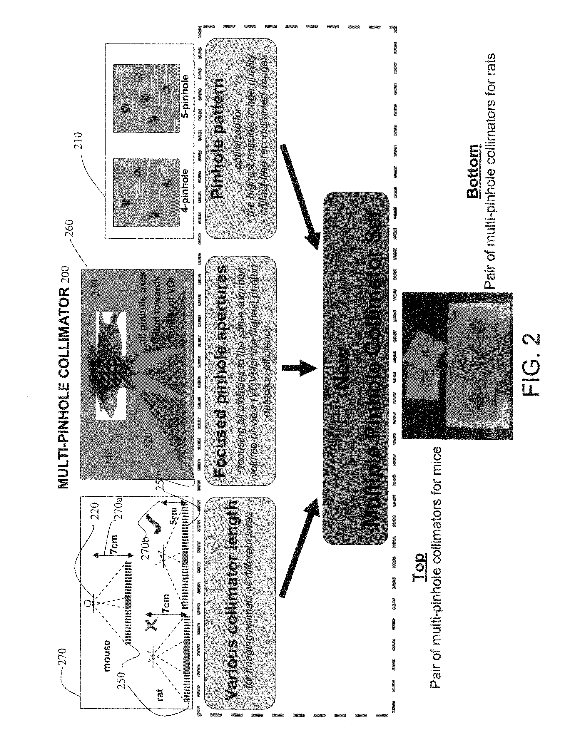
[0011]An aspect of the invention provides a SPECT imaging technique for imaging objects including small animals of different sizes using multi-pinhole (or synthetic) apertures. The imaging technique includes methods, instrumentations, and associated algorithms and software for both high-resolution and high detection efficiency imaging that leads to lower image noise and artifact-free synthetic aperture SPECT images. Also, the technique provides design parameters, hardware settings, and data acquisition methods for optimal imaging of objects with different sizes.

Problems solved by technology

Furthermore, the use of multiple detectors to scan a target may improve the resolution of the scanning by reducing the variance and resulting statistical error produced by a single detector.
However, configuring the multiple detectors and / or multi-hole collimator for precisely scanning throughout a sampling area adds still greater complexity to the design for the nuclear medicine imaging device.
Also, relatively more complicated iterative 3D image reconstruction techniques need to be used with nonstandard pinhole data acquisition geometries versus a standard pinhole SPECT imaging geometries involving the use of a pinhole collimator with a single pinhole aperture, while tomographic projection data are acquired on a planar circular orbit with a flat 2D detector.
Therefore, geometric calibrations and image reconstructions are closely knitted topics in pinhole SPECT imaging, and a reconstruction method implemented without the capability of correcting for geometric misalignments may be of little use in practical molecular imaging studies.
Doing so may potentially speed up the implementation of effective reconstruction algorithms and the investigation of accurate geometric calibration methods for a nonstandard geometry.

Method used


Image

  • Multi-aperture single photon emission computed tomography (SPECT) imaging apparatus
  • Multi-aperture single photon emission computed tomography (SPECT) imaging apparatus
  • Multi-aperture single photon emission computed tomography (SPECT) imaging apparatus

Examples


Embodiment Construction

[0056]In the following detailed description, only certain exemplary embodiments of the present invention are shown and described, by way of illustration. As those skilled in the art would recognize, the described exemplary embodiments may be modified in various ways, all without departing from the spirit or scope of the present invention. Accordingly, the drawings and description are to be regarded as illustrative in nature, and not restrictive.

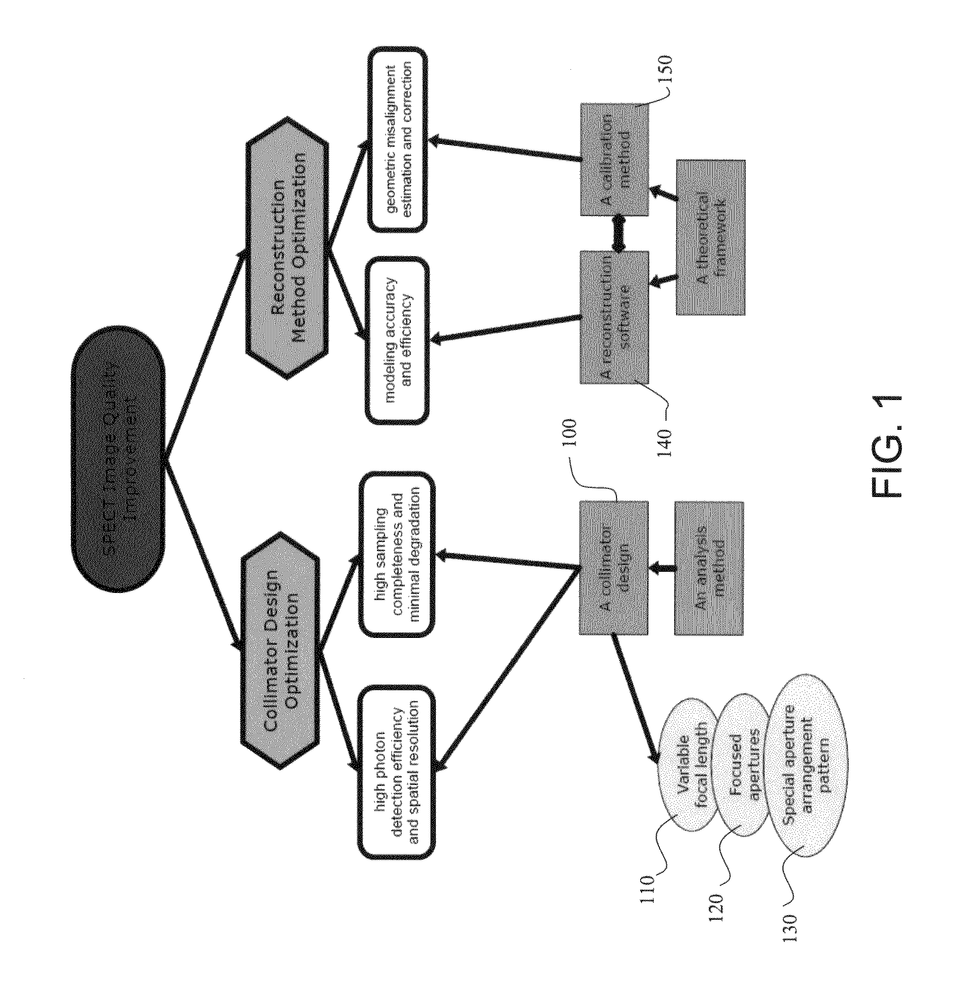
[0057]An embodiment of the invention provides methods, instrumentations, and associated algorithms and software for both high-resolution and high detection efficiency that lead to lower image noise and artifact-free synthetic aperture SPECT images for objects including small animals of different sizes.

[0058]Also, an embodiment of the present invention provides design parameters, hardware settings, and data acquisition methods for optimal imaging of objects with different sizes.

[0059]In one embodiment, a SPECT imaging technique for imaging obj...


PUM


Abstract

Methods and systems for improving image quality of single photon nuclear imaging systems, such as single photon emission computed tomography (SPECT) systems for imaging of an object under study, such as small objects including small animals of different sizes using synthetic apertures. The methods and systems include processes and instrumentations for high-resolution, high detection efficiency leading to lower image noise and artifact-free synthetic aperture single photon nuclear images, such as SPECT images. Also, the method and systems provide design parameters, hardware settings, and data acquisition processes for optimal imaging of objects having different sizes.

Description

CROSS-REFERENCE TO RELATED APPLICATION[0001]This application claims priority to and the benefit of U.S. Provisional Application No. 60 / 859,585, filed Nov. 17, 2006, the entire content of which is incorporated herein by reference.FIELD OF THE INVENTION[0002]The invention relates generally to medical imaging. More particularly, the invention relates to methods and systems for improving image quality of single photon nuclear imaging systems, such as single photon emission computed tomography (SPECT) systems.BACKGROUND OF THE INVENTION[0003]Single photon emission computed tomography (SPECT) is a nuclear medicine tomographic imaging technique that has traditionally used homogeneous area radiation detectors to detect gamma ray emissions. Conventionally, this imaging technique accumulates counts of gamma photons that are absorbed by a scintillation crystal. The crystal scintillates in response to interaction with gamma radiation to produce a flash of light. Photomultiplier tubes (PMTs) beh...

Claims


Application Information

Patent Timeline
IPC IPC(8): G01T1/24
CPCG01T1/249G01T1/1648
Inventor WAGENAAR, DOUGLAS J.PATT, BRADLEY E.TSUI, BENJAMIN M.W.
Owner GAMMA MEDICA IDEAS
Learn More