

Analyzing lesions in a medical digital image
a digital image and lesion technology, applied in the field of digital imaging, can solve the problems of difficult segmentation of voxels in a thoracic ct image, difficulty in distinguishing between the lesion and other normal anatomy structures, and difficulty in distinguishing between abnormal lesion tissue and normal pulmonary structure tissu
- Summary
- Abstract
- Description
- Claims
- Application Information
AI Technical Summary
Benefits of technology
Problems solved by technology
Method used
Image

Examples
Embodiment Construction
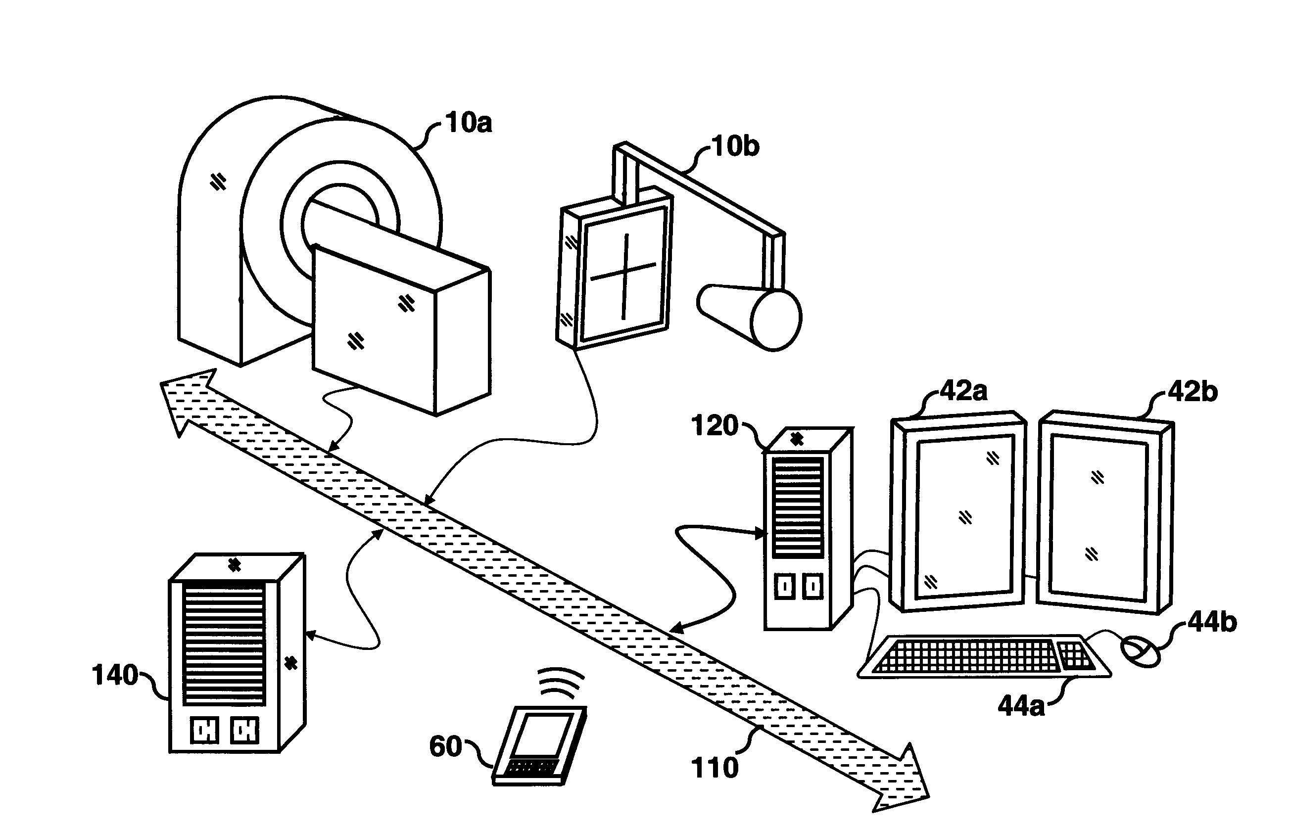
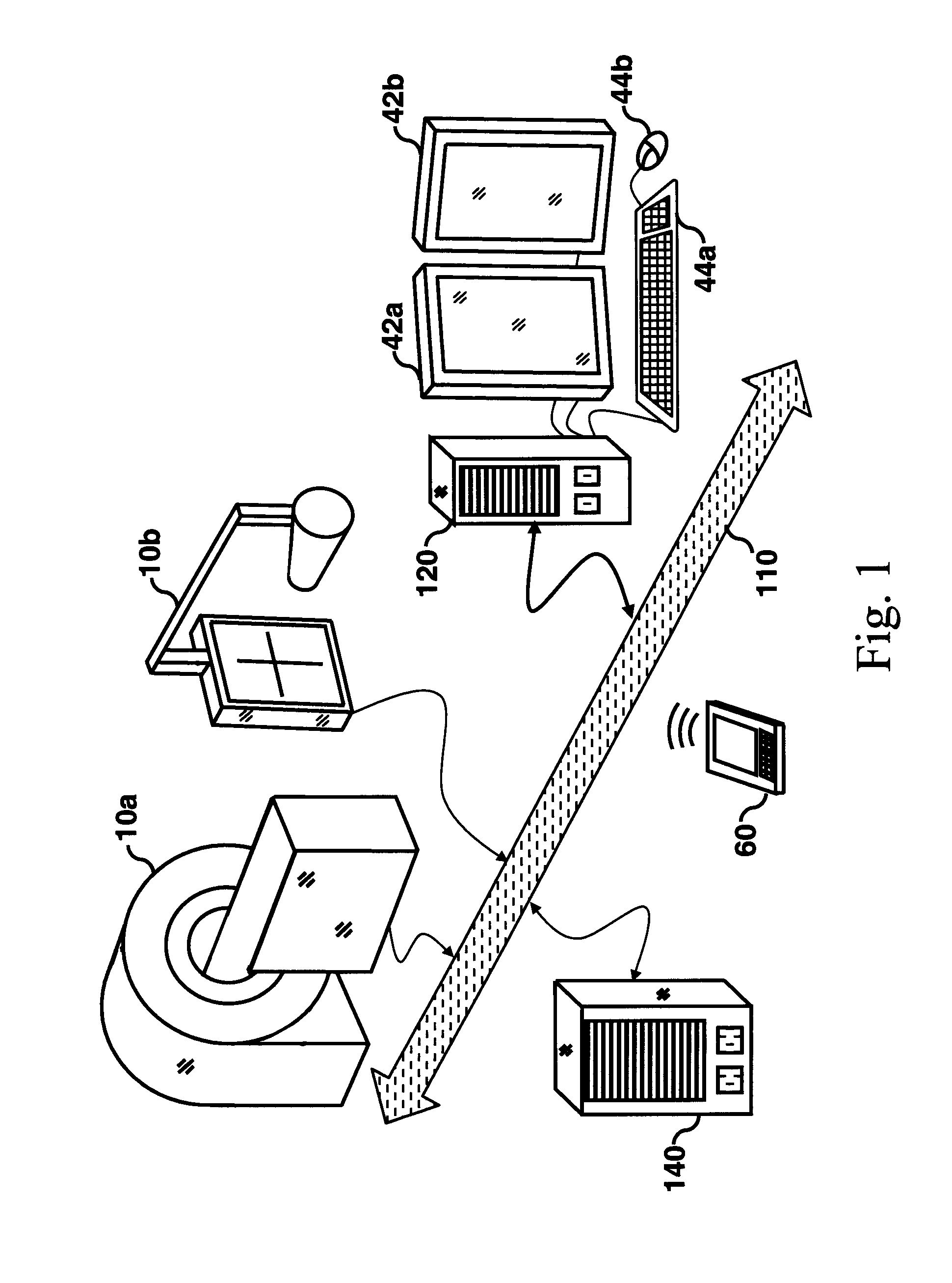
[0025]Many medical imaging applications are implemented via a picture archiving and communications systems (PACS). These systems provide a way for displaying digital images acquired by a wide variety of medical imaging modalities such as, but not limited to, projection radiography (x-ray images), computed tomography (CT images), ultrasound (US images), and magnetic resonance (MR images). Each of the above mentioned medical imaging modalities contain slightly different diagnostic information. In particular, CT and MR images when viewed and studied by radiologist, can reveal much detail about a patient's 3-dimensional internal anatomy. Computer algorithm technology can also be applied to medical images to enhance the rendering of the diagnostic information, to detect an abnormal condition, i.e., computer aided detection (CAD), and to make measurements relating to the patient's condition, i.e., computer aided measurement (CAM).
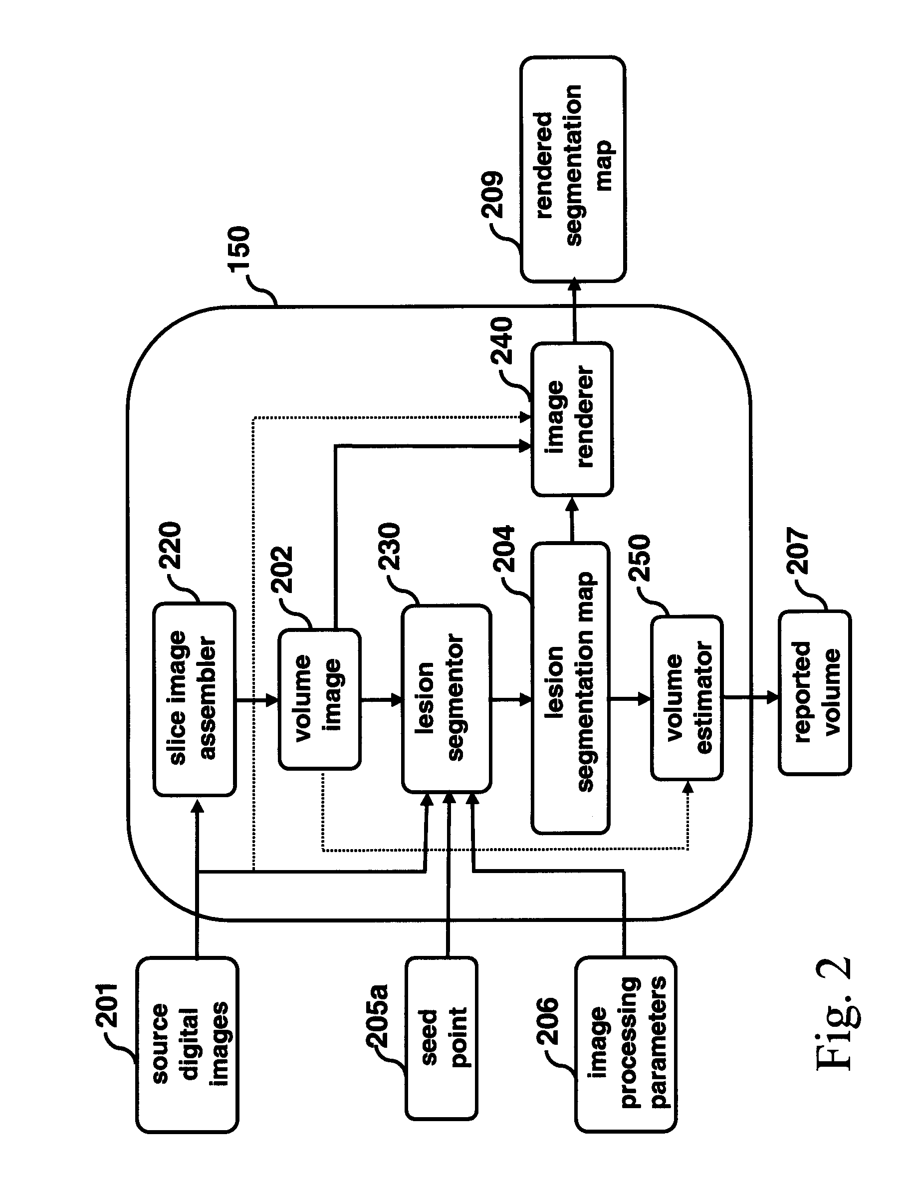
[0026]The present invention represents an algorithmic compu...
PUM
 Abstract
Description
Claims
Application Information