

System, method and apparatus for cancer imaging
a cancer imaging and system technology, applied in tomography, medical science, diagnostics, etc., can solve problems such as inacceptable sensitivity to small cancers
- Summary
- Abstract
- Description
- Claims
- Application Information
AI Technical Summary
Problems solved by technology
Method used
Image

Examples
Embodiment Construction
[0014]In the following detailed description, numerous specific details are set forth in order to provide a thorough understanding of embodiments. However it will be understood by those of ordinary skill in the art that the embodiments may be practiced without these specific details. In other instances, well-known methods, procedures, components and circuits have not been described in detail so as not to obscure the embodiments.
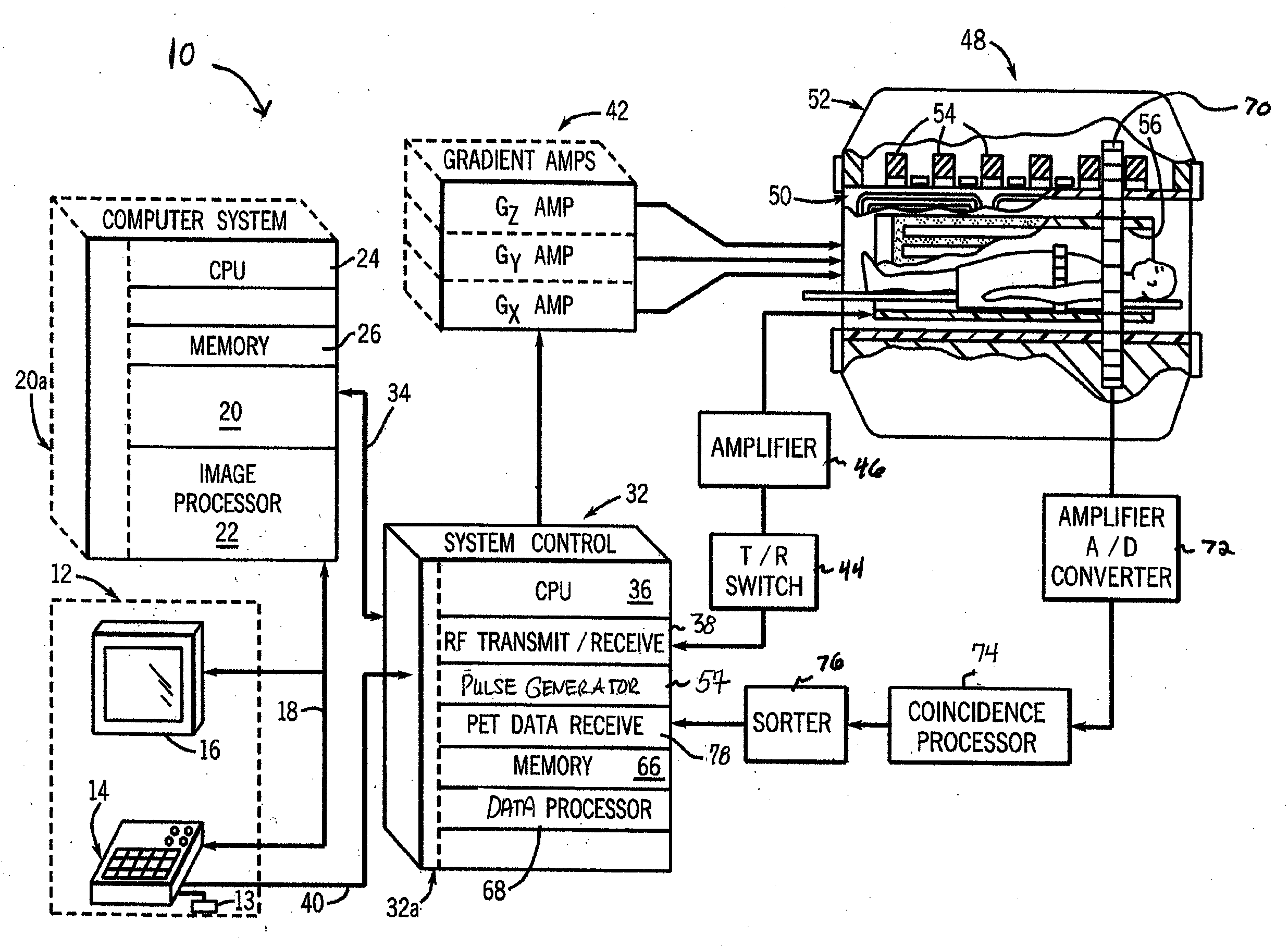
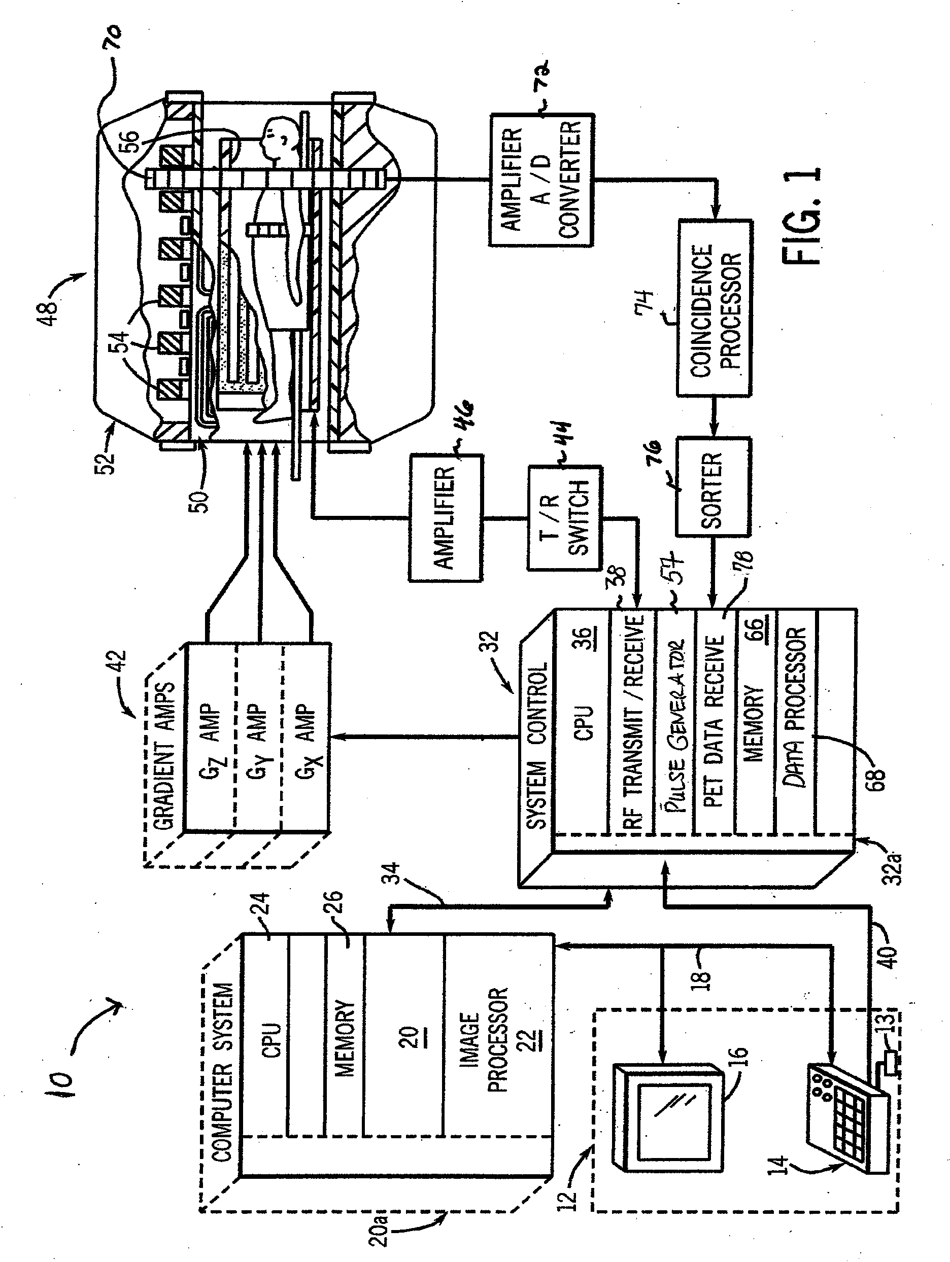
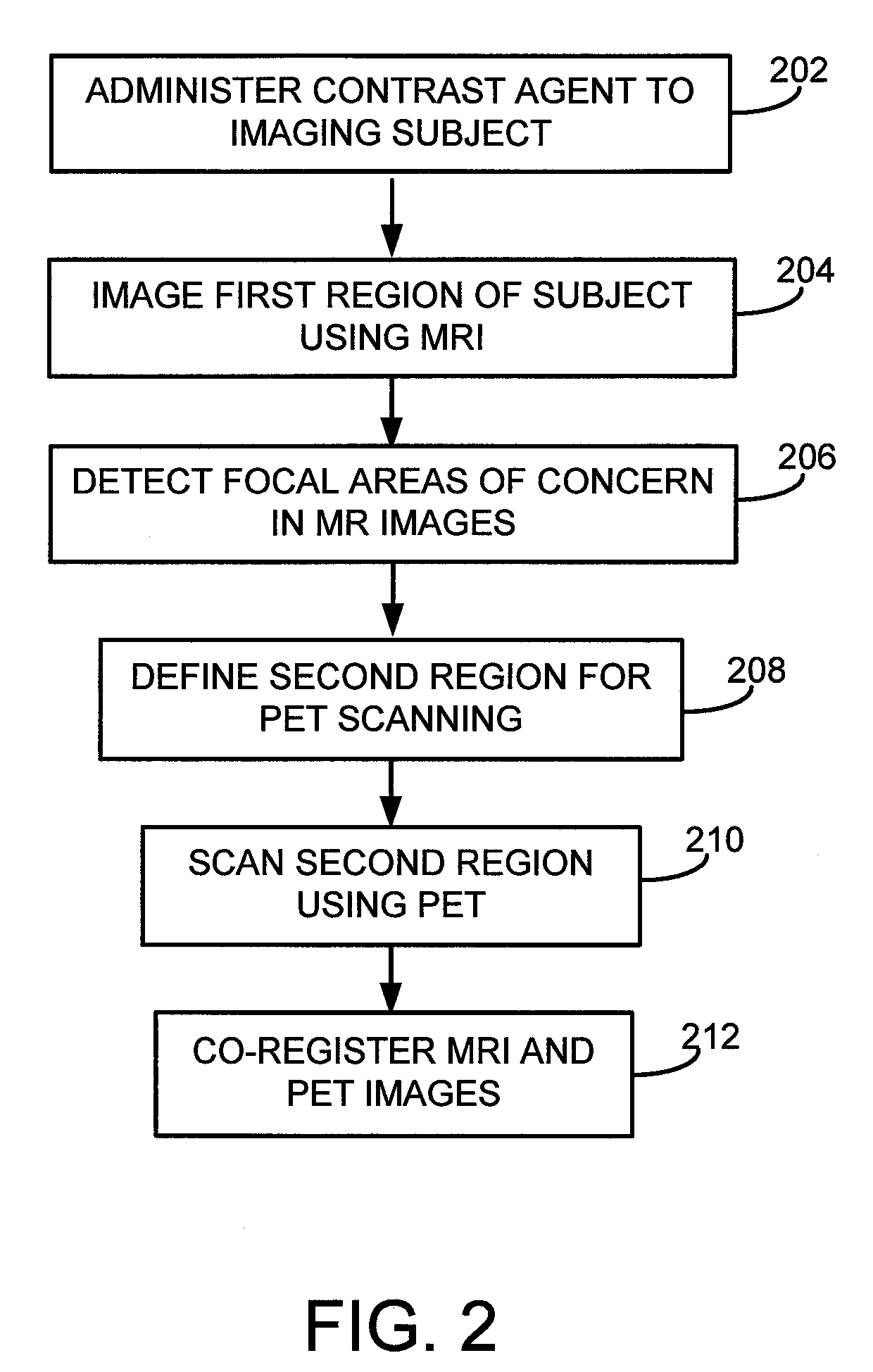
[0015]A combined PET-MRI system may be used for cancer imaging to take advantage of performance characteristics for both PET and MRI in the context of cancer imaging to increase overall imaging effectiveness. MRI may first be used to assess a large area of the body using MR imaging protocols that have high detection sensitivity for cancer. After identifying localized areas of concern on the MR images, PET may then be used to scan a more limited volume encompassing the areas of concern. PET provides metabolic information about the tissue in these smaller region...
PUM
 Abstract
Description
Claims
Application Information