Method for implanting a cardiac implant with real-time ultrasound imaging guidance

a cardiac implant and real-time ultrasound technology, applied in the field of combined ultrasound and computed tomography (ct) image acquisition, can solve the problems of blindly implantation, inability to provide exact guidance in three-dimensional space, and the necessity of exposing the patient to high radiation

Inactive Publication Date: 2008-06-19
SIEMENS AG
View PDF8 Cites 28 Cited by
  • Summary
  • Abstract
  • Description
  • Claims
  • Application Information

AI Technical Summary

Benefits of technology

[0011]An objective of the present invention is to provide a method and apparatus for ultrasound imaging that are suitable for guiding the ...

Problems solved by technology

A primary difficulty with percutaneous implantation of a device within the patient's body, however, is that the implantation must proceed “blindly” because the physician does not have direct visual access to the implantation site or the implantation path to the implantation site.
This information, however, is only in the form of a 2D projection, and thus exact guidance in three-dimensional space is not provided.
Another d...

Method used


Image

  • Method for implanting a cardiac implant with real-time ultrasound imaging guidance
  • Method for implanting a cardiac implant with real-time ultrasound imaging guidance
  • Method for implanting a cardiac implant with real-time ultrasound imaging guidance

Examples


Embodiment Construction

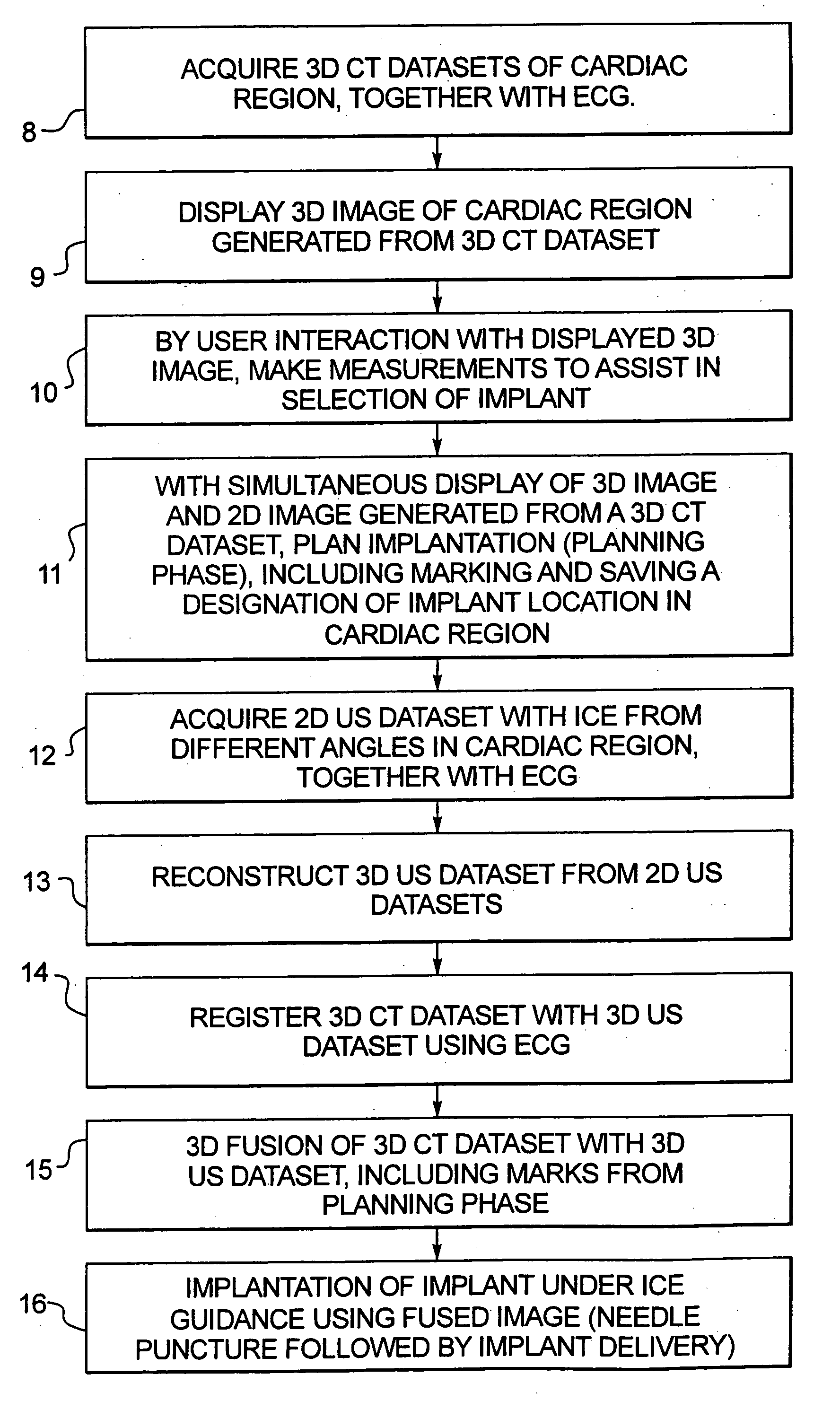
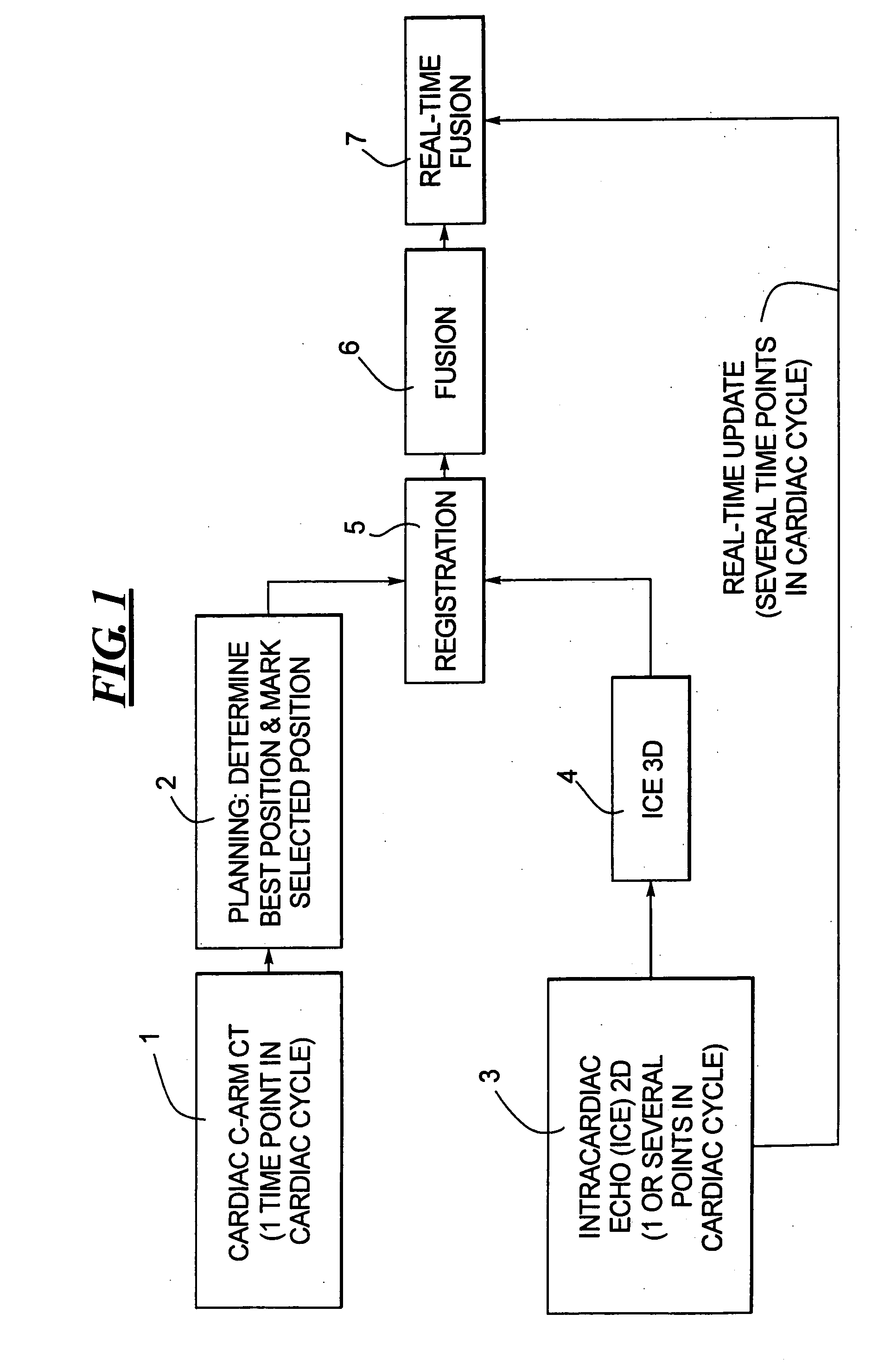
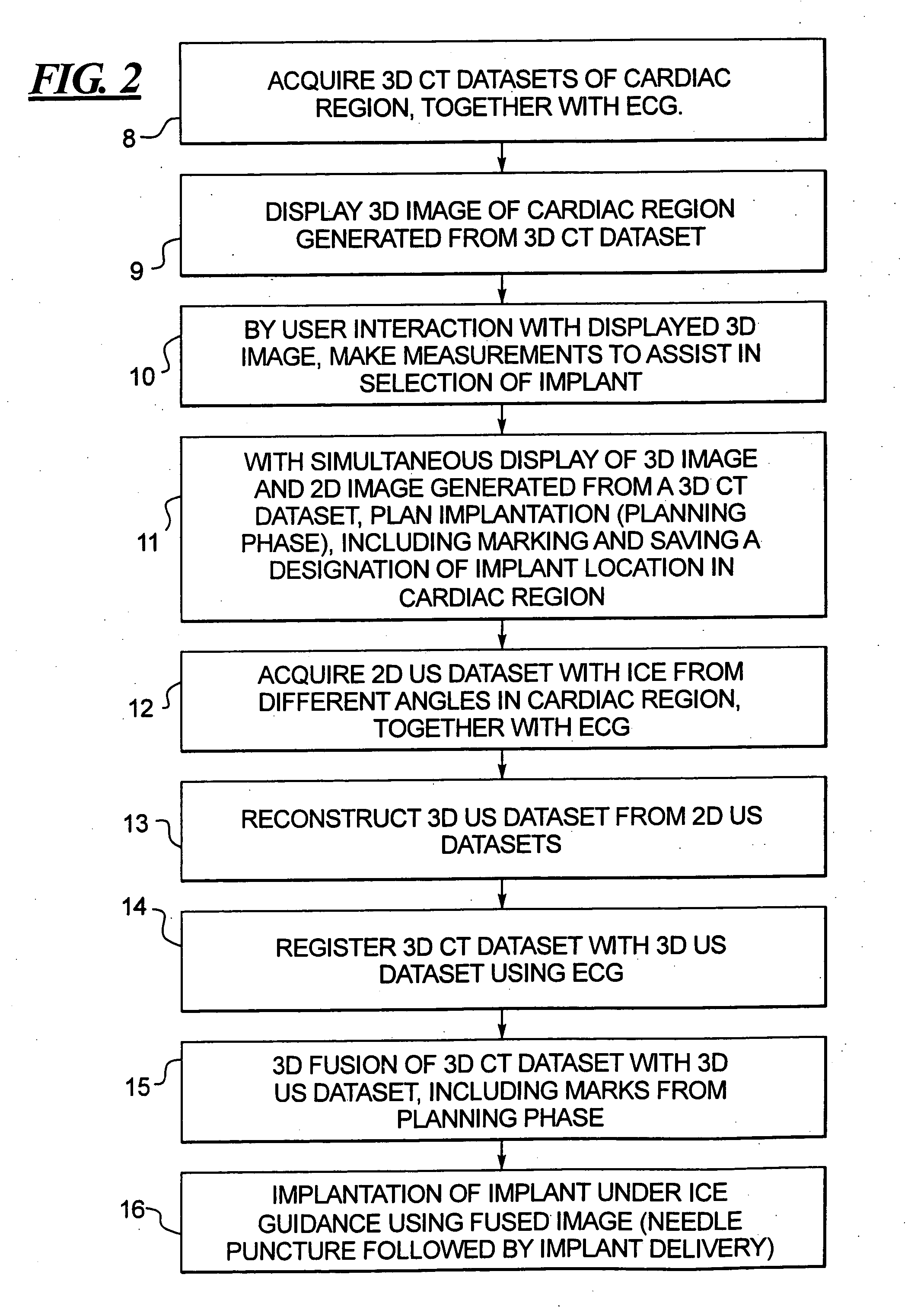
[0019]In the schematic workflow diagram shown in FIG. 1 for illustrating the inventive procedure, computed tomography (CT) data are acquired of the cardiac region of a patient at one point in time of the cardiac cycle. The CT data can be acquired using a C-arm-CT system, which is commercially available from Siemens Medical Solutions. Such a C-arm-CT system delivers CT-like images during angiography and treatment planning procedures.

[0020]The image represented by the CT data encompasses the structures of interest, such as a particular heart chamber and the coronary vein. The CT data are acquired together with ECG signals from the patient. It may be necessary to acquire two or more sets of such CT data in order to display the complete anatomy of interest. If so, all of the acquired CT datasets are then fused together to represent the structures of interest in one 3D CT dataset.

[0021]Block 2 in FIG. 1 schematically illustrates the planning phase wherein, based on the displayed 3D datas...


PUM


Abstract

In a method for implanting a cardiac implant, a 3D CT dataset of a cardiac region of interest at which an implant is to be implanted, is displayed and the implantation procedure is planned, which includes the physician electronically marking a best implantation site in the displayed image. This marking is then included in the 3D CT dataset. A 3D ultrasound dataset of the region of interest is acquired, and is brought into registration with the 3D CT dataset that incorporates the marking, and a fused image is produced therefrom. The fused image is displayed during the implantation procedure, and is updated with multiple real-time 2D ultrasound images obtained using the catheter that is employed to deliver the implant to the implantation site.

Description

BACKGROUND OF THE INVENTION[0001]1. Field of the Invention[0002]The present invention concerns a method and an apparatus for combined ultrasound and computed tomography (CT) image acquisition, particularly in the field of medicine for assisting in procedures conducted in the cath lab.[0003]2. Description of the Prior Art[0004]Implantable devices have recently been developed to improve perfusion of the heart. One such device is called the VPass implant and is commercially available from Percardia, Inc. This device is a stent-like device that is placed within the myocardial wall to create a tunnel between the left ventricle and a coronary vein.[0005]Devices of this type can be implanted percutaneously in the cath lab, so that no open surgery is required. Percutaneous procedures provide many advantages, such as less trauma to the patient's body (less invasive) and a high cost reduction (shorter hospital stay, less medical personnel required), while providing the same benefit to the pat...

Claims


Application Information

Patent Timeline
IPC IPC(8): A61B8/00
CPCA61B6/03A61B6/503A61B6/504A61B6/5247A61B8/0883A61B8/0891A61B8/12A61B8/4416A61B8/483A61B8/0841
Inventor CAMUS, ESTELLEOSTERMEIER, MARTIN
Owner SIEMENS AG
Why Eureka
Try Eureka