Intraoperative electromagnetic apparatus and related technology

Inactive Publication Date: 2008-07-17
THE UNIV HOSPITAL OF NORTH STAFFORDSHIRE NHS TRUST
View PDF14 Cites 3 Cited by
  • Summary
  • Abstract
  • Description
  • Claims
  • Application Information

AI Technical Summary

Problems solved by technology

There are currently no widely available effective intraoperative diagnostic modalities that can reliably inform and guide the surgeon in accurately evaluating tissue status at operation.
Conventional surgical practice is to examine frozen sections removed during surgery, but this typically causes unnecessary operation time delays.
These practices commonly result in removal of more healthy tissue than is necessary, with possible subsequent postoperative structural deformity e.g. in breast, and unnecessary reduction in residual organ function e.g. in lung.
Frozen section analysis which is routinely used in breast tumour surgery to determine diagnosis and extent of resection can be difficult to perform and may provide

Method used


Image

  • Intraoperative electromagnetic apparatus and related technology
  • Intraoperative electromagnetic apparatus and related technology
  • Intraoperative electromagnetic apparatus and related technology

Examples


Embodiment Construction

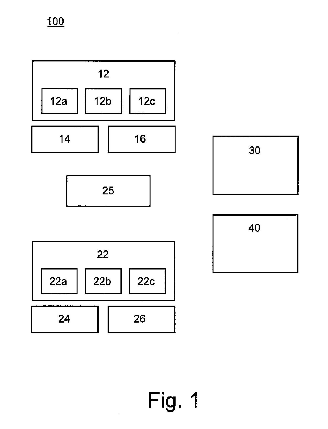
[0025]FIG. 1 shows a schematic block diagram of an apparatus 100 for evaluating physical and biophysical characteristics of a tissue portion according to a first exemplary embodiment of the present invention. The apparatus 100 comprises first and second electromagnetic signal generating units 12,22, first and a second electromagnetic signal detecting units 14,24, and first and second electromagnetic signal analysing units 16,26.

[0026]The first electromagnetic signal generating unit 12 comprises a source of electromagnetic radiation of a first portion of the electromagnetic spectrum, and the first electromagnetic signal detecting unit 14 is operable to detect electromagnetic radiation of the first portion of the electromagnetic spectrum, such as radiation returned from a tissue portion after such radiation has been applied to the tissue portion from the first electromagnetic signal generating unit 12. The first electromagnetic signal analysing unit 16 is operable to give a first eval...


PUM


Abstract

An apparatus capable of applying a first electromagnetic signal, e.g. an optical signal, to a tissue portion is described. The apparatus is also capable of applying a second electromagnetic signal from a different portion of the electromagnetic spectrum, e.g. a microwave signal, to the tissue portion. Signals returned from the tissue portion and related to the first electromagnetic signal are analysed to give a first evaluation of the physical and biophysical characteristics of the tissue portion. Signals returned from the tissue portion and related to the second electromagnetic signal are also analysed, and a final evaluation of the physical and biophysical characteristics of the tissue portion is produced, informed by the first evaluation and the analysed second signal.

Description

[0001]The present invention relates to methods and apparatus for of evaluating physical and biophysical characteristics of a tissue portion, to methods of determining disease state in a tissue portion, to methods of reconstructing a representation of a tissue portion, and to methods of treating a diseased part of a tissue portion, said apparatus and methods suitable for intraoperative diagnosis and treatment relating to tumour resections and general surgery on in vivo and ex vivo tissue portions.BACKGROUND OF THE INVENTION [0002]Healthcare treatment spending continues to rise worldwide as providers invest in better diagnostic technologies and more efficacious therapeutic strategies to enhance disease prognosis and survival rates, whilst reducing adverse incidents.[0003]In the field of healthcare there remains a need for healthcare professionals to obtain real time or near-real time confirmation of the patho-biological status of a particular target tissue, for example a surgeon may n...

Claims


Application Information

Patent Timeline
IPC IPC(8): A61B5/05A61N1/00
CPCA61B5/0084A61B5/0091A61B5/0507A61B18/20A61B5/418A61B5/4312A61B18/18A61B5/415A61B5/0059A61B5/05G01N21/474G01N21/49G01N22/00G01N33/483
Inventor SPITERI, MONICA SILVERSTONESEMENOV, SERGUEI YURIEVICH
Owner THE UNIV HOSPITAL OF NORTH STAFFORDSHIRE NHS TRUST
Why Eureka
Try Eureka