

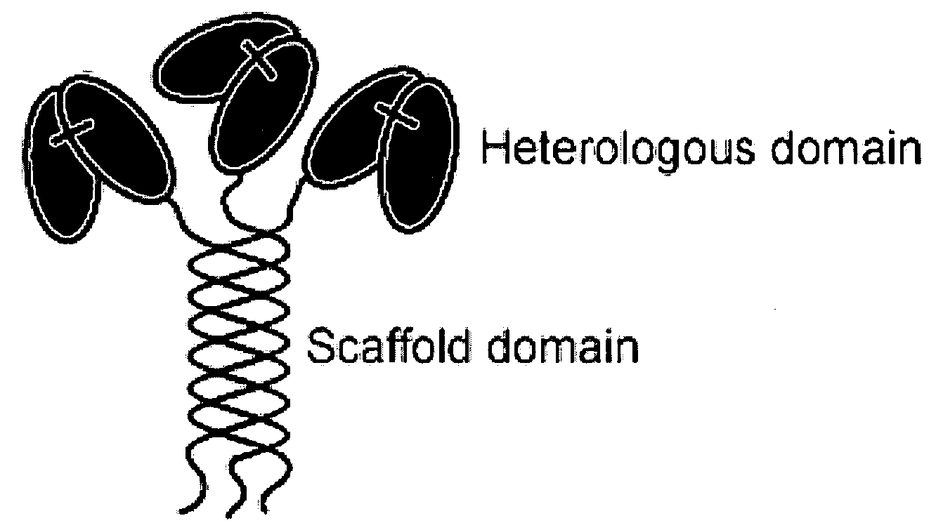
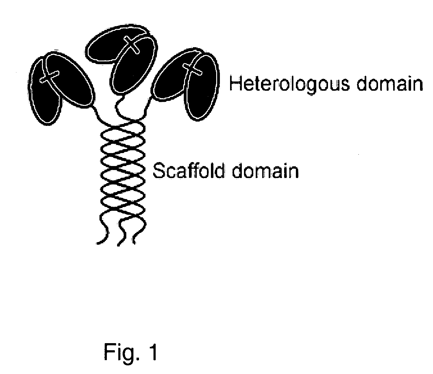
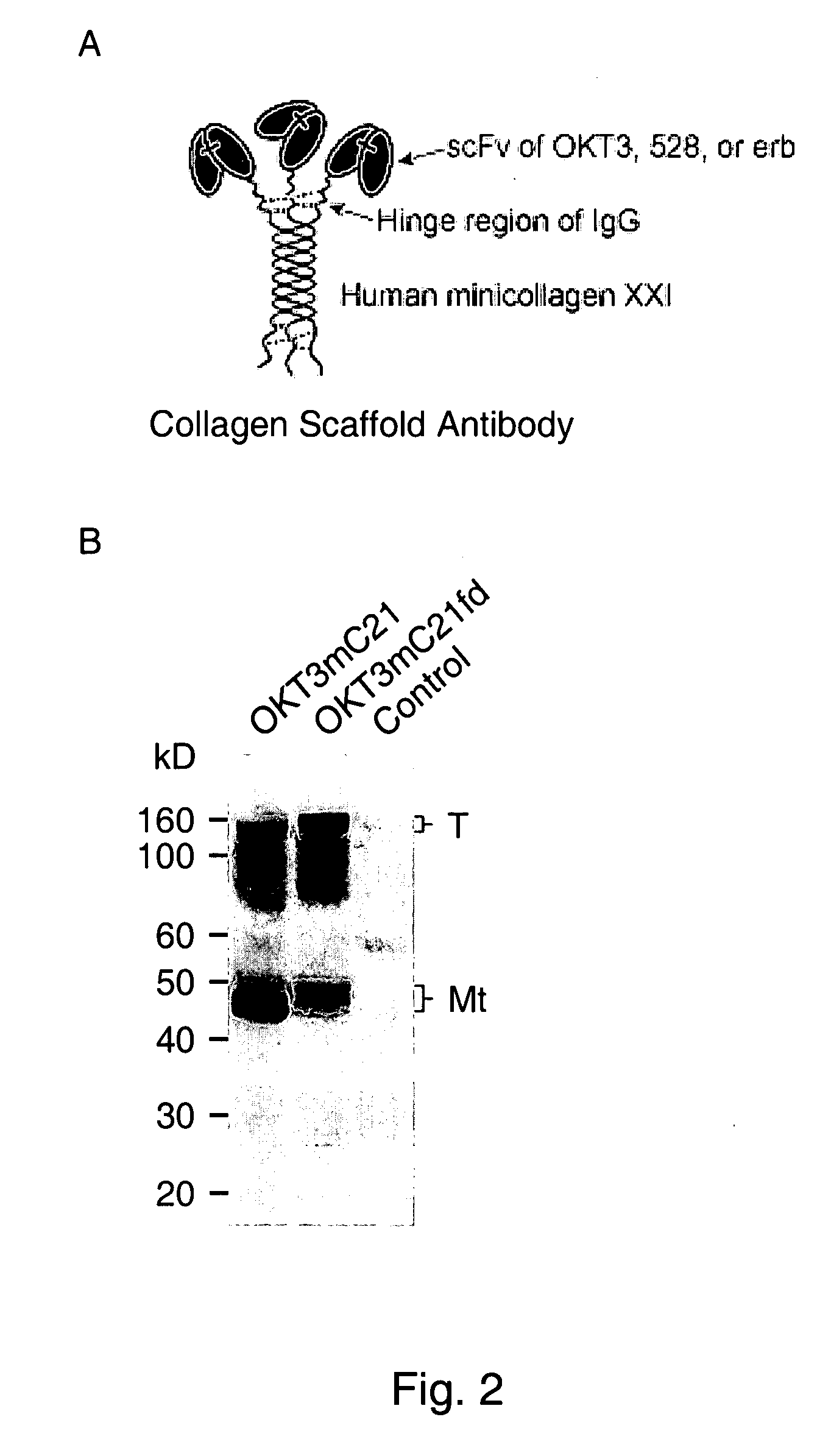
Trimeric collagen scaffold antibodies
a scaffold antibody and collagen technology, applied in the field oftrimeric collagen scaffold antibodies, can solve the problems of limited use of the approaches described above, inability to use conventional and engineered igg for simultaneous binding to more than two different antigens, and severely limit potential therapeutic applications, etc., to achieve low mitogenic effect, high avidity, and high in vivo stability
- Summary
- Abstract
- Description
- Claims
- Application Information
AI Technical Summary
Benefits of technology
Problems solved by technology
Method used
Image

Examples
example 1
Selection of Phage Library for Anti-EGFR Antibody Domain
[0165]An erb phagemid containing an epidermal growth factor receptor extracellular domain (EGFR-ECD)-binding variable fragment (scFv) was isolated by screening a human single fold scFv phage display libraries (Tomlinson I+J; kindly provided by I. M. Tomlinson and G. Winter, MRC Laboratory of Molecular Biology, Cambridge, UK). Selections were performed using immunotubes (Maxisorp; Nunc, Roskilde, Denmark) coated with 10 μg of purified recombinant extracellular domain of EGF receptor (EGFR-ECD; Research Diagnostics, Inc.). Blocking, panning, washing, elution, and reamplification of eluted phage were carried out according to the manufacturer's protocol. A numbers of clones were identified. After confirmation by Western Blotting and ELISA, one clone, erb_scFv, was selected for further experiment. The cDNA encoding erb_scFv was obtained and ligated into an expression vector by a standard method. Listed below are the polypeptide sequ...
example 2
CD3 Antibody Domain
[0167]RT-PCR was conducted to obtain from hybridoma cell lines cDNAs encoding the heavy chain variable region (VH) and light chain variable region (VL) of anti-CD3 monoclonal antibody OKT3. Then, the two cDNAs were ligated to generate a fusion sequence that encodes a fusion protein of VH-VL of OKT3. Listed below are the polypeptide sequence of this fusion protein (SEQ ID NO: 3) and the cDNA sequence encoding it (SEQ ID NO: 4).
SEQ ID NO: 3ValGlnLeuGlnGlnSerGlyAlaGluLeuAlaArgProGlyAlaSerValLysMetSerCysLysAlaSerGlyTyrThrPheThrArgTyrThrMetHisTrpValLysGlnArgProGlyGlnGlyLeuGluTrpIleGlyTyrIleAsnProSerArgGlyTyrThrAsnTyrAsnGlnLysPheLysAspLysAlaThrLeuThrThrAspLysSerSerSerThrAlaTyrMetGlnLeUSerSerLeuThrSerGluAspSerAlaValTyrTyrCysAlaArgTyrTyrAspAspHisTyrCysLeuAspTyrTrpGlyGlnGlyThrThrValThrValSerSerGlyGlyGlyGlySerGlyGlyGlyGlySerGlyGlyGlyGlySerAspIleValLeuThrGlnSerProAlaIleMetSerAlaSerProGlyGluLysValThrMetThrCysSerAlaSerSerSerValSerTyrMetAsnTrpTyrGlnGlnLysSerGlyThrSerProLysArgTr...
example 3
EGFR Antibody Domain
[0168]The same procedures were performed to obtain cDNAs encoding VH and VL of anti-EGFR monoclonal antibody 528 and a fusion sequence that encodes a fusion protein of VH-VL of the anti-EGFR 528. The 528 monoclonal antibody binds to EGFR on cellular membranes, e.g., on human epidermoid carcinoma A431 cells. The polypeptide sequence of this 528 single-chain antibody (SEQ ID NO: 5) and the cDNA sequence encoding it (SEQ ID NO: 6) are listed below.
SEQ ID NO: 5ValLysLeuGlnGIuSerGlySerGIuMetAlaArgProGlyAlaSerValLysLeuProCysLysAlaSerGlyAspThrPheThrSerTyrTrpMetHisTrpVaILysGlnArgHisGlyHisGlyProGluTrpIleGlyAsnIleTyrProGlySerGlyGlyThrAsnTyrAlaGluLysPheLysAsnLysValThrLeuThrValAspArgSerSerArgThrValTyrMetHisLeuSerArgLeuThrSerGluAspPheAlaVaITyrTyrCysThrArgSerGlyGlyProTyrPhePheAspTyrTrpGlyGlnGlyThrThrValThrValSerSerGlyGlyGlyGlySerGlyGlyGlyGlySerGlyGlyGlyGlySerMetThrGlnThrProLeuSerLeuProValSerLeuGlyAspGlnAlaSerIleSerCysArgSerSerGlnAsnIleVaIHisAsnAsnGlyIleThrTyrLeuGluTrpTyrLeuGln...
PUM
| Property | Measurement | Unit |
|---|---|---|
| Angle | aaaaa | aaaaa |
| Solubility (mass) | aaaaa | aaaaa |
| Fluorescence | aaaaa | aaaaa |
Abstract
Description
Claims
Application Information