Device and software for screening the skin

a skin and skin technology, applied in the field of biomedical screening devices and dermoscopy, can solve the problems of difficult processing of acquired pictures, high cost of treatment, and near fatal consequences

Active Publication Date: 2008-09-18
UNIV HOUSTON SYST +1
View PDF10 Cites 71 Cited by
  • Summary
  • Abstract
  • Description
  • Claims
  • Application Information

AI Technical Summary

Benefits of technology

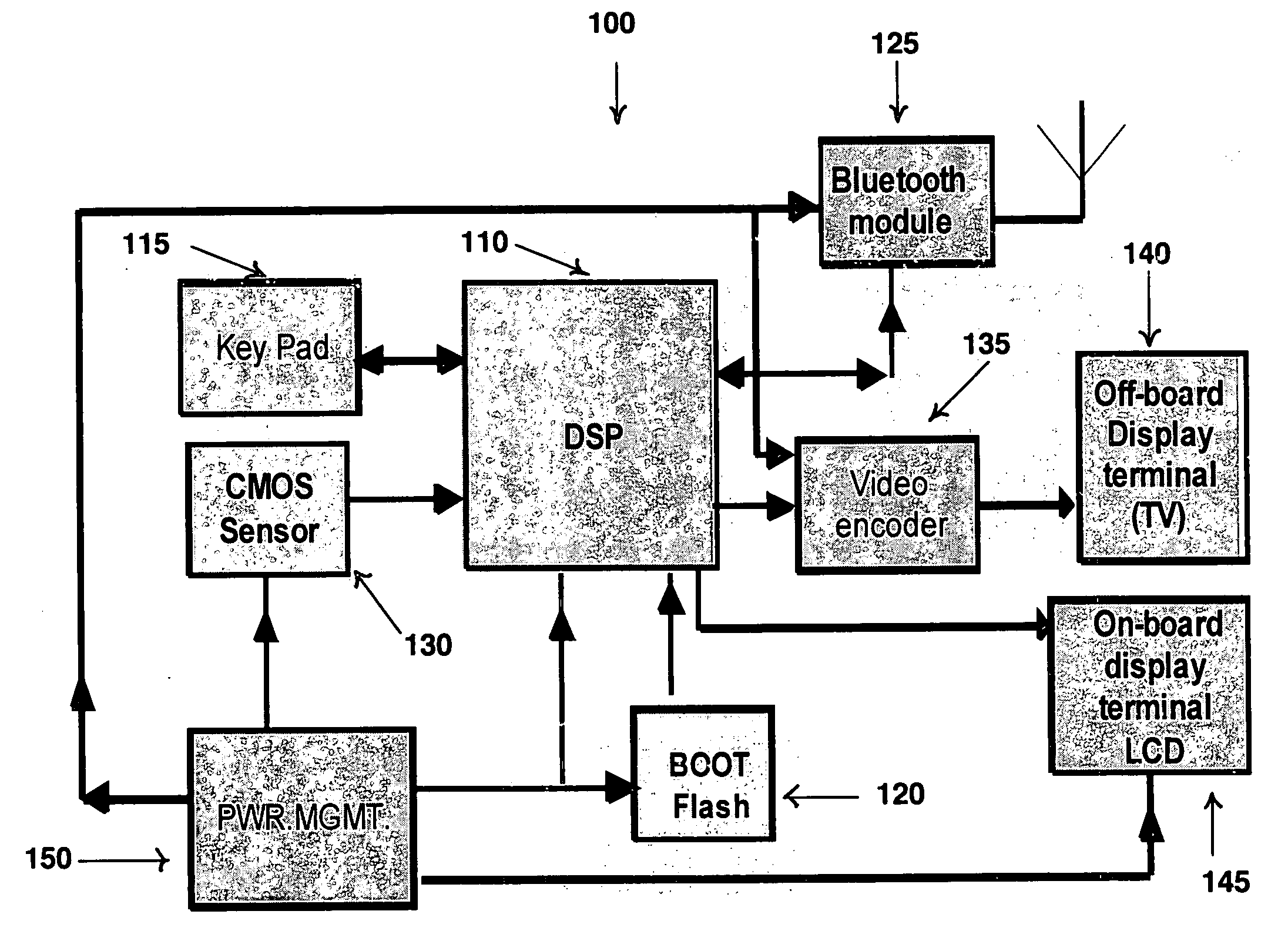
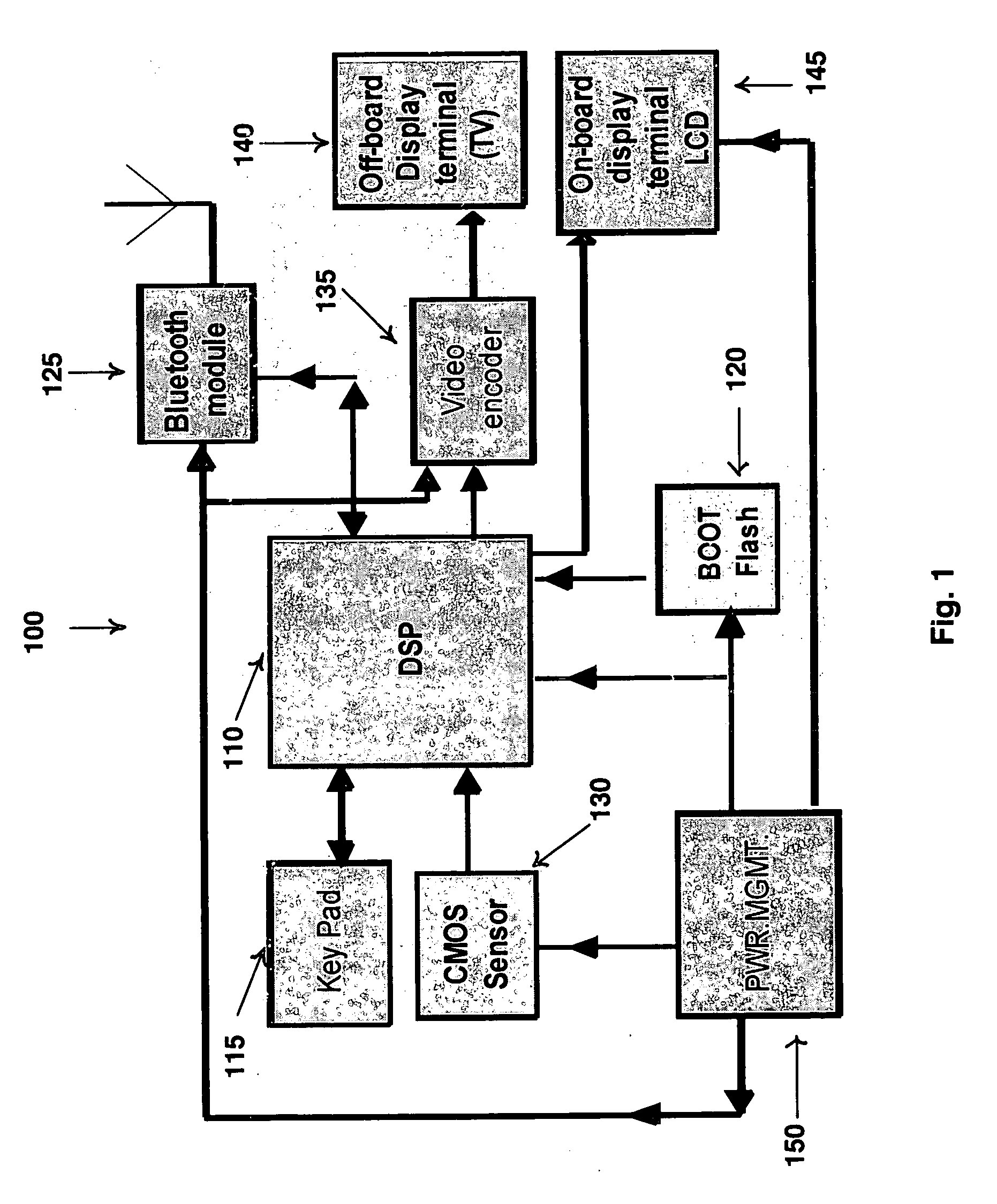
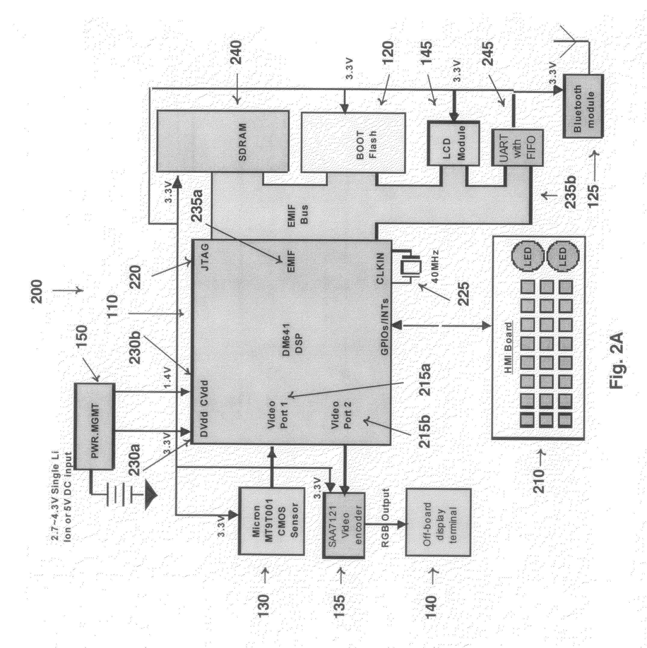
[0014]The present invention also is directed to another device for screening the skin of an individual in real time. The device comprises a programmable digital signal microprocessor with a memory storing processor executable instructions digitally linked to other device components. The other components are a boot flash for parallel flash memory digitally connected to the microprocessor, an input device, a sensor having a combination of one or more light sources and one or more imaging sensors, a digital to analog signal video encoder connected to an off-board display terminal, an on-board display terminal, a wireless device configured to wirelessly communicate between the programmable digital signal microprocessor and a personal computer, including a parallel for a Universal Asynchronous Receiver / Transmitter, and a power management unit. The device also comprises a housing the device that is portable and hand-holdable.
[0015]The present invention is directed further to a method for screening the skin of an individual in real time. The method comprises holding the portable handheld device described herein over a region of interest containing a skin lesion on the individual, acquiring one or more images of the skin lesion in one or more modalities, displaying the acquired image, preprocessing the acquired image(s) using a region-fusion based segmentation with narrow band graph partitioning algorithm, classifying the region of interest on the skin as benign or malignant and displaying the classification results. The present invention is directed to a related method further comprising wirelessly communicating the classification results to a personal computer.
[0016]The present invention is directed further still to a digital processor-implemented system for classifying a region of interest on the skin of an individual. The system comprises a module for pre-processing and image segmentation of the region of interest, a module for image classification of a skin lesion found within the region of interest as a benign or malignant lesion as determined by one or more of a size difference, shape irregularity or texture formation each of which comprises a partial classification, a module for decision voting in terms of confidence based weighted averaging of the partial classifications, and a module for image reconstruction for three-dimensional imaging of the region of interest. The present invention is directed to a related system further comprising a module for wireless communication between the processor and a personal computer.
[0017]The present invention is directed further still to a processor readable medium having processor-executable instructions to perform a method for detecting skin cancers on an individual in real time. The method comprises the steps of acquiring one or more images of a region of interest on the skin in one or more modalities, preprocessing the acquired image(s) comprising extracting the lesion from the background of the image and determining a lesion border using a region-fusion based segmentation with narrow band graph partitioning algorithm, classifying the skin lesion as benign or malignant by a confidence based weighted averaging of a combination of partial classifications based on one or more features of the skin lesion, and displaying the classification results.

Problems solved by technology

Moreover, at an early stage, skin cancers are very economical to treat, while at a later stage, cancerous lesions usually result in near fatal consequences and have extremely high costs associated with treatment.
Existing devices used to observe skin surface can only acquire pictures in one modality at a time, and the acquired pictures can not be readily processed.
Though such devices find interesting uses, they present great limitations to users since the acquired images must be transferred to a personal computer for further analysis.
Existing software only performs limited image analysis and fails to provide physicians with an unambiguous diagnosis.
More, specifically the prior art is deficient in portable handheld devices and software for skin cancer screening in real time.

Method used


Image

  • Device and software for screening the skin
  • Device and software for screening the skin
  • Device and software for screening the skin

Examples


example 1

Curve Evolution of Region-Based and Narrow Band Graph Partitioning Methods Level Set Formulation of Region-Based Curve Evolution

[0087]Given an image I⊂Ω, the region-based active contour model [4] finds the curve C that minimizes the energy based objective function:

E(C→)=λ1∫∫insideI(x,y)-c12xy+λ2∫∫outsideI(x,y)-c22xy+μL(C→)+vA(inside(C→)Eq.(1)

where c1 is the average intensity inside C; c2 is the average intensity outside C; μ, ν≧0, and λ1, λ2>0, are fixed weight defined based on a priori knowledge. L(C) and A(C) are two regulation terms. Following Chan's practice [4], the weights are fixed as λ1=λ2=1, μ=1, and ν=0 without the generality of the following derivation.

[0088]The main drawback of the explicit curve evolution (updating curve C directly) based on difference approximation scheme is that topological changes are di±cult to handle by such a evolving method. The levelset approach [5], however, handles such changes easily by defining the contour of a region as a zero-level set of ...

example 2

Skin Lesion Segmentation Algorithm Based on Region Fusion Narrow Band Graph Partitioning (NBGP)

[0103]Region Fusion Based Segmentation

[0104]At the first stage in Algorithm 1, active contours are applied iteratively inside each segmented regions. Based on the prior knowledge of the application, different strategies can be chosen to further segment only regions with lower intensity, higher intensity, or both. In the skin lesion segmentation, since the skin lesion always have lower intensity than that of the skin and blood volume, regions are always segmented with lower intensity further. The iterative segmentation procedure stops when the number of pixels inside the region is smaller than MinArea % of the total number of pixels in the image. For the skin lesion segmentation, based on extensive test, MinArea=10 was chosen. This makes sure that small lesions can be correctly segmented, while avoiding an over segmentation problem.

[0105]The second stage merges overlapping and / or non-overla...

example 3

Application of Segmentation Algorithm

[0114]Image Acquisition

[0115]XLM and TLM images were acquired with two Nevoscope [34,35] that uses a 5× optical lens (manufactured by Nikon, Japan). An Olympus C2500 digital camera was attached to this Nevoscope. Fifty one XLM images and sixty TLM images were used. The image resolution is 1712×1368 pixels. Region of interests (ROI) was identified in every image and segmentation, manually by dermatologists and automatically using our method, was performed on a 236×236 region obtained from a preprocessing step described in the next section. Results from manual segmentation were used as reference in evaluations. In addition, one hundred ELM images were acquired using oil immersion technique [11-12]. Among them, thirty images were melanoma and the rest were benign. Three dermatologists performed segmentation on the original image scale. The average contour was used as reference in our experiments.

[0116]Pre-Processing and Parameter Settings

[0117]To im...


PUM


Abstract

The present invention provides devices for screening the skin of an individual in real time using a region-fusion based segmentation with narrow band graph partitioning algorithm to analyze and classify a region of interest of the skin as benign or malignant. Also, provided is a method for screening the skin of an individual using the devices described herein. In addition the present invention provides a digital processor-implemented system for classifying a region of interest on the skin and a processor readable medium having processor-executable instructions to perform skin cancer detection.

Description

CROSS-REFERENCE TO RELATED APPLICATIONS[0001]This nonprovisional application claims benefit of priority of provisional U.S. Ser. No. 60 / 905,405, filed Mar. 7, 2007, now abandoned.FEDERAL FUNDING LEGEND[0002]This invention was produced in part using funds obtained through grant 521527 from the National Science Foundation. Consequently, the federal government has certain rights in this invention.BACKGROUND OF THE INVENTION[0003]1. Field of the Invention[0004]This invention relates to the fields of biomedical screening devices and dermoscopy. Specifically, the present invention provides a handheld medical device and software useful for skin cancer screening in real time.[0005]2. Description of the Related Art[0006]Skin cancer is the most common form of cancer, representing about half the number of all cancer types. The most deadly form of skin cancer is melanoma for which incidences increase by 3% annually. Frequent screening of suspicious skin pigmentations and other lesions is of par...

Claims


Application Information

Patent Timeline
Patent Type & Authority Applications(United States)
IPC IPC(8): A61B5/00G06K9/36G16H30/40
CPCA61B5/0059G06T2207/30096A61B5/442A61B5/444A61B5/445A61B5/7264G06F19/321G06F19/345G06K9/342G06K2209/053G06T7/0012G06T7/0081G06T2207/20072G06T2207/20116G06T2207/20141G06T2207/20161G06T2207/30088A61B5/01G06T7/11G06T7/187G16H50/20G16H30/40G06V10/267G06V2201/032
Inventor ZOURIDAKIS, GEORGEYUAN, XIAOJINGCHEN, JISTOTZER, ERICWU, YANMIN
Owner UNIV HOUSTON SYST
Why Eureka
Try Eureka