
Method for treatment of tissue lesion

a tissue lesion and treatment method technology, applied in the field of tissue lesion treatment, can solve the problems of unfavorable patient safety, unintentional clotting, and inability to properly heal connective tissue injuries, and achieve the effects of prolonging the storage time of unactivated platelets, and causing severe pain

Inactive Publication Date: 2008-10-09
BLUE ENGINE BIOLOGICS LLC
View PDF63 Cites 19 Cited by
  • Summary
  • Abstract
  • Description
  • Claims
  • Application Information

AI Technical Summary

Benefits of technology

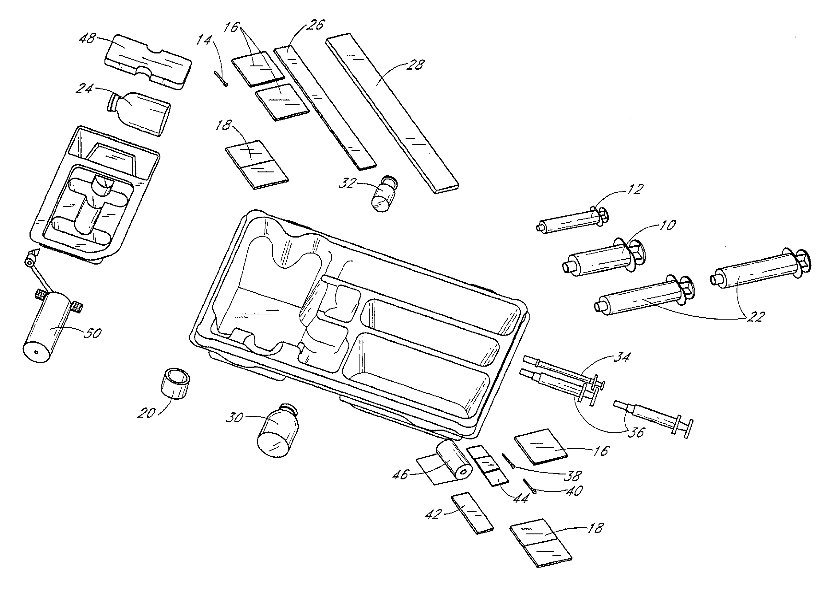
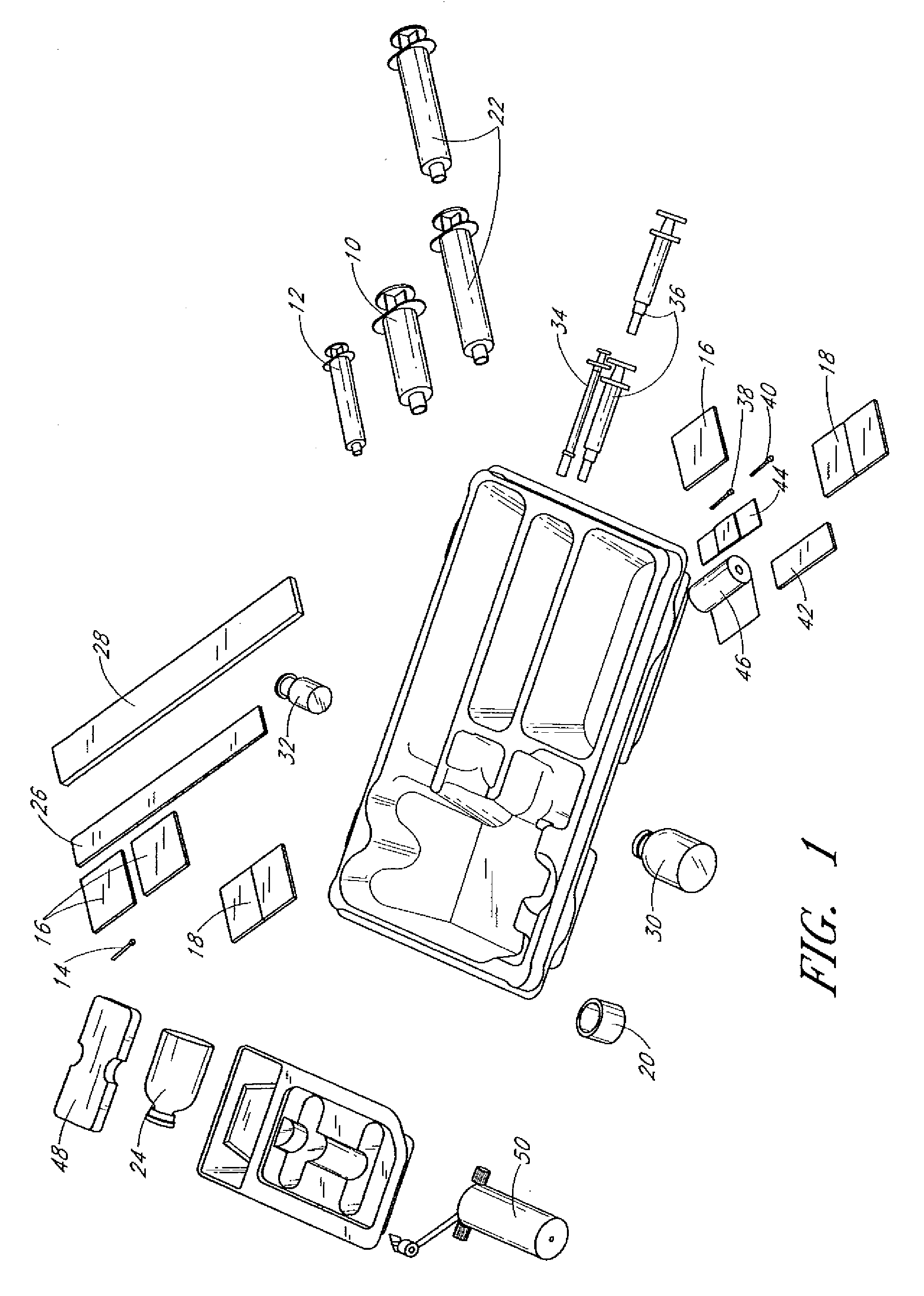
"The invention is a kit and method for treating damaged tissue, such as tendinosis or plantar fasciitis, using a platelet-rich plasma composition. The kit includes an anticoagulant to prevent blood coagulation, a disposable separating tube for isolating platelet fractions, a pH adjusting agent for creating a buffered platelet-rich plasma, and a platelet delivery device for applying the plasma to the affected area. The method involves drawing blood from the patient, adding an anticoagulant, fractioning the blood in the separating tube to isolate platelet fractions, adjusting the pH to create a buffered platelet-rich plasma, and administering the plasma to the affected area to promote vascularization and tissue regeneration. The kit and method provide a safe and effective treatment for damaged tissue with minimal risk of complications."

Problems solved by technology

Injuries or other damage to flexible, relatively avascular connective tissues (hereafter “connective tissue” or “connective tissues”) are known to take a very long time to heal (months or even years).
In many cases, injuries to connective tissues may never heal properly, necessitating surgical intervention.
Tendinosis is an accumulation over time of small-scale injuries that don't heal properly; it is a chronic injury of failed healing.
Tendinosis can result from repetitive activities such as playing sports, using computers, playing musical instruments, or doing manual labor.
The body attempts to repair these microtears but the healing process is incomplete in many cases.
This incomplete attempt at repair results in degenerative, immature and avascular tissue.
Incompletely repaired tissue is weaker than normal tendon tissue and lacks the strength to function normally.
This tissue also limits the patient by causing pain and negatively impacting the patient's quality of life.
Unfortunately, plantar fasciitis usually takes many months to resolve.
Rest and activity modification may help patients with some of these conditions, but there remains a significant clinical population that are not reachable with these therapies.
Despite widespread use, oral anti-inflammatory medications have not proven to be useful in controlled studies.
Some studies further suggest that non-steroidal medication may actually have an adverse effect on the healing process for ligament injuries.
Cortisone injections are frankly controversial in the treatment of tendinoses and are contraindicated in acute ligament injuries.
Results beyond one year, however, reveal a high symptom recurrence rate and only an equivocal efficacy rate.
These injections also carry the risk of tendon rupture, infection, skin depigmentation, subdermal atrophy and hyperglycemia in diabetic patients.
Physical therapy is commonly used, but expensive and it is not clear that such treatment is helpful (Smidt 2003).
Furthermore, the average cost is high: $1200-1500.
However, open or arthroscopic surgery has many potential complications such as deep infection, damage to neurovascular structures, and scar formation.
The surgery is also expensive and carries the additional risks associated with regional or general anesthesia.
None of the prior art teach treatment of connective tissue with PRP to promote tissue repair.

Method used


Image

  • Method for treatment of tissue lesion
  • Method for treatment of tissue lesion
  • Method for treatment of tissue lesion

Examples


example 1

[0091]PRP was prepared using a centrifuge unit made by Harvest (Plymouth, Mass.). (Similar units are available as The Cell Factor Technologies GPS system, the Depuy Symphony machine and the Medtronic Magellan machine.) Approximately 55 cc of blood was drawn from the patient using a standard sterile syringe, combined with 5 cc of a citrate dextrose solution for anticoagulation, and then spun down to isolate the platelets according to the manufacturer's protocol. These platelets were then resuspended in approximately 3 cc of plasma. The resulting platelet-rich plasma solution (PRP) was quite acidic and was neutralized with using approximately 0.05 cc of an 8.4% sodium bicarbonate buffer per cc of PRP under sterile conditions to approximately physiologic pH of 7.4. The PRP was not activated through addition of exogenous activators. This PRP composition is referred to herein as autologous platelet extract (APEX).

example 2

[0092]Fifty cc of whole blood is drawn from a patient, and then prepared according to the method of Knighton, U.S. Pat. No. 5,165,938, column 3. The PRP is activated according to Knighton using recombinant human thrombin. The degranulated platelets are spun down and the releasate containing supernatant is recovered. The releasate may be optionally pH adjusted to a pH of 7.4 using sodium bicarbonate buffer.

example 3

[0093]Thirty ml of whole blood were drawn from a patient. A platelet composition was prepared according to Example 1 of U.S. Pat. No. 5,510,102 to Cochrum, incorporated herein by reference in its entirety, except that no alginate is added to the platelet composition.


PUM

PropertyMeasurementUnit
pHaaaaaaaaaa
molecular weightaaaaaaaaaa
timeaaaaaaaaaa

Abstract

Methods are disclosed to promote vascularization of tissue in mammals using an autologous platelet-rich plasma composition. In particular, a method of treating a cardiac tissue lesion is disclosed.

Description

RELATED APPLICATIONS[0001]This application is a continuation of U.S. application Ser. No. 11 / 085,987, filed Mar. 22, 2005 which claims priority under 35 U.S.C. § 119(e) to provisional application No. 60 / 655,668, filed Feb. 23, 2005 and is a continuation-in-part of U.S. application Ser. No. 10 / 941,124, filed Sep. 15, 2004 which is a continuation of U.S. application Ser. No. 10 / 412,821, filed Apr. 11, 2003, now U.S. Pat. No. 6,811,777, which claims priority under 35 U.S.C. § 119(e) to U.S. Provisional Application No. 60 / 372,682, filed Apr. 13, 2002. All of the above are incorporated by reference in their entirety.BACKGROUND OF THE INVENTION[0002]1. Field of the Invention[0003]Embodiments of the invention are directed to methods and kits for the treatment of damaged tissue in a human or non-human animal using autologous platelet-rich plasma. In a preferred embodiment, methods and kits are described for the treatment of lateral epicondylitis, plantar fascists, and rotator cuff injury.[0...

Claims


Application Information

Patent Timeline
Patent Type & Authority Applications(United States)
IPC IPC(8): A61F2/04A61K35/16A61P9/00A61K35/12
CPCA61K35/16A61K38/1841A61K38/1858A61K38/30A61K38/363A61K38/39A61K38/4833A61K2300/00A61P9/00
Inventor MISHRA, ALLAN
Owner BLUE ENGINE BIOLOGICS LLC
Learn More