
Diagnostic Primers and Method for Detecting Avian Influenza Virus Subtype H5 and H5n1

a technology of avian influenza virus and primers, applied in chemical libraries, combinational chemistry, sugar derivatives, etc., can solve the problems of high mortality rate of affected poultry populations, massive culling of millions of poultry, and severe economic repercussions, and achieves rapid, specific and sensitive effects

Inactive Publication Date: 2008-10-23
AGENCY FOR SCI TECH & RES
View PDF3 Cites 2 Cited by
  • Summary
  • Abstract
  • Description
  • Claims
  • Application Information

AI Technical Summary

Benefits of technology

The patent describes a method for detecting avian influenza virus subtype H5 or H5N1 in a sample using conserved regions within the HA and NA genes. The method involves amplifying DNA reverse-transcribed from RNA obtained from the sample using primers that are specific to these genes and detecting the presence of the amplified product. The method is rapid, specific, and sensitive, and can be used for early detection and diagnosis of infection. The primers can also be used in other amplification methods and in nucleic acid microarrays for detecting the presence of the virus. The patent also provides a kit for detecting the virus.

Problems solved by technology

However, the virus can have high pathogenic effect in poultry, with sudden outbreaks causing high mortality rates in affected poultry populations.
(1998) Lancet 351: 467-471), leading to the death of some patients.
350: 1179-1188) that resulted in massive culling of millions of poultry which had severe economic repercussions.
Symptoms of avian flu infection include fever, respiratory difficulties including shortness of breath and cough, lymphopenia, diarrhea and difficulties regulating blood sugar levels.
Due to the high pathogenicity of H5 subtypes, particularly H5N1, and their demonstrated ability to cross over to infect humans, there is a significant economic and public health risk associated with these viral strains, including a real epidemic and pandemic threat.
As a result, H5N1 avian influenza A virus represents a potential danger to human health not only in Asia but to the world.
However, each of these methods uses genetic information derived from only a few isolates or variants of H5 or H5N1 to confirm the presence of virus.
Furthermore, these assays are reported to be low in specificity and sensitivity.
Clinically, the low sensitivity of these diagnostics may limit the usefulness for reliable detection of influenza A (H5N1) virus in humans.

Method used


Image

  • Diagnostic Primers and Method for Detecting Avian Influenza Virus Subtype H5 and H5n1
  • Diagnostic Primers and Method for Detecting Avian Influenza Virus Subtype H5 and H5n1
  • Diagnostic Primers and Method for Detecting Avian Influenza Virus Subtype H5 and H5n1

Examples


example 1

Detection of Avian Influenza Virus H5 and H5N1 Using Gel-Based Detection Platform

[0145]The following is a general protocol for detection of avian influenza virus subtype H5 or H5N1.

[0146]Generally, RNA is extracted from samples according to the manufacturer's instructions, using either TRIzol™ or RNA extraction kits (Qiagen).

[0147]The first-strand cDNA synthesis is performed on extracted RNA using the relevant reverse primer(s) (2 μl of 10 μM stock) in a 20 μl reaction volume. A first round PCR reaction is set up using 2.5 μl of the CDNA reaction, containing cDNA product as template with relevant forward and reverse primer(s) (1.25 μl total volume for each of forward and reverse) in a 25 μl reaction volume. The PCR conditions are set up as follows: incubation at 94° C. for 2 min; 35 cycles of 94° C. for 10 sec, 50° C. for 30 sec, 72° C. for 1 min; followed by an incubation at 72° C. for 7 min. A second round of PCR is performed using the product of the first round PCR (2.5 μl) as te...

example 2

Detection of Avian Influenza Virus H5 and H5N1 Using Real-Time RT-PCR Detection Platform

[0152]The following reactions are performed in a LightCycler™ instrument.

[0153]The reaction master mixture is prepared on ice by mixing the following reagents in order, to a volume of 20 μl: water (volume adjusted as necessary), 50 mM manganese acetate (1.3 μl), ProbeNPrimer mix containing forward primer and reverse primer to a final concentration of 0.2 to 1 μM and fluorescently labelled probes (2.6 μl ), LightCycler RNA Master Hybridization Probes (7.5 μl), which contains buffer, nucleotides and enzyme.

[0154]The reactions are transferred to glass capillary tubes suitable for use in the LightCycler™. 5 μl of extracted RNA template is added to each reaction and briefly centrifuged. The RT-PCR reactions are run using the following programs (Tables 5-8):

TABLE 5Program 1-Reverse TranscriptionCycle Program DataValueCycles1Analysis ModeNoneTemperature TargetsSegment 1Target T° C.61Incubation time20 mi...

example 3

Detection of Avian Influenza Virus H5N1 Using Real-Time RT-PCR with Various Primer Sets

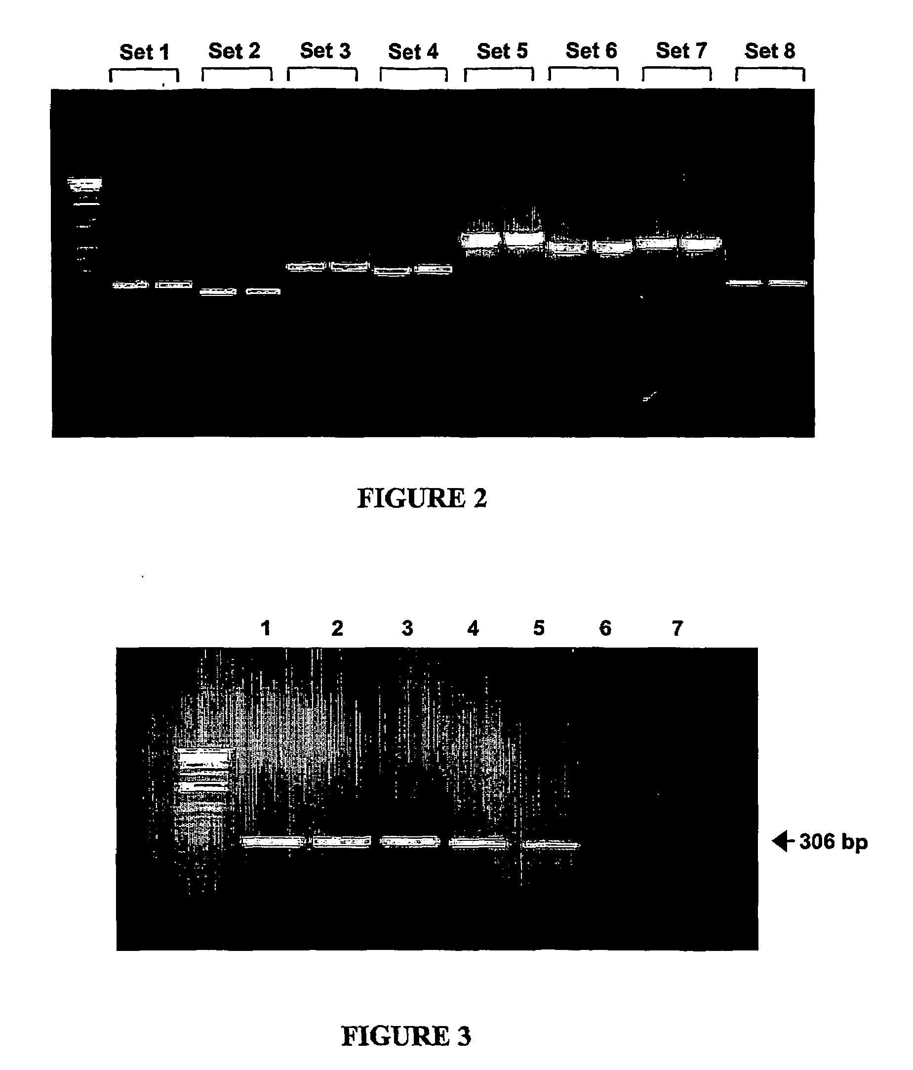
[0155]Real time PCR reactions were performed using the 8 primer sets described in Example 2 above. The reactions were performed using SYBR green fluorescent detection kit, in accordance with standard protocols and commercially available reagent kits (Roche). FIG. 6 displays the amplification products obtained for the reactions performed with each of the 8 primer sets as visualized on a 1.5% agarose gel stained with ethidium bromide.

[0156]To confirm the sensitivity of the primers using the real time PCR protocol, amplification curves were generated to monitor the production of amplification product. Results are shown in FIGS. 7 to 14 for each of primer sets 1 to 8, respectively. Melting curves of the amplified product were performed at the end of the amplification reaction. Generally, specific amplification products will have a higher melting temperature than non-specific products, and the melting ...


PUM

PropertyMeasurementUnit
thermal melting pointaaaaaaaaaa
volumeaaaaaaaaaa
volumeaaaaaaaaaa

Abstract

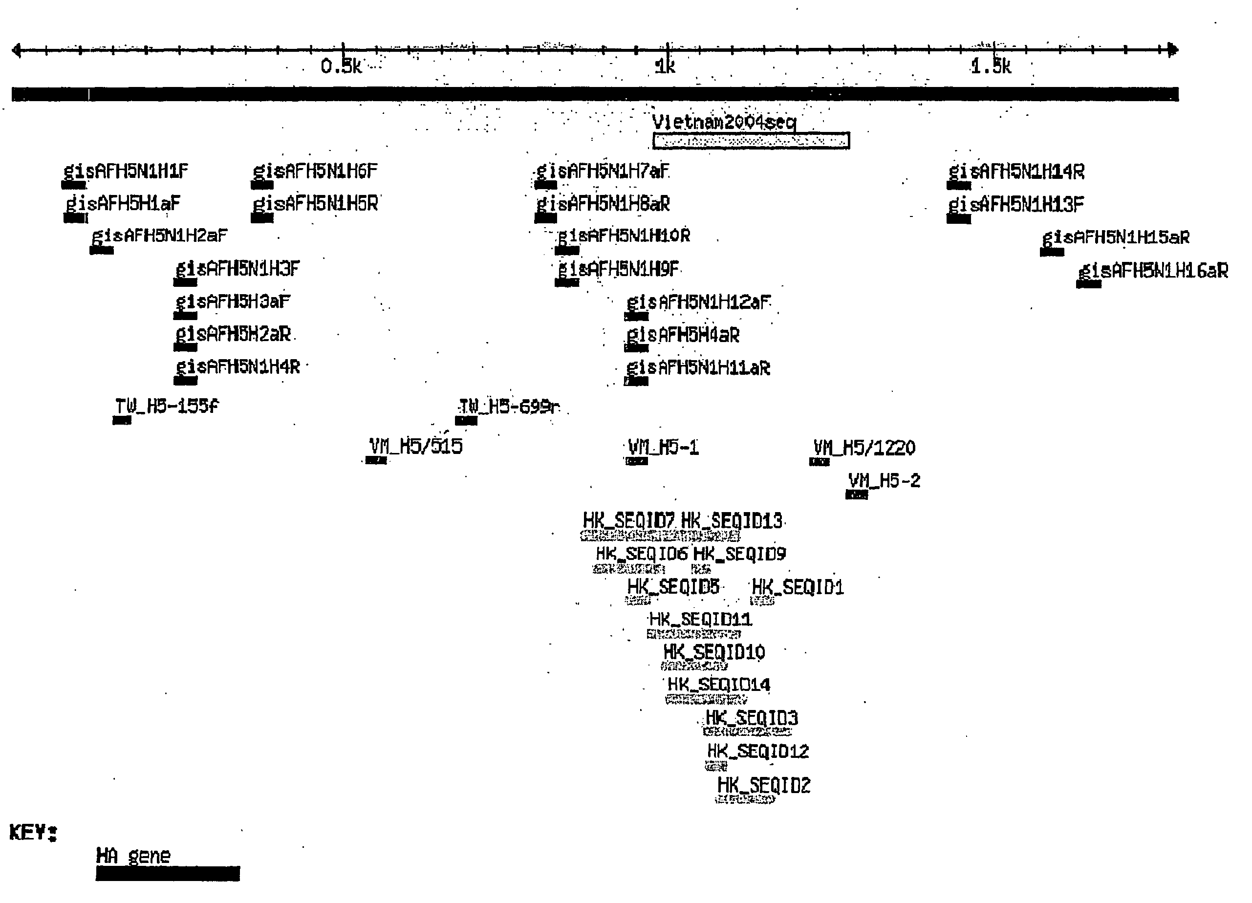
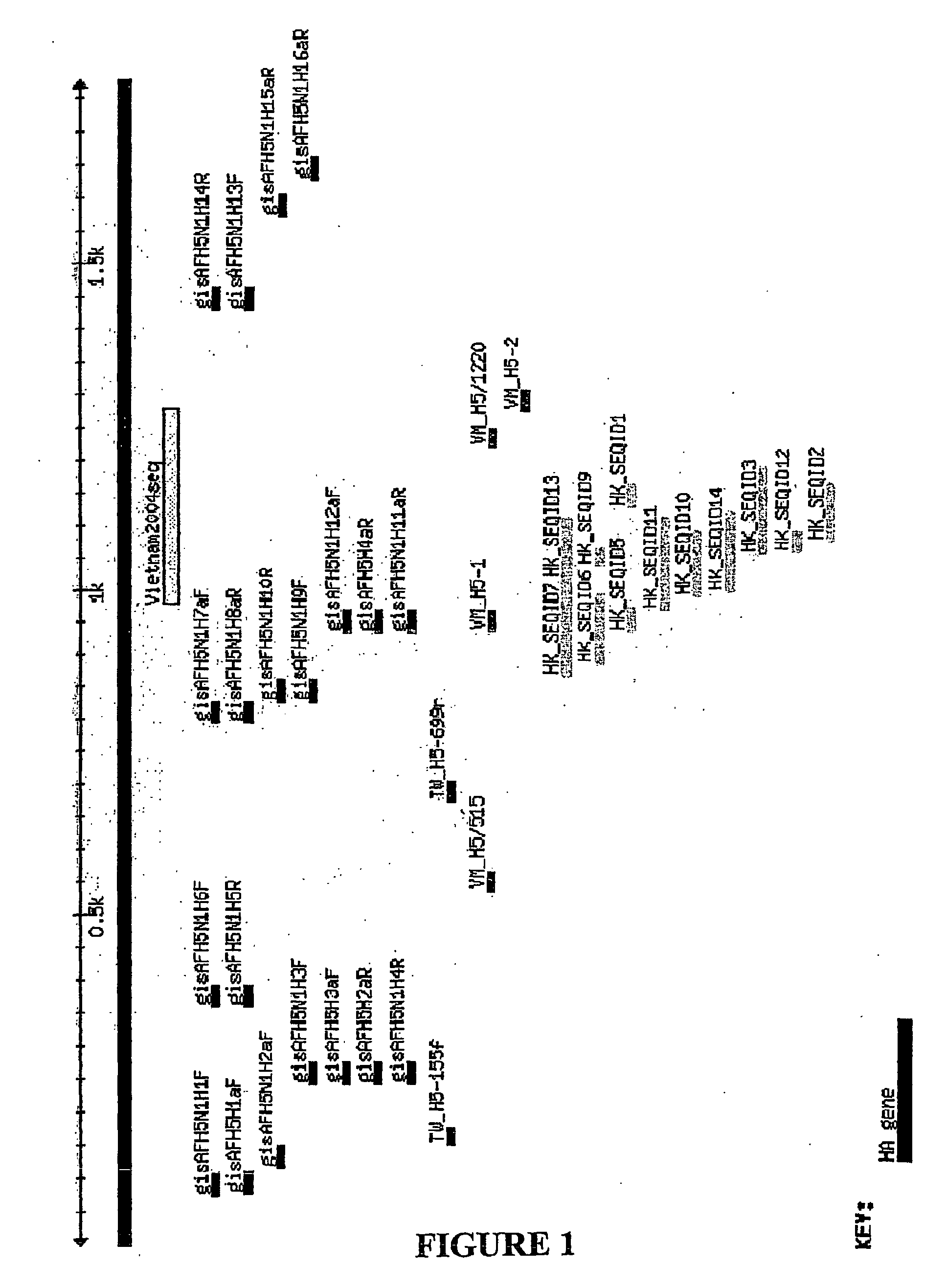
The present invention provides primers directed to conserved regions of the HA and NA genes of avian influenza virus subtypes H5 or H5N1, and provides a method for detecting avian influenza subtype H5 or H5N1.

Description

CROSS-REFERENCE TO RELATED APPLICATION[0001]This application claims benefit and priority from U.S. provisional patent application No. 60 / 578,353, filed on Jun. 10, 2004, the contents of which are incorporated herein by reference.FIELD OF THE INVENTION[0002]The present invention relates to a nucleic acid based detection method, more particularly, to primers and a method of detecting avian influenza virus.BACKGROUND OF THE INVENTION[0003]Three types of influenza viruses, types A, B, and C are known and they belong to a family of single-stranded negative-sense enveloped RNA viruses called Orthomyxoviridae (Swayne, D. E., and D. L. Suarez (2000) Rev. Sci. Tech. 19:463-482). The viral genome is approximately 12 000 to 15 000 nucleotides in length and comprises eight RNA segments (seven in Type C).[0004]Influenza A virus infects many animals such as humans, pigs, horses, marine mammals, and birds (Nicholson, K. G., et al. (2005) Lancet 362:1733-1745). Its natural reservoir is in aquatic b...

Claims


Application Information

Patent Timeline
Patent Type & Authority Applications(United States)
IPC IPC(8): C12Q1/70C07H21/00C40B40/06C12N15/11C12N15/44C12Q1/68
CPCC12Q1/701
Inventor REN, EE CHEENG, LISA FONG POHCHIA, JER-MING
Owner AGENCY FOR SCI TECH & RES
Learn More