Image system for retaining contrast when merging image data

a technology of image data and contrast, applied in the field of imageprocessing, imagevisualizing, imagearchiving system, can solve the problems of loss of information about the exact spatial positioning of implants or medical instruments in relation to the surrounding tissue area, failure of methods, and loss of information about the spatial relationship between segmented image objects bo in the foreground bv of the merged aggregate image b, etc., to achieve the effect of increasing the precision and safety of minimally invasive medical interventions

Inactive Publication Date: 2008-11-06
SIEMENS HEALTHCARE GMBH
View PDF19 Cites 23 Cited by
  • Summary
  • Abstract
  • Description
  • Claims
  • Application Information

AI Technical Summary

Benefits of technology

[0011]Proceeding from the above-cited prior art, the object of the present invention is to retain the contrast definition when fluoroscopic 2D x-ray recordings and reconstructed 2D projections or, as the case may be, 3D views of recorded image objects are overlaid, in order to increase the precision and safety of minimally invasive medical interventions or operations performed under x-ray control using merged and co-registered image data featuring the same imaging modality or, as the case may be, using merged and co-registered image data featuring different or different kinds of imaging modalities.

Problems solved by technology

What, though, is disadvantageous about that method is that image objects (such as ends of catheters and cardiovascular stent implants etc.) shown in the fluoroscopy image F with a low contrast definition will upon mixing-over of a 3D reconstruction M that is projected into the projection plane Exy of the relevant fluoroscopy image F and has a high contrast definition be virtually obscured by said reconstruction when a blending factor close to one is employed.
That, though, has the disadvantage in some applications that the information indicating the spatial relationship between the segmented image object BO in the foreground BV of the merged aggregate image B and the areas of tissue and objects shown in the image background BH of the merged aggregate image B can be lost.
Because the background FH of the fluoroscopy image F is with that method subtracted as a mask when segmenting has been performed, the areas of tissue imaged therein can no longer be shown in the merged aggregate image B. With a few exceptions that is very disadvantageous in most applications because the information about the exact spatial positioning of the implants or medical instruments that are shown in relation to the surrounding areas of tissue is lost.
That method will, moreover, fail if the spatial positioning of the implants or medical instruments shown in the foreground FV of the fluoroscopy image F changes relative to the position of the areas of tissue shown in said image's background FH, for example because the mobile C-arm of a multidirectional C-arm x-raying system or the table on which the patient being examined is lying has been moved or, as the case may be, owing to the patient's moving or breathing or because his / her moving organs (such as the lungs or heart) have moved through pulsating.

Method used


Image

  • Image system for retaining contrast when merging image data
  • Image system for retaining contrast when merging image data
  • Image system for retaining contrast when merging image data

Examples


Embodiment Construction

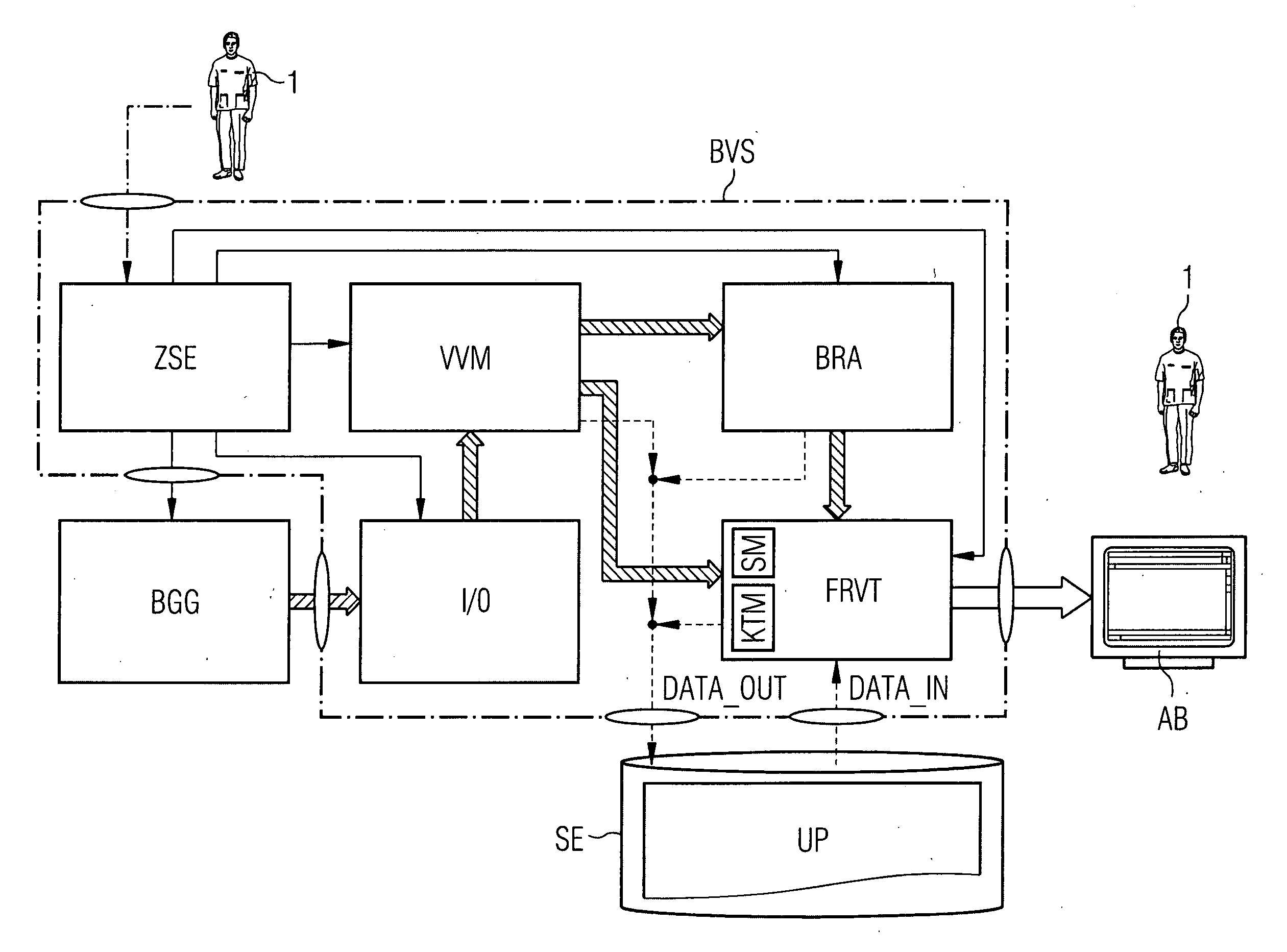
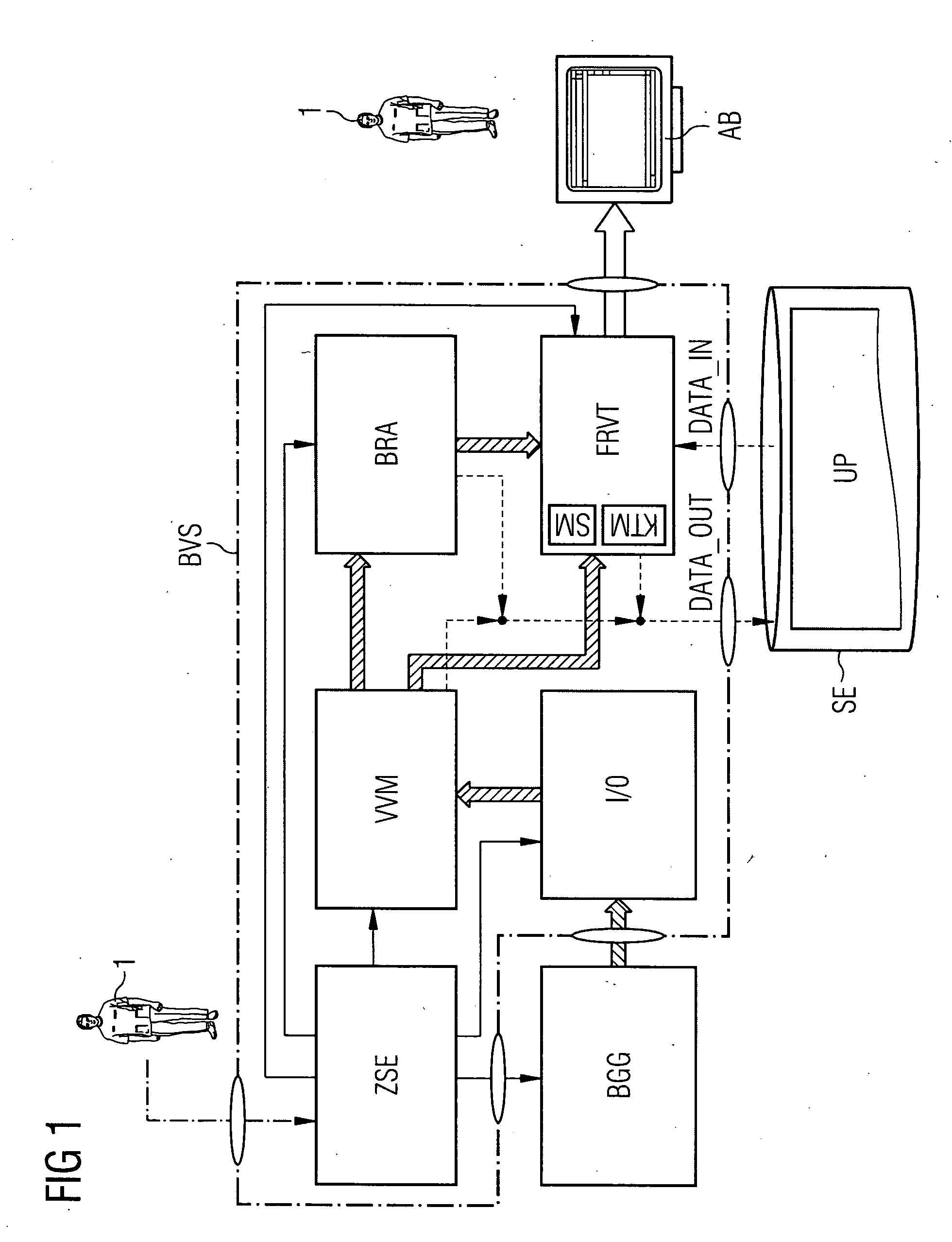
[0024]The system components of the inventive image-processing, image-visualizing, and image-archiving system and the steps of the associated inventive method are described in detail in the following sections with the aid of the attached drawings.

[0025]FIG. 1 is a schematic block diagram of an image-processing, image-visualizing, and image-archiving system according to the present invention, which system makes it possible to jointly log, register, and archive image data—generated by a medical imaging device BGG such as, for example, an angiography system—relating to internal organs, areas of tissue of interest, pathological structures, medical implants, and other objects in the body of a patient being examined, in the form on the one hand of fluoroscopic 2D x-ray recordings and on the other of reconstructed 2D projections shown from any projection angles or reconstructed 3D views and to visualize it in the form of merged graphical presentations on a monitor terminal's display screen ...


PUM


Abstract

The invention relates to a method for the joint of registering, archiving, contrast-retaining merging, and visualizing of 2D x-ray images and reconstructed 2D projections or 3D views of generated image data in minimally invasive medical interventions or operations performed under x-ray control on internal organs, areas of tissue, lesions, or pathological structures in a patient's body. A first image and the image data of a second image being mixed over the first image are mutually linked, co-registered, and merged using an alpha-channel-based mixing technique, whereby the color values or gray-scale values of the individual pixels of the first image are prior to being mixed-over with the segmented foreground of the second image brightened through the application of a color-value or gray-scale-value for compensating or lessening the reduction in contrast that occurs during mix-over in the first image before being displayed graphically on a monitor terminal.

Description

CROSS REFERENCE TO RELATED APPLICATIONS[0001]This application claims priority of German application No. 10 2007 021 035.5 filed May 4, 2007, which is incorporated by reference herein in its entirety.FIELD OF THE INVENTION[0002]The present invention relates to a method performed using an image-processing, image-visualizing, and image-archiving system that can be employed particularly in the area of diagnostic and interventional radiology for the joint registering, archiving, contrast-retaining merging, and visualizing of 2D x-ray images and reconstructed 2D projections or, as the case may be, 3D views of generated image data required within the scope of, for instance, minimally invasive medical interventions or operations performed under x-ray control on internal organs, areas of tissue, lesions, or pathological structures in a patient's body.BACKGROUND OF THE INVENTION[0003]For navigating the medical instruments required during a medical intervention, 2D x-ray images generated in re...

Claims


Application Information

Patent Timeline
Patent Type & Authority Applications(United States)
IPC IPC(8): A61B6/03G06K9/00
CPCG06F19/321G06T5/50G06T15/503G06T2207/10072G06T2207/10121G06T2207/20221G06T2207/30021G06T2207/30101G06T5/009G06T2207/20144G06T7/194G16H30/20
Inventor PFISTER, MARCUS
Owner SIEMENS HEALTHCARE GMBH
Why Eureka
Try Eureka