
Ischemic stroke therapy

a stroke and ischemic technology, applied in the field of ischemic stroke therapy, can solve the problems of reducing the likelihood of recovery, affecting the patient's clinical sequel, and permanent deficit, and achieve the effect of reducing the risk of infection

Inactive Publication Date: 2008-12-25
PENUMBRA
View PDF11 Cites 63 Cited by
  • Summary
  • Abstract
  • Description
  • Claims
  • Application Information

AI Technical Summary

Benefits of technology

[0010]It is another object of the present invention to provide improved methods for delivering ultrasound energy to a patient's intracranial space to treat ischemic stroke.
[0015]According to yet another embodiment, an acoustically conductive film can be placed between the transmitter and the patient's duramater to provide a sterile barrier and reduce the risk of infection.

Problems solved by technology

Such a bleed after the onset of an ischemic stroke can further worsen the patient's clinical sequel and reduce his / her likelihood for recovery.
In such a situation, the physician is presented with a conundrum; if the patient is not treated, it is almost guaranteed to result in a permanent deficit for the patient.
On the other hand, the treatment options available today are limited to endovascular approaches, which have their own limitations.
However, during an ischemic stroke, the blood vessels that are affected can become leaky over time or as a result of the treatment protocol.
Endovascular treatment protocols for opening up occluded intracranial blood vessels face access challenges due to the tortuous nature of the intracranial blood vessels.
Also, endovascular devices are at risk of causing a vessel perforation during navigation since typical fluoroscopy imaging techniques are inhibited by the occluded vessels not filling during contrast injections.
In addition, opening an occlusion using endovascular devices will typically result in instantaneous blood flow to the effected blood vessels.

Method used


Image

  • Ischemic stroke therapy
  • Ischemic stroke therapy
  • Ischemic stroke therapy

Examples


example 1

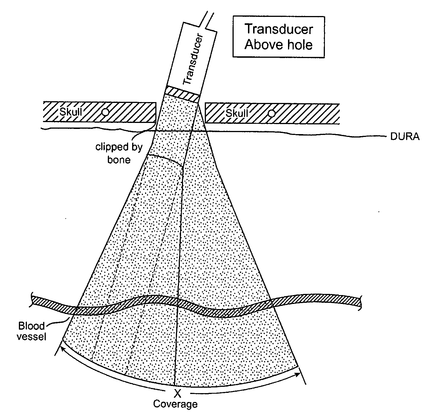
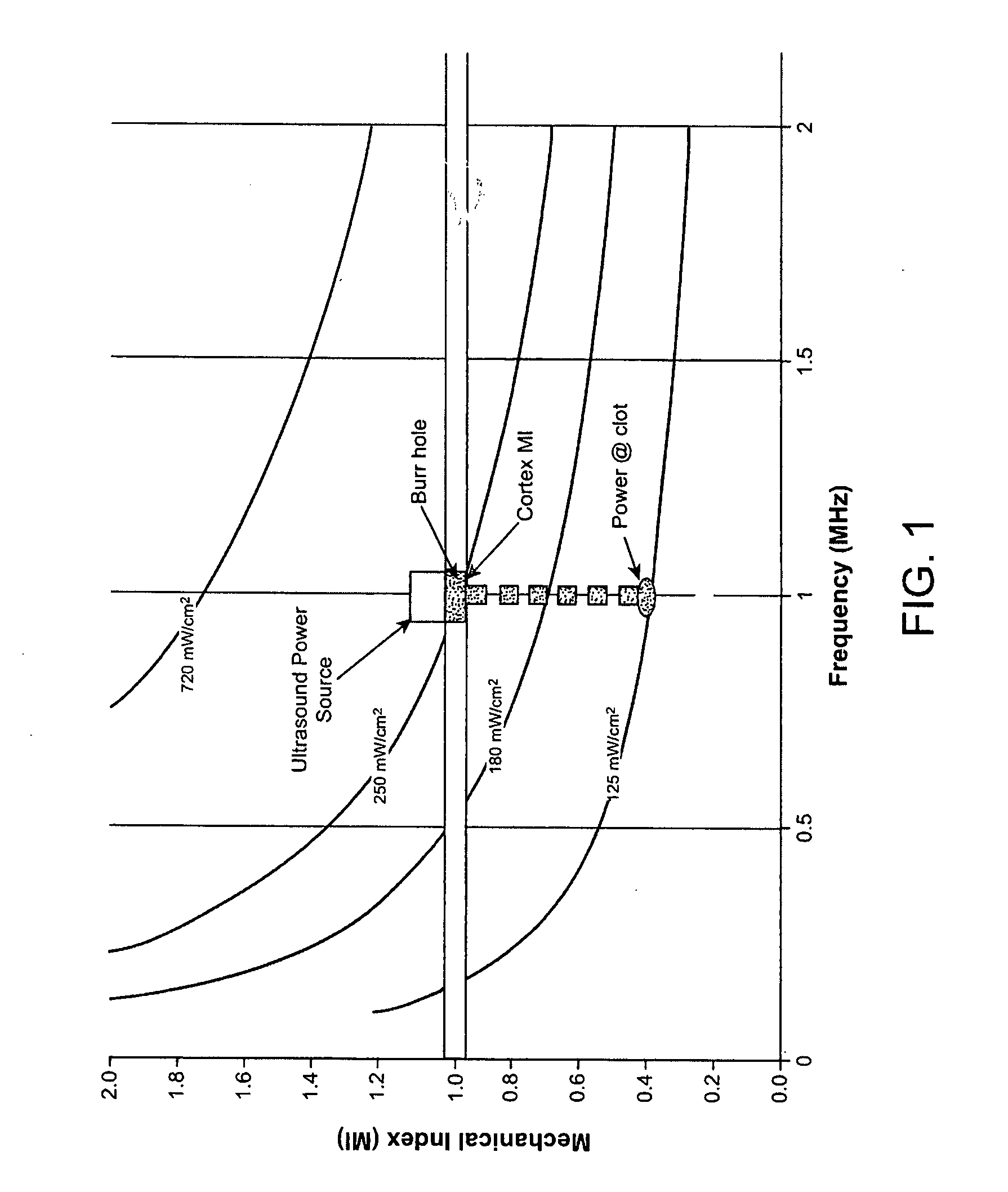
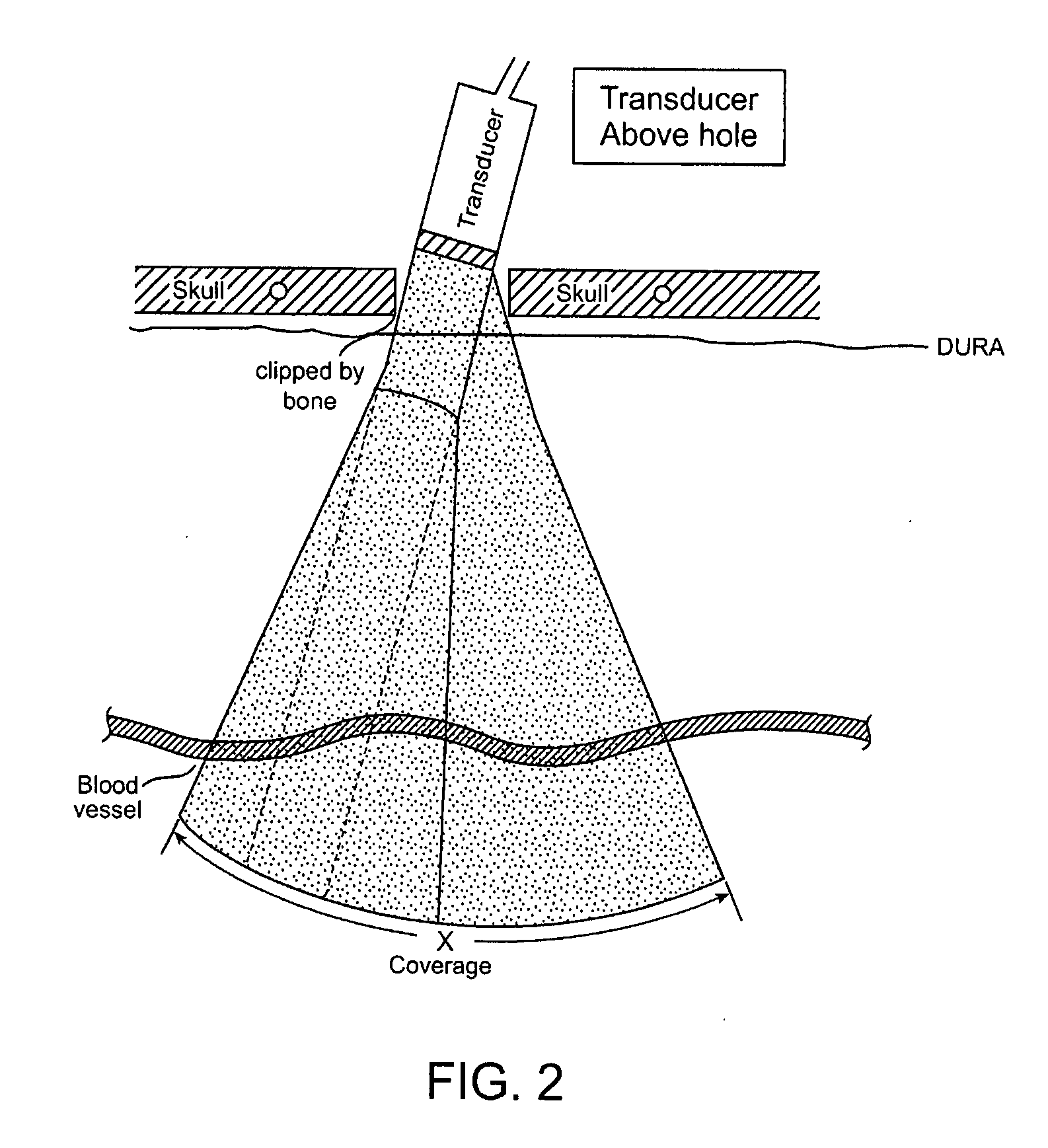
[0046]At least one hole is formed in the patient's skull, and at least one ultrasound probe is positioned near but preferably at least partially within the hole. Next, microbubbles are delivered through IV or IA delivery. An ultrasound diagnostics procedure is carried out by using low power and sweeping the ultrasound probe (rotating and / or angling and / or moving the probe towards and away from the brain cortex) about or within the hole to locate the clot location in the brain. During this procedure, it is preferable to use ultrasound probe power levels that minimize the number of microbubbles that are imploded to less than 80%. As described above, the diagnostic algorithm comprises Power Intensity (PI) at clots: 50 mw / cm22, and Frequency (F): 500 kHz<F<2 MHz with the resultant Mechanical Index (MI): 0.2<MI<1.0. Also, the power level used for the diagnostic treatment will either be equivalent to or lower than the value used during the therapeutic phase. Once the clot is located, the ...

example 2

[0048]At least one hole is formed in the patient's skull, and at least one ultrasound probe is positioned near but preferably at least partially within hole. Next, microbubbles and aspirin are delivered sequentially, through any known delivery method, including orally, or through IV or IA. Next, an ultrasound diagnostics procedure is performed in the manner described above for Example 1 to locate a clot, and then therapeutic ultrasound is delivered to the clot in the manner described above for Example 1.

example 3

[0049]At least one hole is formed in the patient's skull, and at least one ultrasound probe is positioned near but preferably at least partially within hole. Next, a mixture of microbubbles and aspirin is delivered, through any known delivery method, including orally, or through IV or IA. Next, an ultrasound diagnostics procedure is performed in the manner described above for Example 1 to locate a clot, and then therapeutic ultrasound is delivered to the clot in the manner described above for Example 1.


PUM


Abstract

A method for delivering ultrasound energy to a patient's intracranial space includes the steps of forming a hole in a patient's skull, locating an ultrasound transmitter near or into the hole, and transmitting ultrasound from the transmitter into the intracranial space, wherein the Mechanical Index of ultrasound energy traveling through cerebral tissue in the intracranial space is less than 1.0, the power intensity delivered to a target tissue in the intracranial space is greater than 50 mW / cm2 and less than 200 mW / cm2, and the frequency of the transmitted ultrasound is within the range between 500 kHz and 2 MHz. Microbubbles, aspirin, both microbubbles and aspirin, and a mixture of microbubbles and aspirin, can also be delivered into the intracranial space.

Description

INCORPORATION BY REFERENCE[0001]Applicant expressly incorporates herein by this reference the entire disclosures in pending application Ser. No. 11 / 203,738 filed Aug. 15, 2005, Ser. No. 11 / 165,872 filed Jun. 24, 2005, Ser. No. 11 / 274,356 filed Nov. 15, 2005 and Ser. No. 11 / 490,971 filed Jul. 20, 2006.BACKGROUND OF THE INVENTION[0002]1. Field of the Invention[0003]The present invention relates to ischemic stroke therapy, and in particular, to methods for delivering ultrasound energy to a patient's intracranial space.[0004]2. Description of the Prior Art[0005]After the onset of an ischemic stroke, affected blood vessels can leak blood and / or the bloods' constituents into the intra-cerebral space if (i) the occluded vessels are revascularized too late, (ii) the vessels are damaged during the revascularization process, and (iii) the blood vessels are opened too quickly.. Bleeding into the intra-cerebral space is a result of a breakdown of the blood brain barrier (BBB) and is also known ...

Claims


Application Information

Patent Timeline
Patent Type & Authority Applications(United States)
IPC IPC(8): A61N7/00
CPCA61N7/00A61N7/022A61B8/0816A61N2007/0021A61N2007/0039A61N2007/0043
Inventor NITA, HENRY
Owner PENUMBRA
Learn More