Method of Three-Dimesionally Culturing Chondrocytes

a chondrocyte and three-dimesion technology, applied in the field of producing chondrocytes, can solve the problems of chondrocytes not being able to easily migrate from a healthy site to the injured site, granulation is available, and the restoration capacity of chondrocytes is extremely low

Inactive Publication Date: 2009-01-01
OTSUKA PHARM CO LTD
View PDF4 Cites 3 Cited by
  • Summary
  • Abstract
  • Description
  • Claims
  • Application Information

AI Technical Summary

Benefits of technology

[0015]The present invention of three-dimensional chondrocyte culture system made it possibles to grow the cells with keeping the

Problems solved by technology

That is, as the joint chondrocytes are highly differentiated, scarcely proliferate and differentiate, their restoration capacity is extremely low.
Since a cartilage tissue lacks a nerve and a blood vessel, no restoration mechanism by hemorrhage, inflammation and granulation is available.
Since a periphery of the cartilage tissue is surrounded by extracellular matrix, the chondrocytes can not easily migrate from a healthy site to the injured site.
The injured site in the joint cartilage induces degradation of its periphery and adjacent cartilage with time, finally advances to osteoarthritis, and causes the reduc

Method used


Image

  • Method of Three-Dimesionally Culturing Chondrocytes
  • Method of Three-Dimesionally Culturing Chondrocytes
  • Method of Three-Dimesionally Culturing Chondrocytes

Examples


example 1

Culture of Chondrocytes

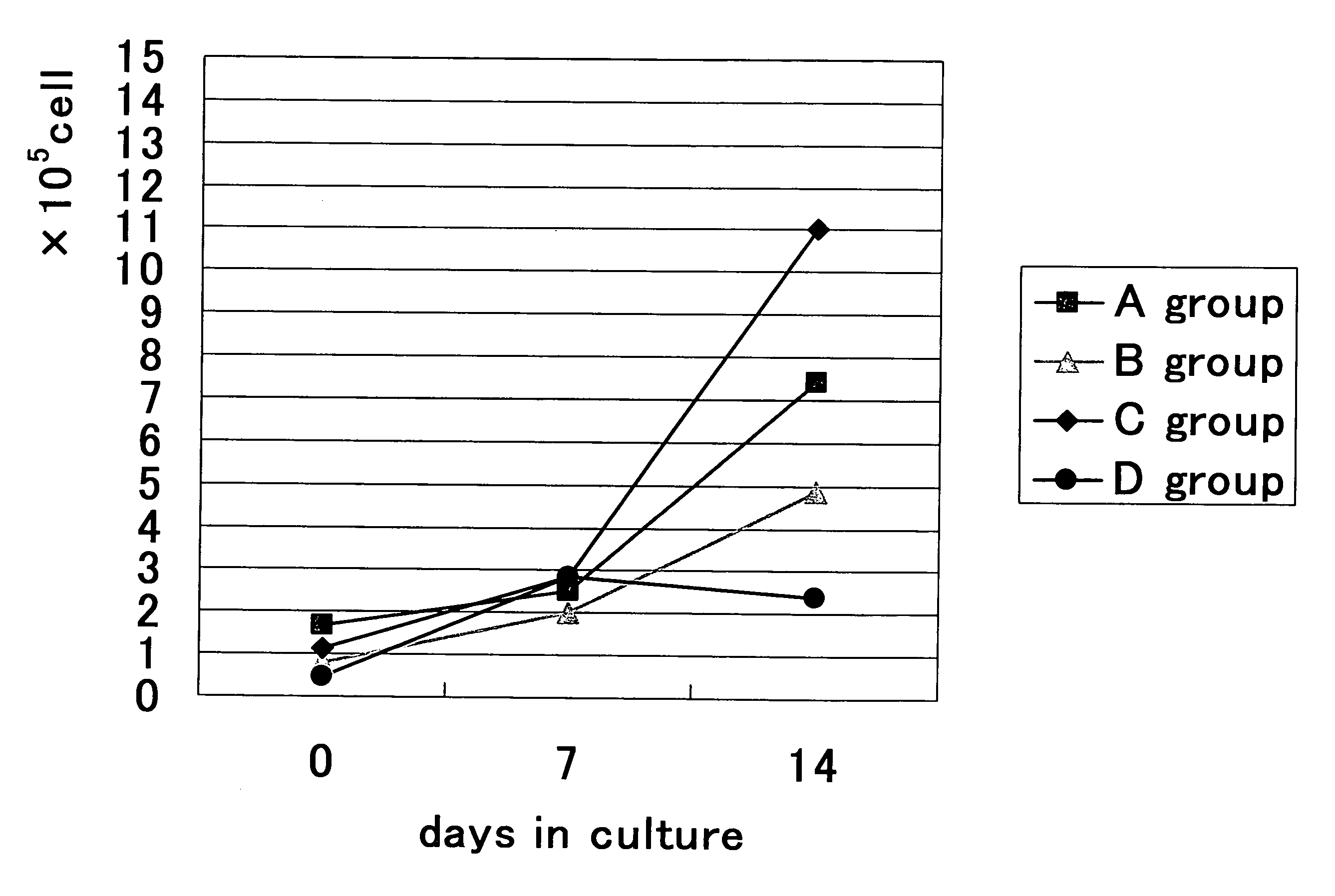
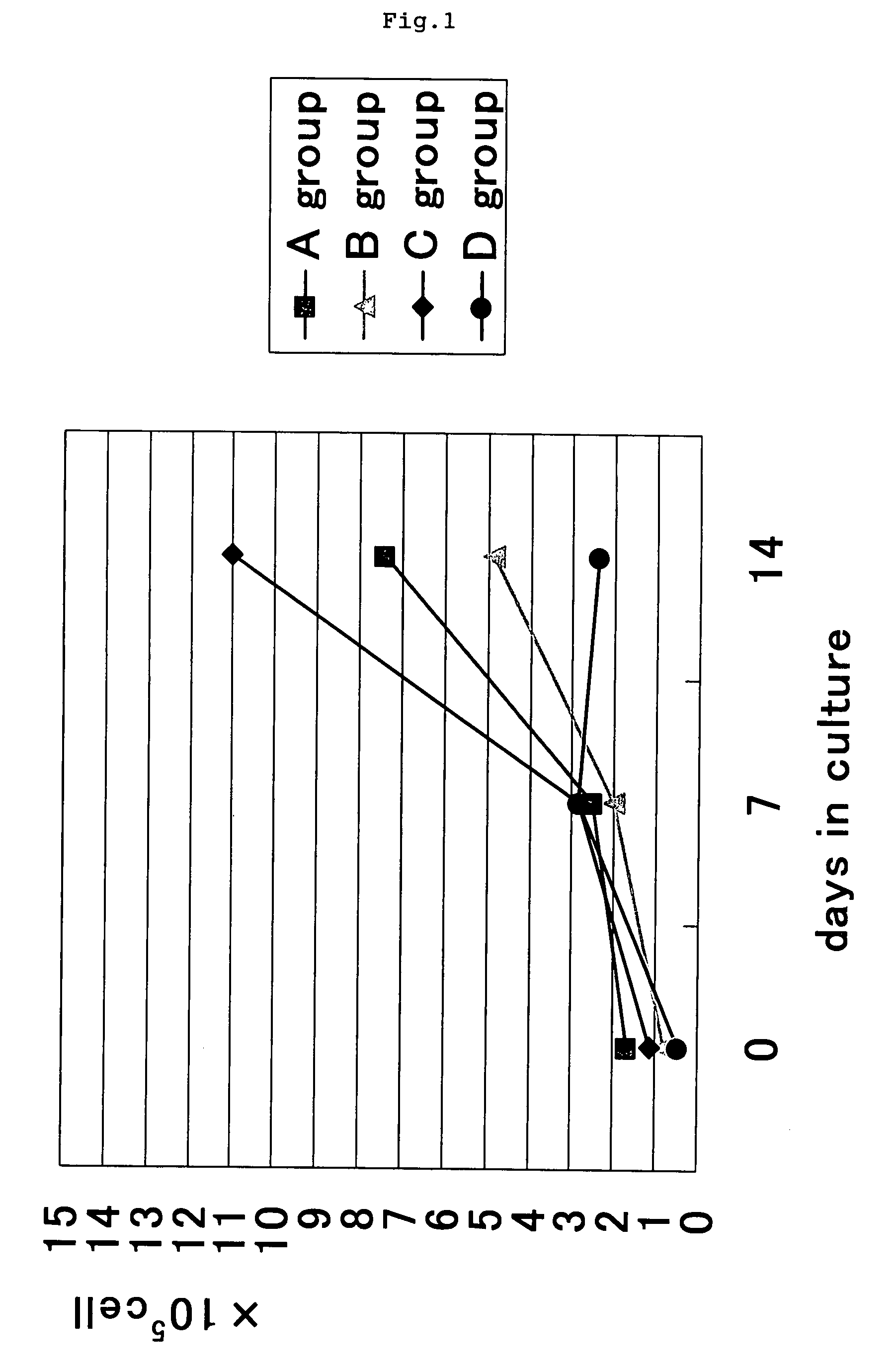
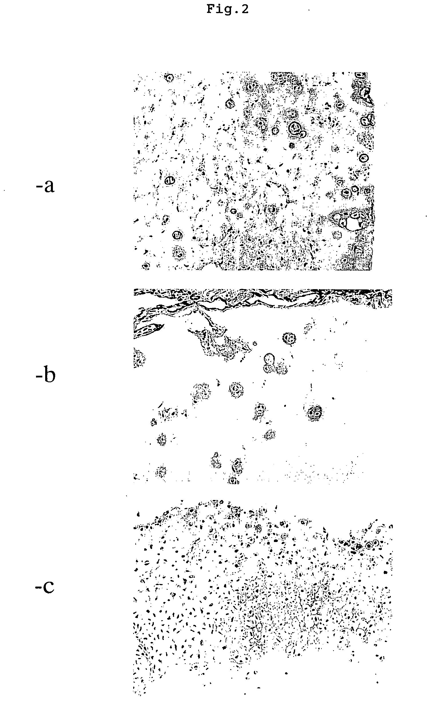
[0037]Chondrocytes (purchased from Hokudo Co., Ltd.) isolated from a rabbit knee joint cartilage were used. The chondrocytes obtained after the first passage were used for the experiment. A culture medium RPMI 1640 containing 10% FBS and 100 μM ascorbic acid (Hokudo Co., Ltd.) was used. A mixture containing following ratio, 3% Type I collagen:(proteoglycan+culture medium)=65 μL:60 μL was prepared and the chondrocytes ( about 1×10⁵)were added thereto (weight ratios of atelocollagen proteoglycan, A group [1:0], B group [1:0.05], C group [1:0.51] and D group [1:2.56]). Then the mixture was thoroughly stirred, dropped on a 35 mm dish, and gelled by incubating at 37° C. for 10 minutes. After confirming that the gel was formed well, 2.5 mL of the culture medium was added. The cells were cultured in 5% CO2 at 37° C. The medium was changed twice a week, and the culture was performed for 21 days.

[0038]In the meantime, the cell number was counted on the 7th and 14th day...


PUM

PropertyMeasurementUnit
Weight ratioaaaaaaaaaa
Structureaaaaaaaaaa

Abstract

It is intended to provide a method of three-dimensionally culturing normal joint chondrocytes; the production and supply of chondrocytes; and a transplantation material to be used in an injured site in a joint tissue.

Description

TECHNICAL FIELD[0001]The present invention mainly relates to a method of producing chondrocytes, and to providing a transplantation material to an injured site in a cartilage tissue. The present invention also relates to a kit for studying the cartilage tissue.BACKGROUND TECHNOLOGY[0002]When a damage or a defect occurs in a joint cartilage due to some cause, it is rare that the joint cartilage is completely restored and regenerated. One of the reasons includes a nature of chondrocyte. That is, as the joint chondrocytes are highly differentiated, scarcely proliferate and differentiate, their restoration capacity is extremely low. Another reason includes a peripheral inherent environment surrounding the chondrocytes. Since a cartilage tissue lacks a nerve and a blood vessel, no restoration mechanism by hemorrhage, inflammation and granulation is available. Since a periphery of the cartilage tissue is surrounded by extracellular matrix, the chondrocytes can not easily migrate from a he...

Claims


Application Information

Patent Timeline
IPC IPC(8): A61K35/12C12N11/10A61P19/00A61L27/00A61L27/22A61L27/24A61L27/38C12N5/07C12N5/077C12N11/04
CPCA61L27/3817A61L27/3852A61L27/3895C12N2533/70C12N5/0655C12N2533/54A61L2430/06A61P19/00A61P19/02
Inventor OHSHIKA, SHUSATAKAGAKI, KEIICHITOH, SATOSHIISHIBASHI, YASUYUKIYOKO, TAKAGAKI
Owner OTSUKA PHARM CO LTD
Why Eureka
Try Eureka