Method and Apparatus for Providing Depth-of-Interaction Detection Using Positron Emission Tomography (PET)

a technology of positron emission tomography and depth-of-interaction detection, which is applied in the field of nuclear medicine imaging, can solve the problems of reducing active area, negatively affecting imaging, and resolution, and achieves the effect of increasing resolution and minimizing the edge

Active Publication Date: 2009-01-08
SIEMENS MEDICAL SOLUTIONS USA INC
View PDF7 Cites 16 Cited by
  • Summary
  • Abstract
  • Description
  • Claims
  • Application Information

AI Technical Summary

Benefits of technology

[0006]According to certain embodiments, a detector is provided that employs a positron emission tomography (PET) scintillation block, which supplies depth-of-interaction (DOI) information. The DOI information is acquired by reading out t

Problems solved by technology

Unfortunately, this approach has the drawback of a reduced active area due to “edge effects.” That is, near edges (or corners) photons are not easily directed and can result in misinformation, thereby negatively impact imaging; notably the resolution is reduced.
That is, when gamma interactions o

Method used


Image

  • Method and Apparatus for Providing Depth-of-Interaction Detection Using Positron Emission Tomography (PET)
  • Method and Apparatus for Providing Depth-of-Interaction Detection Using Positron Emission Tomography (PET)
  • Method and Apparatus for Providing Depth-of-Interaction Detection Using Positron Emission Tomography (PET)

Examples


Embodiment Construction

[0015]A method and apparatus for providing depth-of-interaction detection using position emission tomography (PET) are described. In the following description, for the purposes of explanation, numerous specific details are set forth in order to provide a thorough understanding of the embodiments of the invention. It is apparent, however, to one skilled in the art that the embodiments of the invention may be practiced without these specific details or with an equivalent arrangement. In other instances, well-known structures and devices are shown in block diagram form in order to avoid unnecessarily obscuring the embodiments of the invention.

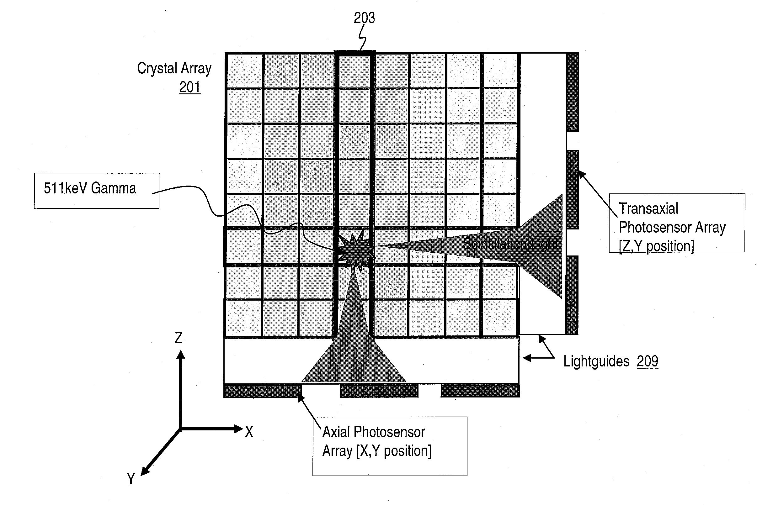
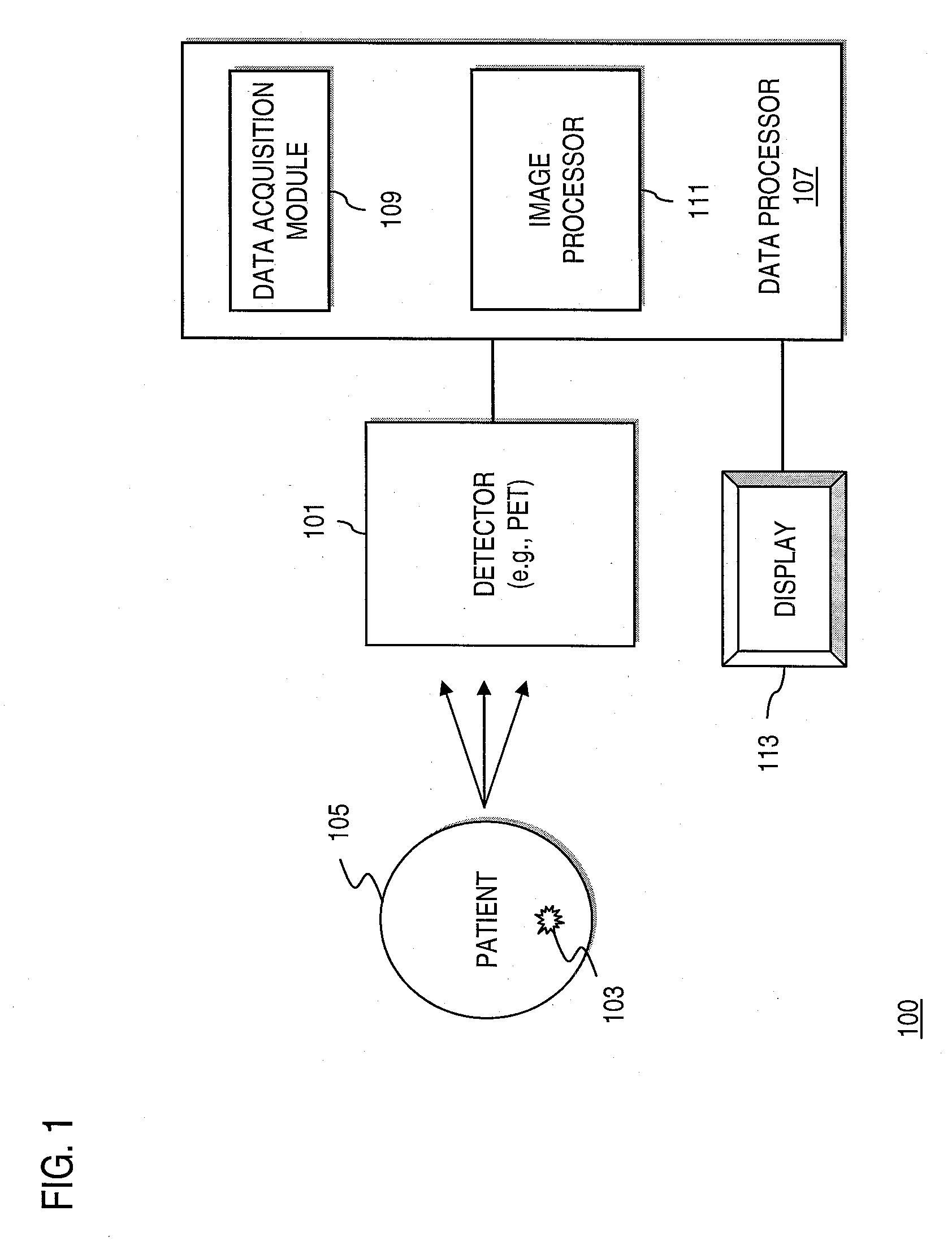
[0016]FIG. 1 is a diagram of a detection system utilizing a PET scintillation block for generating depth-of-interaction information, according to various embodiments. As shown, a detection system 100 includes a detector 101 to observe events stemming from a radiation source 103 emitting radiation (e.g., gamma rays) from a subject (patient) 105. Th...


PUM


Abstract

A detector is provided for nuclear medicine imaging. Scintillator pixels form an axial array and a transaxial array. A first photosensor is positioned along the axial array; and a second photosensor is positioned along the transaxial array, wherein the first photosensor and the second photosensor provide dual event localization for nuclear medicine imaging.

Description

FIELD OF THE INVENTION[0001]The present invention, according to certain embodiments, relates to nuclear medicine imaging.BACKGROUND OF THE INVENTION[0002]Medical radionuclide imaging, commonly referred to as nuclear medicine, is a significant diagnostic tool that involves the use of ionizing radiation to obtain accurate imaging of an in vivo patient. Typically, one or more biologically appropriate radiopharmaceuticals are administered to a patient, as by ingestion, inhalation, or injection. Tracer amounts of these radioactive substances emanate gamma quanta while localizing at specific organs, bones, or tissues of interest within the patient's body. One or more radiation detectors (e.g., positron emission tomography (PET) detector) are then used to record the internal spatial distribution of the radiopharmaceutical as it propagates from the study area. Known applications of nuclear medicine include: analysis of kidney function, imaging blood-flow and heart function, scanning lungs f...

Claims


Application Information

Patent Timeline
IPC IPC(8): G01T1/20
CPCA61B6/037G01T1/1644G01T1/2002
Inventor GRAZIOSO, RONALDAYKAC, MEHMET
Owner SIEMENS MEDICAL SOLUTIONS USA INC
Why Eureka
Try Eureka