

System and Method for Estimating Cardiac Pressure Based on Cardiac Electrical Conduction Delays Using an Implantable Medical Device
a technology of electrical conduction delay and cardiac electrical pressure, which is applied in the field of implantable medical devices, can solve the problems of long delay and associated with worsening cardiac status, and achieve the effects of reducing venous return, rapid pacing, and precise calibration
- Summary
- Abstract
- Description
- Claims
- Application Information
AI Technical Summary
Benefits of technology
Problems solved by technology
Method used
Image

Examples
Embodiment Construction
[0041]The following description includes the best mode presently contemplated for practicing the invention. The description is not to be taken in a limiting sense but is made merely for the purpose of describing the general principles of the invention. The scope of the invention should be ascertained with reference to the issued claims. In the description of the invention that follows, like numerals or reference designators will be used to refer to like parts or elements throughout.
Overview of Implantable Medical System
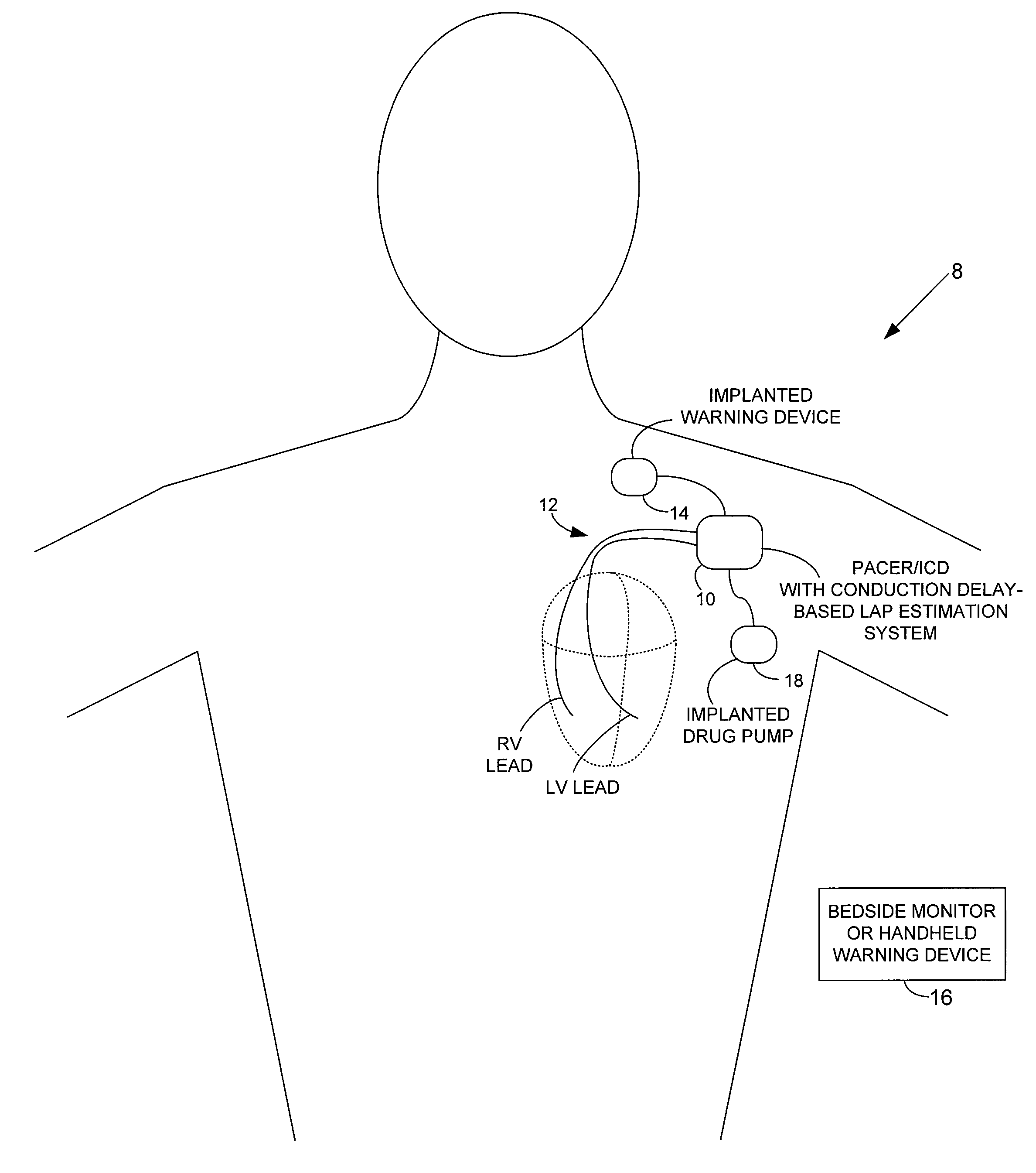
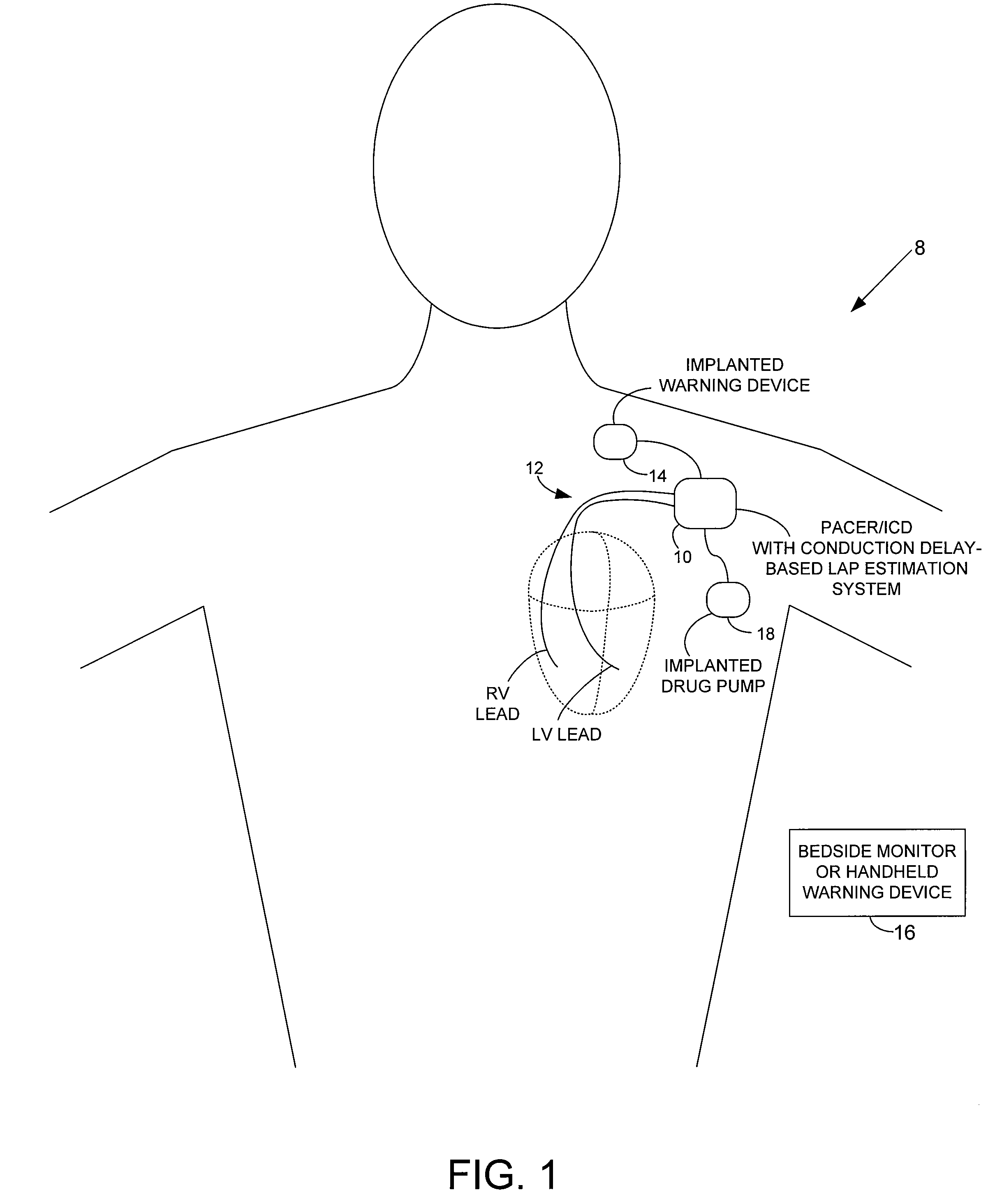
[0042]FIG. 1 provides a stylized representation of an exemplary implantable pacing medical system 8 capable of detecting electrical conduction delays within the heart of the patient and estimating LAP based on the conduction delays. To this end, implantable system 8 includes a pacer / ICD 10 or other cardiac stimulation device that incorporates internal components (shown individually in FIG. 15, and discussed below) for detecting one or more conduction delays using elec...
PUM
 Abstract
Description
Claims
Application Information