

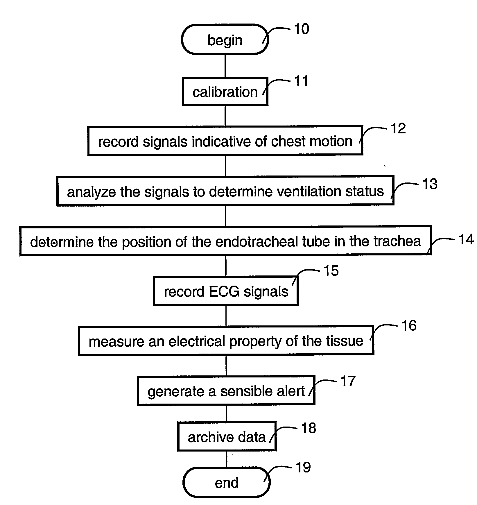
Method device and system for monitoring lung ventilation
a technology for monitoring devices and lung ventilation, applied in the field of lung ventilation, can solve problems such as inability to fix the inner end, misplacement of the tube, both during intubation or during ventilation, and can have dire consequences, and achieve the effects of reducing the number of patients
- Summary
- Abstract
- Description
- Claims
- Application Information
AI Technical Summary
Benefits of technology
Problems solved by technology
Method used
Image

Examples
example 1
Ventilation Indices
[0144]Following is a list of ventilation indices which can be used to characterize the ventilation status, according to various exemplary embodiments of the present invention. Each of the following ventilation indices is preferably calculated as a function of time and for each sensing location. An averaging procedure can also be employed either over a predetermined period of time and / or over two or more sensing locations. Additionally, an interpolation procedure can be employed so as to obtain spatial distribution of the ventilation index. Thus, each of the following ventilation indices can be a function of both time and space, a function of time, a function of space or a number.
Tidal Motion Index (TMI)
[0145]The TMI is preferably defined to describe the local peak-to-peak amplitude changes of the chest wall displacement during ventilation. The TMI can be calculated by subtracting the lowest amplitude of the chest wall from the highest amplitude of the chest wall o...
example 2
Ventilated Lung Model
[0155]A simplified scheme of a ventilated lung is illustrated in FIG. 4a.
[0156]Vin represents the mechanical ventilation machine generating a pulsatile pressure or flow.
[0157]R_T represents the impedance to flow through the endotracheal tube. The thin tubes (3-4 millimeters in diameter for neonates) impose high resistance, especially at high velocity.
[0158]R_L represents the air leak through the upper air ways (mouth and nose), especially when the tip of the tube in place in the oropharynx. It is described by a variable resistor. When the tube is displaced to the oropharynx this resistance decreases to simulate the large leak. When the tube is placed in the appropriate position and a balloon is inflated to fix the position of the tube and to prevent leak this resistance approach infinity.
[0159]R_SS, R_SP and C_S represent the components of the impedance to flow into the stomach. This impedance include a serial resistive element (flow in the esophagus) R_SS, and...
example 3
Experimental Trial
[0166]Following is a description of an experiment in which chest wall motion in a ventilated rabbit model was monitored according to preferred embodiments of the present invention using a prototype system.
Methods
[0167]The experiments were performed according to the guidelines approved by the Institutional Animal Experimental Ethics Committee of the Technion-Israeli Institute of Technology. The experimental study was carried in 7 adult male rabbits, 2.3±0.2 Kg of weight. The experimental subjects were anesthetized by intramuscular injection of Xylazine (10 mg / kg), Ketamine (90 mg / kg) and Fentanyl (0.2-0.6 ml / kg) with repeated doses during the experiments. Endotracheal intubation was performed by direct laryngoscopy and a 3 mm diameter endotracheal tube (ETT—Portex Ltd., UK) was introduced into the trachea.
[0168]Mechanical ventilation was initiated and ventilatory parameters were standardized in order to keep the blood PH, PO2, and PCO2 within physiological limits. E...
PUM
 Abstract
Description
Claims
Application Information