

Imaging facial signs of neuro-physiological responses
a neuro-physiological response and facial sign technology, applied in the field of peripheral sympathetic response detection via imaging, can solve the problems of limited time resolution of sympathetic activation thereof, difficult to determine the activation of sympathetic activation through vital sign monitoring, and often affected vital signs, so as to reduce the restrictions on a subject
- Summary
- Abstract
- Description
- Claims
- Application Information
AI Technical Summary
Benefits of technology
Problems solved by technology
Method used
Image

Examples
example 1
Data Collection
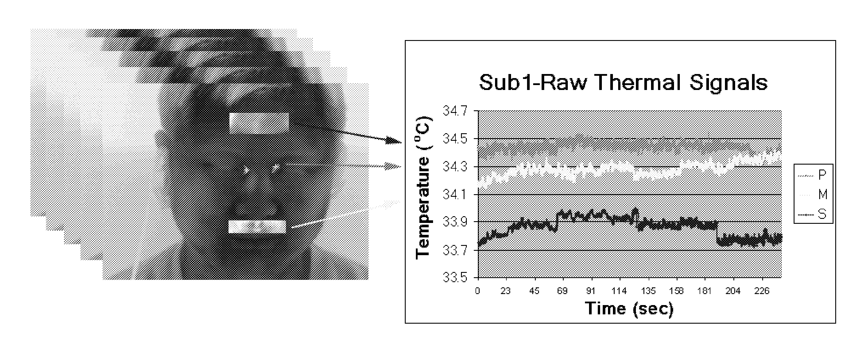
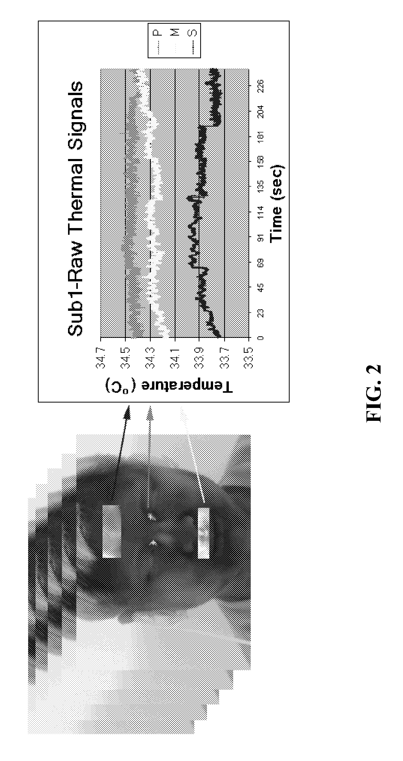
[0059]A high quality Thermal Imaging (TI) system was used for data collection. The centerpiece of the TI system was a ThermoVision SC6000 Mid-Wave Infrared (MWIR) camera9 (NEDT=0.025C). Ten (10) thermal clips were recorded from the faces of ten (10) subjects while resting in an armchair. Concomitantly, ground-truth GSR, palm thermistor, and piezo-respiratory signals were recorded with the PowerLab 8 / 30, ML870 data acquisition system.10 The data set featured subjects of both genders, different races, and with varying physical characteristics. The subjects were focused on a mental task while they were measured using the thermal imaging and contact sensors. The experiment duration was 4 minutes. Exclusion criteria were the presence of any overt peripheral neuropathy or psycho-physiological disorder. The subjects were asked to abstain from consuming vasomotor substances (e.g., caffeine and nicotine) for at least three hours prior to participating in the experiment. All pa...
example 2
Applying Modeling Methodology to GSR Waveforms
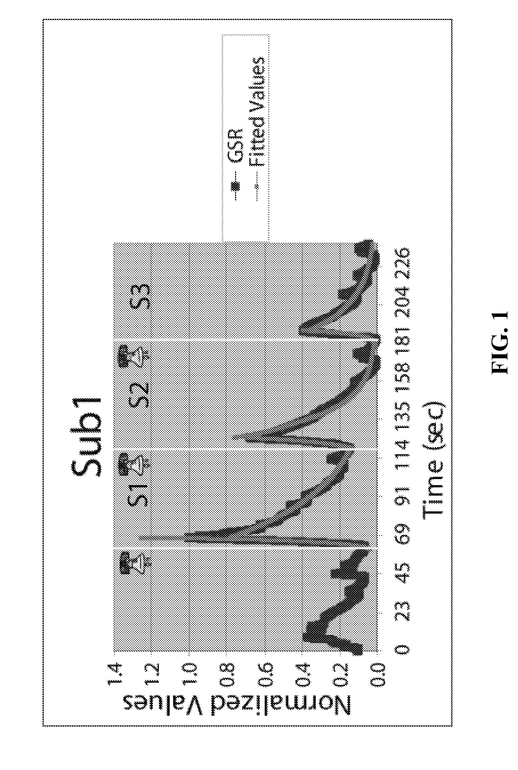
[0061]The GSR modeling methodology disclosed in Section IIA above was applied to each segment of every GSR waveform obtained as described in Example 1 hereinabove. Specifically, the disclosed modeling scheme was tested to verify that it shows that individuals tend to habituate and therefore, GSR amplitudes tend to reduce, latencies tend to increase, and wave-shapes tend to remain unaltered. These well-established and understood patterns of repeated arousals in normal subjects were used to validate the experimental design and execution.
[0062]Subjects were stimulated with 3 auditory startles spaced at least 1 minute apart. Therefore, 3 segments (S1, S2, S3)×3 sub-segments (LS, RS, LSOS)×10 subjects=90 cases were obtained, for which the scale parameter β (Laplace fitting for LS and RS) or the slope (linear fitting for LSOS) had to be estimated. Table 1 presents the estimated β parameters for the LS and RS Laplace distributions along with th...
example 3
Comparative Wavelets Analysis Results
[0068]The wavelets analysis methodology detailed in Section IIB hereinabove was used for all 6 sympathetic signals from all 10 subjects tested as described in Example 1. FIG. 3a shows the wavelet energy curves in lower scales of subject Sub1; FIG. 3b shows the wavelet energy curves in higher scales for subject Sub1. In lower scales (i.e., 50-250) the piezo-respiratory signal (Brt) appears to have a dominant component, as it is manifested by the high bell-shaped bulge. This is in accordance with its expected function. The second most prominent component is featured by the maxillary signal (M). This verifies the hypothesis of breathing modulation for this signal, as it is sampled in proximity to the nostrils.
[0069]In higher scales, (see FIG. 3b), for example from about 1000-3000, the GSR signal (GSR) appears to have a dominant component, as it is manifested by the high bell-shaped bulge. This is the phasic component as the scale is about ⅓ of the t...
PUM
 Abstract
Description
Claims
Application Information