
Papillary Muscle Attachment for Left Ventricular Reduction

a left ventricular and phlebotomy technology, applied in the field of phlebotomy for left ventricular reduction, can solve the problems of inability to efficiently pump blood around the body, weak muscle of the heart, inability to etc., to reduce the transventricular dimension of said heart, reduce the transventricular size and geometry, and reduce the length of said heart.

Inactive Publication Date: 2009-04-16
MIAMI THE UNIV OF
View PDF99 Cites 104 Cited by
  • Summary
  • Abstract
  • Description
  • Claims
  • Application Information

AI Technical Summary

Benefits of technology

[0004]The present invention proposes the surgical implantation of a link, which may be in the form of a tether or a looped band, to connect papillary muscles in the left ventricle to reduce dilation and improve heart function by reducing left ventricular failure and decreasing mitral valvular regurgitation.
[0006]Thus, the invention may be embodied in a method of treating dilated cardiomyopathy comprising: securing at least one tether structure to opposed, facing portions of first and second papillary muscles within a ventricle of the heart of a patient having dilated cardiomyopathy; and reducing a length of said at least one tether structure so as to draw said facing portions of said papillary muscles towards each other to reduce a transventricular dimension of said heart.
[0007]The invention may also be embodied in a method of reducing a transventricular size and geometry in a patient having dilated cardiomyopathy comprising: securing at least one tether structure to opposed, facing portions of first and second papillary muscles within the left ventricle of said patient's heart; and reducing a distance between said papillary muscles by drawing said facing portions of said papillary muscles towards each other with said at least one tether structure to reduce a transventricular size and geometry of the patient's heart, thereby to mitigate the affects of the dilated cardiomyopathy. Decreasing the distance between the papillary muscle will also more appropriately align the chordal apparatus to decrease mitral regurgitation.

Problems solved by technology

Ischemic and Non Ischemic Dilated Cardiomyopathy causes the heart to become enlarged and to function poorly.
As a result, the muscle of the heart becomes weak, thin or floppy and is unable to pump blood efficiently around the body.
This typically causes fluid to build up in the lungs which therefore become congested, resulting in a feeling of breathlessness.
Often there is also right heart failure which causes fluid to accumulate in the tissues and organs of the body, usually the legs and ankles, and the liver and abdomen.
Left ventricular dilation can also lead to secondary Mitral valvular regurgitation, further worsening cardiac performance.

Method used


Image

  • Papillary Muscle Attachment for Left Ventricular Reduction
  • Papillary Muscle Attachment for Left Ventricular Reduction
  • Papillary Muscle Attachment for Left Ventricular Reduction

Examples


Embodiment Construction

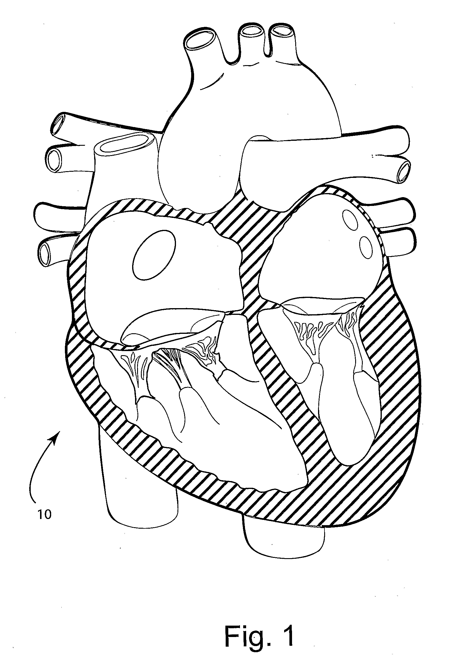
[0016]As noted above, Dilated Cardiomyopathy is a condition wherein the heart has become enlarged and too weak to efficiently pump blood around the body causing a build up of fluid in the lungs and / or tissue. FIG. 1 illustrates a normal four chamber heart 10 whereas FIG. 3 illustrates the enlarged, thin walled heart 110 of a patient having Dilated Cardiomyopathy.

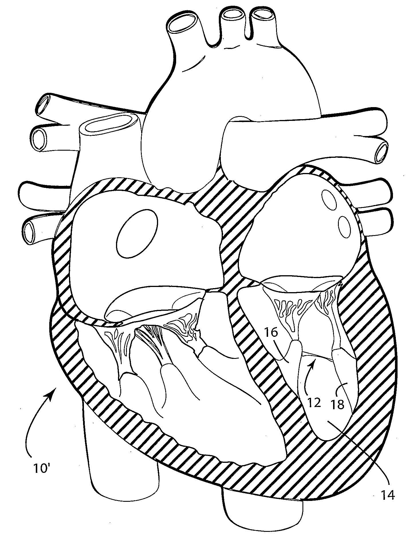
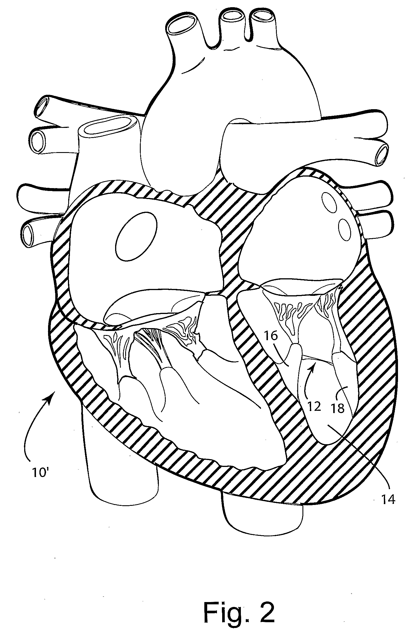
[0017]Referring to FIG. 2, some individuals have a congenital malformity of the heart in the form of a false tendon, more specifically, a left ventricular abnormal tendon 12 spanning the ventricular cavity 14 between the two papillary muscles 16, 18. This congenital malformation has no apparent affect on the function of an otherwise normal heart 10′. The inventor has observed, however, that patients with Dilated Cardiomyopathy that have this congenital false tendon appear to maintain a more favorable ventricular geometry, i.e., have less ventricular dilation, and consequently a more favorable clinical course than patients wi...


PUM


Abstract

The surgical implantation of a link, which may be in the form of a tether or a looped band, is proposed to connect and reduce the spacing between papillary muscles, to reduce dilation of the left ventricle. The implanted link thus improves heart function by reducing left ventricular failure.

Description

CROSS-REFERENCE TO RELATED APPLICATION[0001]This application claims the benefit of U.S. Provisional Application No. 60 / 688,730, which was filed on Jun. 9, 2005, the disclosure of which is incorporated herein by this reference.BACKGROUND OF THE INVENTION[0002]Ischemic and Non Ischemic Dilated Cardiomyopathy causes the heart to become enlarged and to function poorly. Some people have stable disease and there is little worsening of their condition. Others have progressive disease. As a result, the muscle of the heart becomes weak, thin or floppy and is unable to pump blood efficiently around the body. This typically causes fluid to build up in the lungs which therefore become congested, resulting in a feeling of breathlessness. This is referred to as congestive (left) heart failure. Often there is also right heart failure which causes fluid to accumulate in the tissues and organs of the body, usually the legs and ankles, and the liver and abdomen. Left ventricular dilation can also lea...

Claims


Application Information

Patent Timeline
IPC IPC(8): A61F2/02
CPCA61B17/00234A61B17/0401A61B17/0487A61F2/2457A61B17/10A61B2017/00243A61B2017/0496A61B17/083
Inventor DE MARCHENA, EDUARDO
Owner MIAMI THE UNIV OF
Learn More