Percutaneously deliverable heart valve

a prosthetic heart valve and implantable technology, applied in the field of prosthetic heart valves, can solve the problems of affecting the function of the mitral valve apparatus, the valve itself will decay, degenerate or disrupt, and require repair or replacement, and achieve the effect of minimal invasive delivery

Inactive Publication Date: 2005-07-28
3F THERAPEUTICS
View PDF20 Cites 476 Cited by
  • Summary
  • Abstract
  • Description
  • Claims
  • Application Information

AI Technical Summary

Benefits of technology

[0026] It is still another object of the present invention to provide a method for minimally invasively delivering a foldable heart valve prosthesis into a patient. The method comprises folding the support structure with the attached tissue heart valve inside a lumen of a delivery apparatus; delivering the delivery apparatus to a target valvular annulus of the patient; unfolding the support structure to deploy the foldable heart valve prosthesis in place; and coupling the crossbars by sliding the slidable ring connectors to an appropriate location of the crossbars. In one embodiment, the method may further comprise a shrinking step after the coupling step, wherein the shrinking step reduces a circumferential length of the slidable ring connector.

Problems solved by technology

Similarly, they close under the pressure exerted on the same leaflet elements when blood flow is retrograde, thus impeding return of blood flow to the chamber it has just left.
When disease conditions affect the structure of the materials of the components of the valve apparatus, the valve itself will decay, degenerate or disrupt and require repair or replacement to restore proper function necessary for the continuation of life.
Those valves used in the aortic position cannot alone replace the mitral valve apparatus without anatomical and functional compromise.
In such aortic diseases, patients can have syncope, chest pain, and difficulty in breathing.
The evolution of such a disease is disastrous when symptoms of cardiac failure appear and many patients die in the year following the first symptoms of the disease.
Unfortunately, the result is sub-optimal with a high restenosis rate.
Until recently, the implantation of a valve prosthesis for the treatment of aortic stenosis is considered unrealistic to perform since it is deemed difficult to superimpose another implantable valve on the distorted stenosed native valve without excising the latter.
However, the support structure is a generally fixed expandable-collapsible structure with little flexibility of modifying configuration at the collapsed shape for easy delivery percutaneously.
This type of design is inherently fragile, and such structures are not strong enough to be used in the case of aortic stenosis because of the strong recoil that will distort this weak structure and because they would not be able to resist the balloon inflation performed to position the implantable valve.
However, the expandable frame is a generally fixed expandable-collapsible structure with little flexibility of modifying configuration or minimizing the profile at the collapsed stage for easy delivery percutaneously.
In some aspects, this intravascular procedure is not only dangerous and tedious because of the long tortuous route, but also of limited use because of the catheter size suitable for insertion intravascularly.
However, the presence of the support structure consisting of a stent and sewing ring prevents the tissue valve from being anatomically accurate in comparison to a normal heart valve.

Method used


Image

  • Percutaneously deliverable heart valve
  • Percutaneously deliverable heart valve
  • Percutaneously deliverable heart valve

Examples


Embodiment Construction

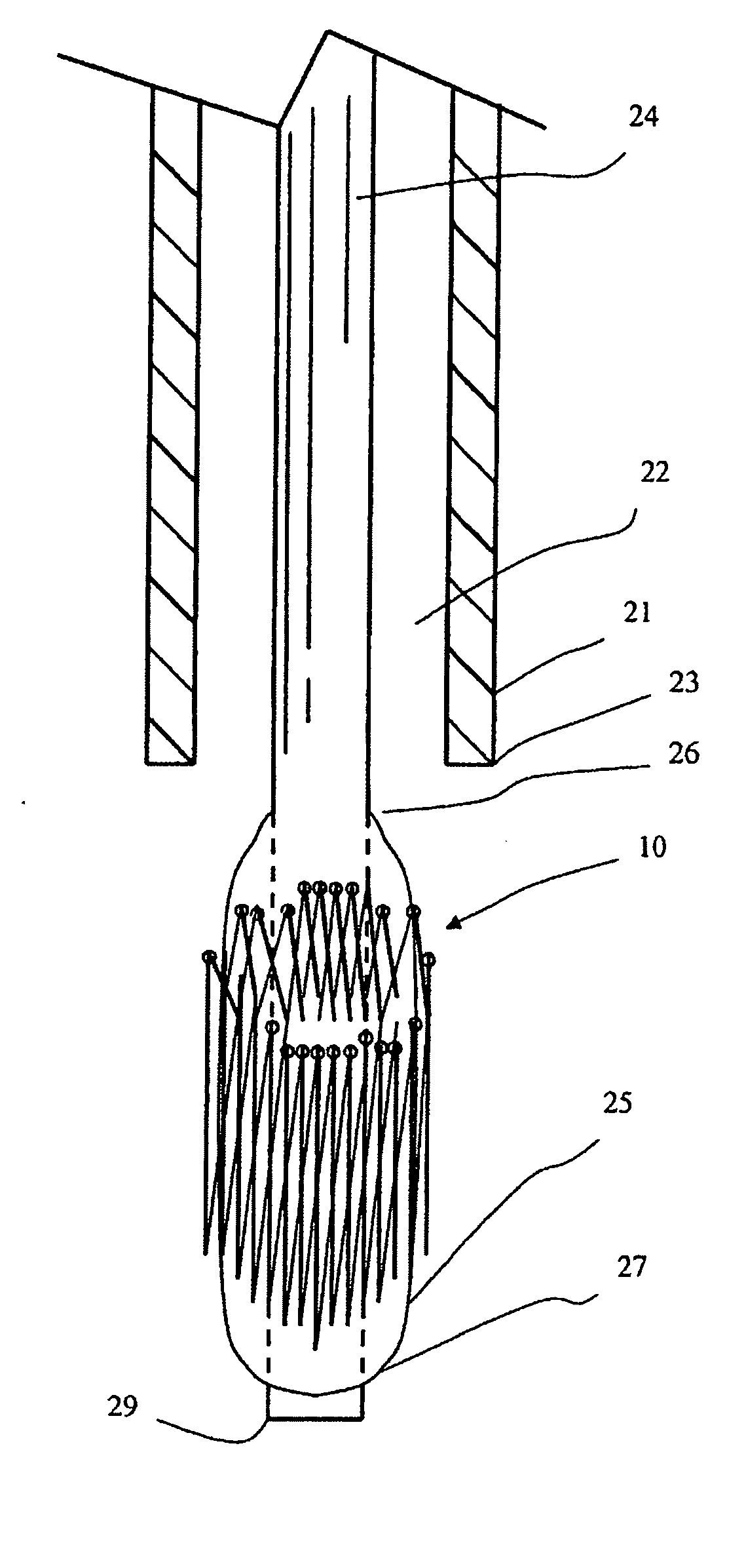
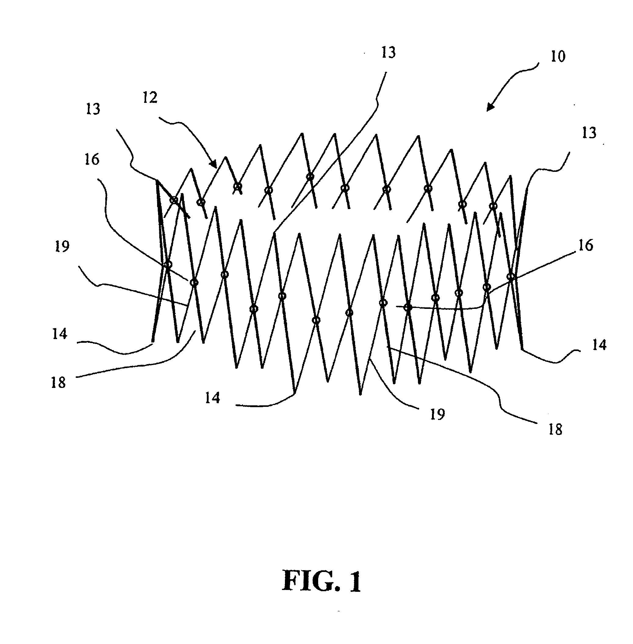
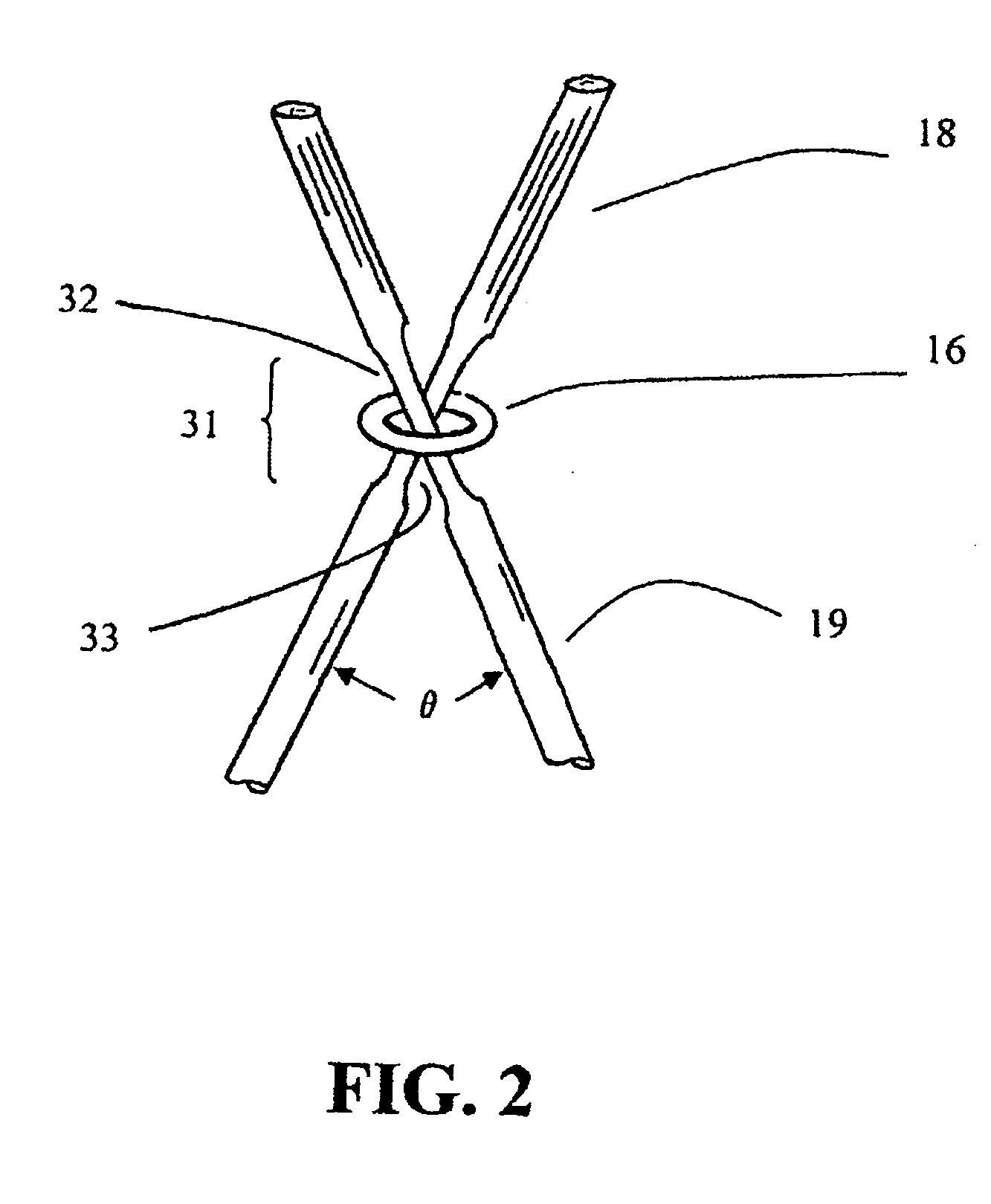
[0037] Referring to FIGS. 1 to 8, what is shown is an embodiment of a percutaneously deliverable heart valve prosthesis and delivery means thereof, including an illustrative cardiac valve. The same principles of percutaneously implantable valves could also apply to implantation of a venous valve, an esophagus valve, ureter valve, a biliary valve, and a valve in the intestines or in the lymphatic systems. While the description sets forth various embodiment specific details, it will be appreciated that the description is illustrative only and should not to be construed in any way as limiting the invention. Furthermore, various applications of the invention, and modifications thereto, which may occur to those who are skilled in the art, are also encompassed by the general concepts described below.

[0038] Andersen et al. in U.S. Pat. No. 6,168,614, No. 5,840,081 and No. 5,411,552 discloses a valve prosthesis for implantation in the body by use of a catheter comprising a stent made from ...


PUM


Abstract

This invention discloses a percutaneously deliverable heart valve and delivery means thereof, wherein the percutaneously deliverable heart valve is a foldable heart valve prosthesis to replace a diseased valve of a patient comprising: a support structure with a diameter, wherein the support structure is foldable to a smaller diameter, the support structure comprising a plurality of crossbar frames, wherein each crossbar frame has a plurality of crossbars connected at an end of each crossbar; a flexible tissue heart valve with a plurality of valvular leaflets attached to the support structure; and a plurality of slidable ring connectors, wherein at least a slidable ring connector encircles a first crossbar from a first crossbar frame and a second crossbar from a second crossbar frame configured to coupling the first and the second crossbars.

Description

RELATED APPLICATION [0001] The present application is a continuation of U.S. patent application Ser. No. 10 / 241,718, the entirety of which is hereby incorporated by reference.FIELD OF THE INVENTION [0002] The present invention relates to a prosthetic valve for implantation in a body channel, more particularly, to a percutaneously implantable prosthetic heart valve suitable for replacement of a defect or diseased human heart valve. BACKGROUND OF THE INVENTION [0003] Replacement heart valves or heart valve prostheses have been fabricated or manufactured for the last forty years. Such devices have been assembled from a variety of materials. Specifically the materials have been of the biologic or artificial nature, generally leading to two distinct categories of the prostheses as biological or mechanical replacement heart valves. [0004] The prosthetic heart valves are fabricated to replace the natural heart valves that, because of disease, congenital malformations, aging or trauma have ...

Claims


Application Information

Patent Timeline
Patent Type & Authority Applications(United States)
IPC IPC(8): A61F2/24
CPCA61F2/2418A61F2/2475A61F2230/0054A61F2220/005A61F2220/0075A61F2220/0016
Inventor ANDUIZA, JAMES PETERQUIJANO, RODOLFO C.TU, HOSHENG
Owner 3F THERAPEUTICS
Why Eureka
Try Eureka