Method of efficient extraction of protein from cells

a cell and protein technology, applied in the field of cell protein extraction, can solve the problems of false negative results, up to 40% of conventional pap tests are compromised, and tests are prone to errors

Active Publication Date: 2009-05-14
ARBOR VITA CORP +1
View PDF31 Cites 18 Cited by
  • Summary
  • Abstract
  • Description
  • Claims
  • Application Information

AI Technical Summary

Problems solved by technology

Despite the success of such cytological tests, the tests are prone to error.
For example, it has been estimated that up to 40% of conventional Pap tests are compromised by the presence of contaminants such as mucous, blood cells and obscuring inflammatory cells.
These contaminants lead to false negative results, false positive results, and a significant amount of follow-up work.
Such methods are not straightforward, however, because it is not always possible to perform such methods on fixed cells.
For example, certain fixatives (e.g., those transport media employed in THINPREP™ or SUREPATH™ test systems) may cause particular cellular proteins to precipitate or aggregate, thereby making those proteins insoluble and difficult or impossible to reliably detect using conventional means, e.g., using an enzyme-linked immunosorbancy assay (ELISA) or another immunological test.

Method used


Image

  • Method of efficient extraction of protein from cells
  • Method of efficient extraction of protein from cells
  • Method of efficient extraction of protein from cells

Examples


example 1

Extraction of Spiked Clinical Samples

[0142]Cells transfected with the HPV16 E6 gene (C33A+) were fixed with THINPREP™ medium and added (i.e., “spiked”) into portions of the THINPREP™-fixed clinical samples as listed below. The cells were spiked into half of each of five clinical samples (each half clinical sample having 20 million C33A+ cells).

[0143]Extraction Scheme:

[0144]C33A(+) ThinPrep cells / 20M cells per ml

[0145]1-20M C33A(+) ThinPrep cells into ½ clinical negative #229 (1.0 ml extraction)

[0146]2-20M C33A(+) ThinPrep cells into ½ clinical negative #230 (1.0 ml extraction)

[0147]3-20M C33A(+) ThinPrep cells into ½ clinical negative #231 (1.0 ml extraction)

[0148]4-20M C33A(+) ThinPrep cells into ½ clinical negative #232 (1.0 ml extraction)

[0149]5-20M C33A(+) ThinPrep cells into ½ clinical negative #233 (1.0 ml extraction)

[0150]6-20M C33A(+) ThinPrep cells (1.0 ml extraction)

[0151]Extraction Reagent:

[0152]Triton X-100 / Lot 092K0171—(1%=250 ul)

[0153]5M NaCl / Lot 5701-53—(0.15M=750 ul)...

example 2

Effect of High pH Buffer Plus Additives on MBP-E6 Detection

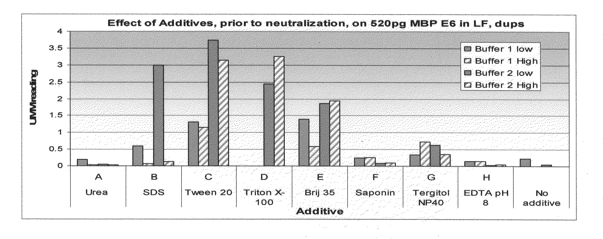
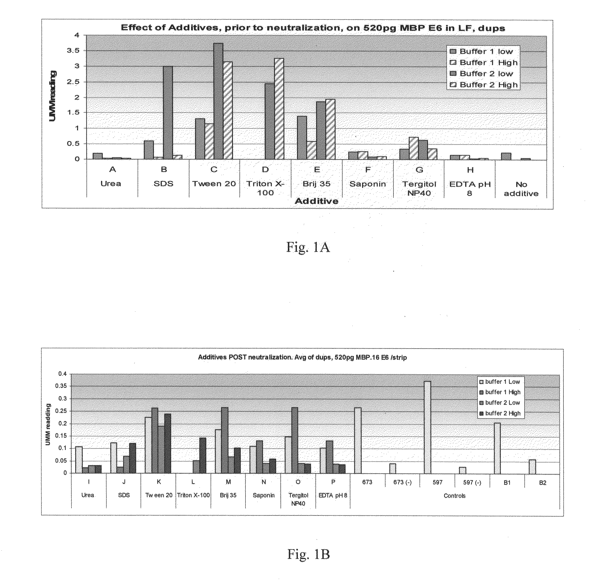
[0209]The example described in FIG. 1 illustrates the effect of buffers of differing composition on the detection of recombinant maltose-binding protein (MBP)-E6 protein. Ordinarily, the extraction buffers described herein would be used to extract protein from cells. Here, however, previously-purified recombinant protein was used in order to optimize conditions. Briefly, the experiments described in FIG. 1A involved suspending the MBP-E6 protein in freshly-prepared extraction buffers differing in additive content and pH. This “extraction” step was followed by a neutralization step in which the pH of the sample was adjusted to a neutral pH. MBP-E6 protein was then detected using a lateral flow assay.

[0210]Composition of the Buffers

[0211]The buffers used in the experiments described herein were derived from one of two buffers, either a lower pH buffer, “Buffer 1” or a higher pH buffer, “Buffer 2.” Buffer 1 (pH of about 11.5) c...

example 3

Effect of Additives on the Extraction of E6 Protein From Cells

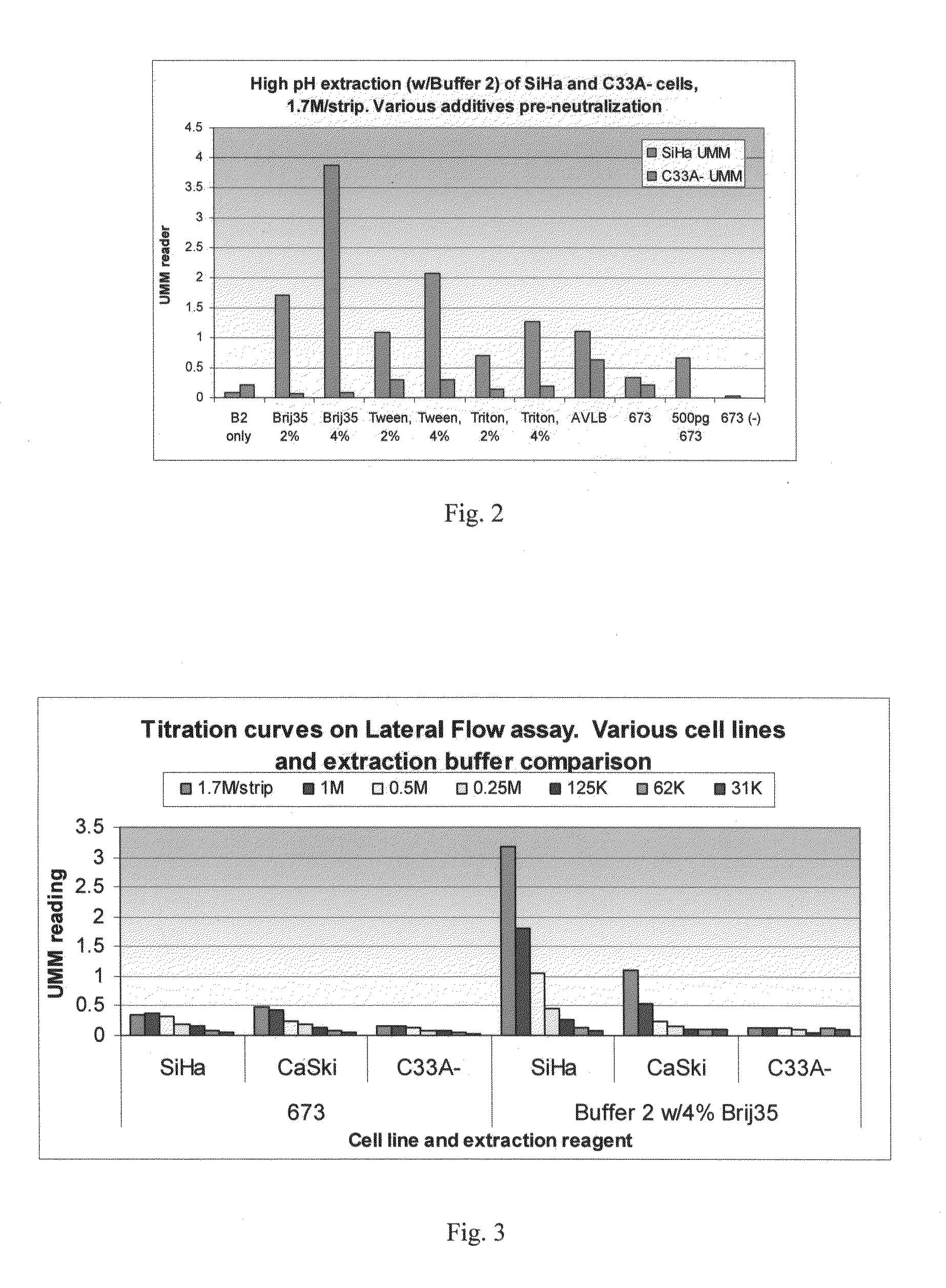
[0226]The example illustrated by FIG. 2 evaluated the effect of different percentages of buffer additives on the extraction of E6 protein from cells. In this example, protein was extracted either from SiHa cells expressing the HPV16 E6 gene or from C33A-cells, which are a non-HPV infected cervical cancer cell line.

[0227]Extraction Step

[0228]For the extraction step, cell pellets containing about 10 million cells were first removed from a −80° C. freezer and allowed to thaw at RT for about 10 minutes. Then, about 750 μl of Buffer 2, described in Example 2, was added to most of the samples. Additives such as Brij™ 35, Tween™-20, or Triton™X-100 were also introduced to the indicated samples. These additives were present at a final concentration of either 2% or 4% (v / v). As a control, some samples were extracted with Buffer 673, a neutral pH buffer containing 20 mM Tris pH 8, 2% BSA, 1% Triton™-X 100, 2% Tween™-20, 0.2% Sarcos...


PUM

PropertyMeasurementUnit
pHaaaaaaaaaa
pHaaaaaaaaaa
pHaaaaaaaaaa

Abstract

Methods for producing a protein extract from cells, such as cells or cellular samples containing viral proteins, are provided. In general terms, the methods may involve: increasing the pH of the cells to a pH of at least about pH 10.0 to produce an intermediate composition, and then, in the presence of a non-ionic detergent such as a polyoxyethylene alkyl ether, neutralizing the pH of the intermediate composition to produce the protein extract. Such methods can be used in conjunction with methods for detecting one or more target proteins in a sample, such as viral proteins. Systems, kits and compositions for practicing the subject methods are also provided.

Description

BACKGROUND OF THE INVENTION[0001]In many diagnostic methods, cells are taken from a subject and deposited into a liquid medium containing a fixative. The cells are fixed in the medium and examined cytologically in order to provide a diagnosis. For example, detection of precancerous or cancerous cells in cervical tissue is routinely performed by microscopic assessment of exfoliated cervical cells. This method, developed by George N. Papanicolaou and known as the “Pap” test, involves exfoliating cells from a woman's cervix using a sampling device, depositing the exfoliated cells into a transport medium that contains a fixative, and then depositing the cells onto a slide. The cells are then stained and examined by light microscopy for cellular abnormalities by a trained medical professional. Over 55 million Pap tests are performed each year in the United States alone.[0002]Despite the success of such cytological tests, the tests are prone to error. For example, it has been estimated th...

Claims


Application Information

Patent Timeline
Patent Type & Authority Applications(United States)
IPC IPC(8): C12Q1/70C12P21/00G01N33/50C12Q1/00G01N33/567
CPCC07K1/145C12N2710/20022G01N2469/10G01N2333/16G01N33/56983C12N15/52G01N1/4044G01N33/54393G01N2333/025
Inventor MALICK, ADRIEN P.CREWS, VIRGINIA M.ROSALES, JULIE L.FERGUSON, CARRIE S.BRUTON, JEFF H.BEADENKOPF, ROBERT J.
Owner ARBOR VITA CORP
Why Eureka
Try Eureka