
Percutaneous Devices for Separating Tissue, Kits and Methods of Using the Same

Inactive Publication Date: 2009-06-04
VERTOS MEDICAL
View PDF3 Cites 47 Cited by
  • Summary
  • Abstract
  • Description
  • Claims
  • Application Information

AI Technical Summary

Benefits of technology

[0016]Another aspect of the invention is directed to a kit for tissue separation. Suitable kits comprise, for example, the devices disclosed herein and and packaging. Additional components of suitable kits include, but are not limited to, for example, one or more injectable media, such as a contrast medium; one or more hydrophillic-lipophillic block copolymer gels; one or more guides adaptable for use with the device.
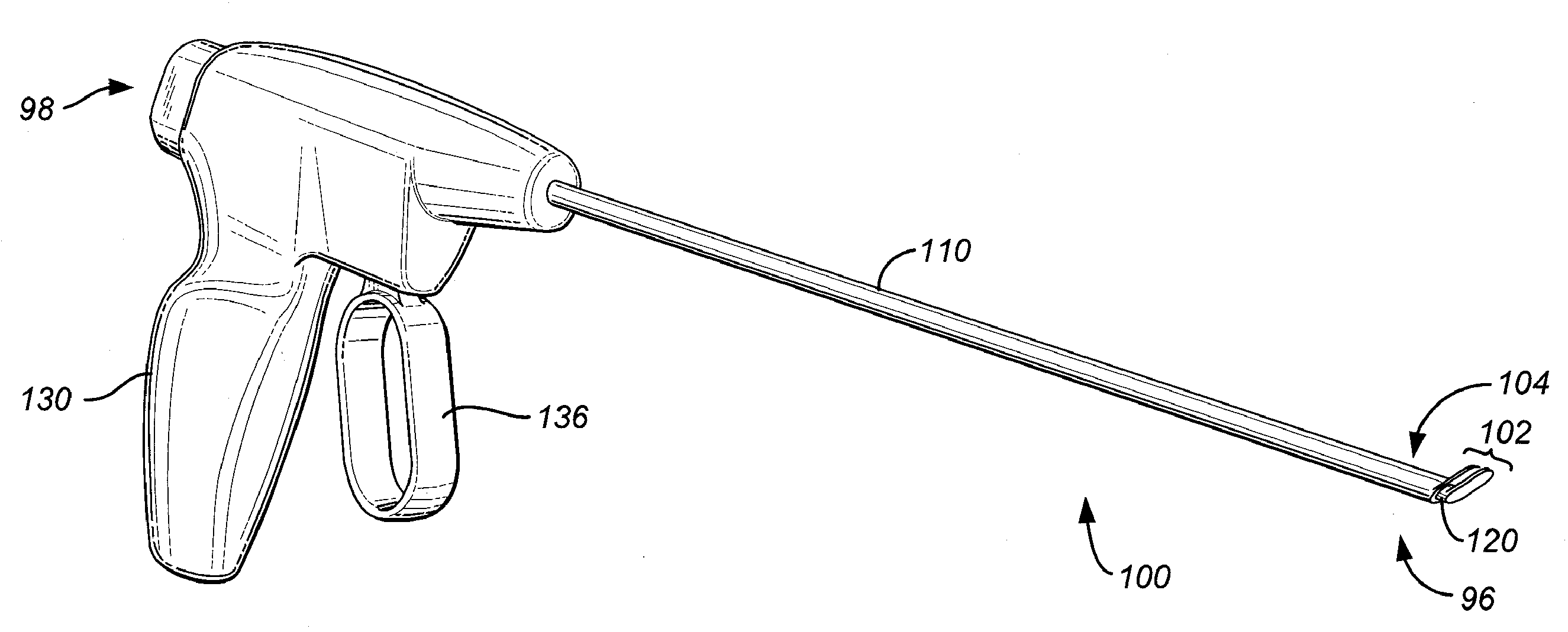
[0017]Yet another aspect of the invention is directed to methods for treating stenosis in a spine of a patient with reference to a median plane of the patient. The methods comprise, for example, the steps of: compressing a dural sac in the region of interest by injecting a fluid to form a safety zone and establish a working zone in the region of interest, the safety zone lying between the working zone and the dural sac; percutaneously accessing an epidural space in the region of interest on a first lateral side of a median plane; and inserting a device for separating a first tissue from a second tissue at a surgical site, comprising, a hollow body having a distal end and a proximal end, wherein the distal end further comprises an upper separation member extendable laterally from hollow body at a first angle; an inner member having a distal end and a proximal end and slidably disposed within the hollow body, wherein the distal end of the inner member has a lower separation member extendable laterally from inner member at a second angle, wherein the upper separation member and the lower separation member h

Problems solved by technology

In many cases, the pain severely limits a person's functional ability and quality of life.
Back pain interferes with work, routine daily activities, and recreation.
It is the most common cause of job-related disability and a leading contributor to missed work.
Spinal stenosis, a condition that results from narrowing of the spinal canal causing nerve pinching, leads to persistent pain in the buttocks, limping, lack of feeling in the lower extremities, and decreased physical activity.
The excessive epidural fat compresses the dural sac, nerve roots and blood vessels contained therein and resulting in back, leg pain and weakness and numbness of the legs.
These conservative treatment options frequently fail.
The risks associated with surgery include bleeding, blood clots and dural tears.
In some cases surgical intervention fails to relieve symptoms, or the symptoms return over time.
Also, because the spine stabilizing back muscles and ligaments are stripped and detached from the spine during the laminectomy, these patients frequently develop spinal instability post-operatively.
Percutaneous discectomy devices with fluoroscopic guidance have been used successfully to treat disorders of the disc but not to treat spinal stenosis or the ligamentum flavum directly.
These devices and techniques are limited by the small size of the canal and these operations are difficult to perform and master.
In addition, these procedures are painful and often require general anesthesia.
Further, the arthroscopy procedures are time consuming and the fiber optic systems are expensive to purchase and maintain.
Still further, because the nerves of the spinal cord pass through the spinal canal directly adjacent to and anterior to the ligamentum flavum, any surgery, regardless of whether open or percutaneous, includes a risk of damage to the nerves of the spinal cord.

Method used


Image

  • Percutaneous Devices for Separating Tissue, Kits and Methods of Using the Same
  • Percutaneous Devices for Separating Tissue, Kits and Methods of Using the Same
  • Percutaneous Devices for Separating Tissue, Kits and Methods of Using the Same

Examples


Embodiment Construction

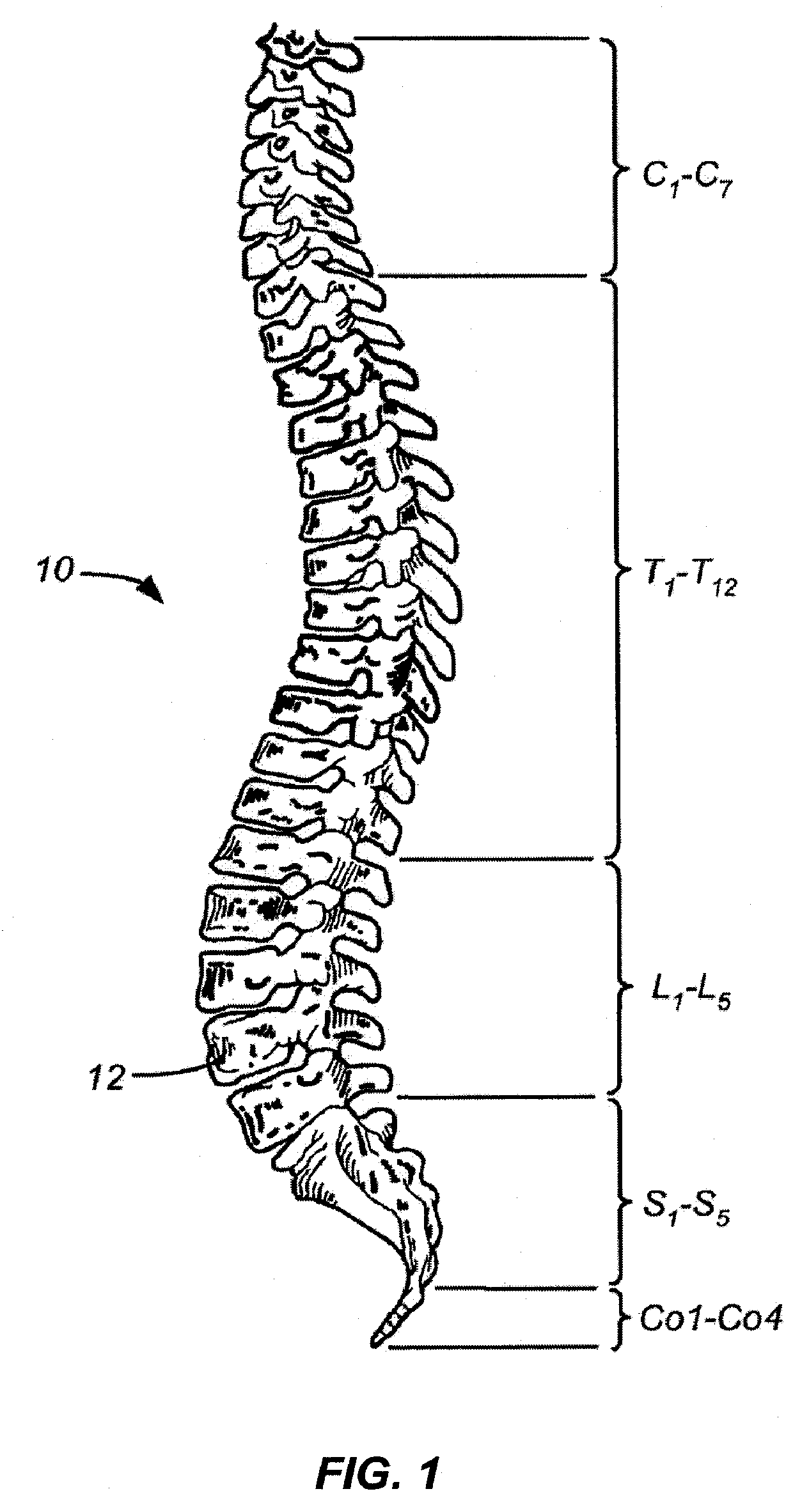
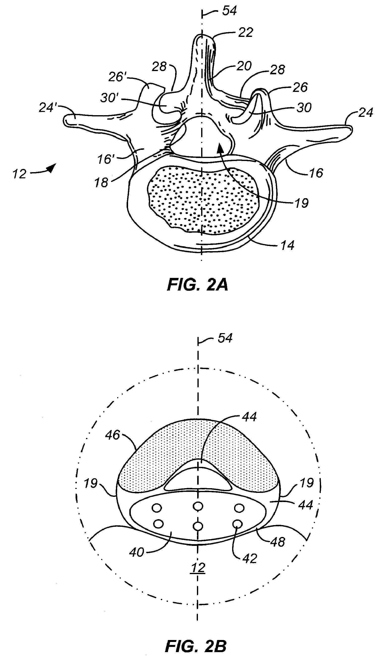
[0035]The invention relates generally to devices, apparatus or mechanisms that are suitable for use within a human body to restore, modify and / or augment tissue, and systems therefor. As will be appreciated by those skilled in the art, tissue is an aggregation of morphologically similar cells and associated intercellular matter acting together to perform one or more specific functions in the body. There are four basic types of tissue: muscle, nerve, epidermal, and connective (which includes bone and ligament).

[0036]For purposes of illustrating the usefulness of the invention, the invention is described in the context of treating spinal pathologies. However, persons of skill in the art will appreciate that the devices can be used in conjunction with other pathologies without departing from the scope of the invention. In some instances the devices can include devices designed to separate body parts or structure. The devices, apparatus or mechanisms are configured such that the devices...


PUM


Abstract

The disclosure describes devices for separating a first tissue from a second tissue at a surgical site, comprising, a hollow body having a distal end and a proximal end, wherein the distal end further comprises an upper separation member extendable laterally from hollow body at a first angle; an inner member slidably disposed within the hollow body, wherein the distal end of the inner member has a lower separation member extendable laterally from inner member at a second angle, wherein the upper separation member and the lower separation member have a first configuration and a second configuration into a tissue in the working zone on the first lateral side of the median plane. Additionally, methods for using the device and kits containing the device are disclosed.

Description

CROSS-REFERENCE[0001]This application claims the benefit of U.S. Provisional Application No. 60 / 991,946 filed Dec. 3, 2007, which application is incorporated herein by reference.BACKGROUND[0002]1. Field of the Invention[0003]The present invention relates generally to minimally invasive devices, systems methods and kits for treating spinal disorders; including devices, systems, methods and kits that use imaging guidance. More particularly, the present invention relates to devices, systems, methods and kits to reduce stenosis and increase the cross-sectional area of the spinal canal available for the spinal cord. Still more particularly, the present invention relates to devices, systems, methods and kits to percutaneously separate tissue such as portions of an enlarged ligamentum flavum from the lamina of a spine.[0004]2. Background of the Invention[0005]Back pain is a common ailment. In many cases, the pain severely limits a person's functional ability and quality of life. Back pain ...

Claims


Application Information

Patent Timeline
IPC IPC(8): A61B17/32A61B19/00
CPCA61B17/0206A61B17/0218A61B17/3415A61B2017/320044A61B2017/0256A61B2017/2925A61B2017/00867
Inventor SAND, PAUL
Owner VERTOS MEDICAL
Learn More