Capsule endoscope system and endoscopic image filing method

a technology of endoscope and endoscope, which is applied in the field of endoscope system and endoscope image filing method, can solve the problem that the treatment according to the result of diagnosis cannot be rapid, and achieve the effect of high efficiency

Inactive Publication Date: 2009-08-13
FUJIFILM CORP
View PDF7 Cites 148 Cited by
  • Summary
  • Abstract
  • Description
  • Claims
  • Application Information

AI Technical Summary

Benefits of technology

[0007]In view of the foregoing problems, an object of the present invention is to provide a capsule endoscope system and endoscopic image filing method in which image reading of endoscopic images can be carried out with high efficiency.
[0016]The receiver includes a data size reduction device for reducing a data size of the image received from the capsule endoscope before transmission in the first wireless interface.
[0027]Furthermore, a data size of the image received from the capsule endoscope to the receiver is reduced before the transmitting step.
[0032]Consequently, image reading of endoscopic images can be carried out with high efficiency, because an image is wirelessly transmitted from the receiver toward an information manager for image filing during imaging with the capsule endoscope.

Problems solved by technology

There is a problem in that treatment according to a result of the diagnosis cannot be rapid, as considerably long time is required for the diagnosis.

Method used


Image

  • Capsule endoscope system and endoscopic image filing method
  • Capsule endoscope system and endoscopic image filing method
  • Capsule endoscope system and endoscopic image filing method

Examples


first embodiment

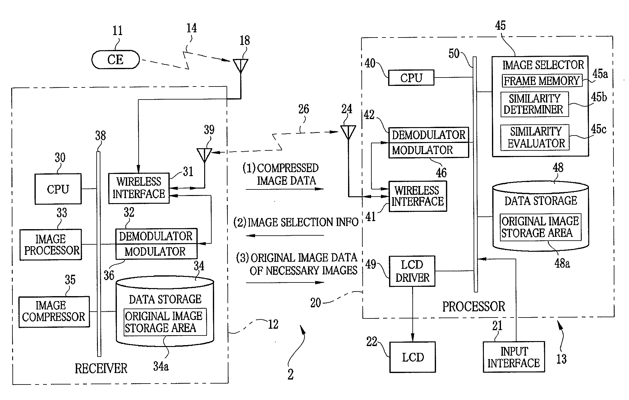
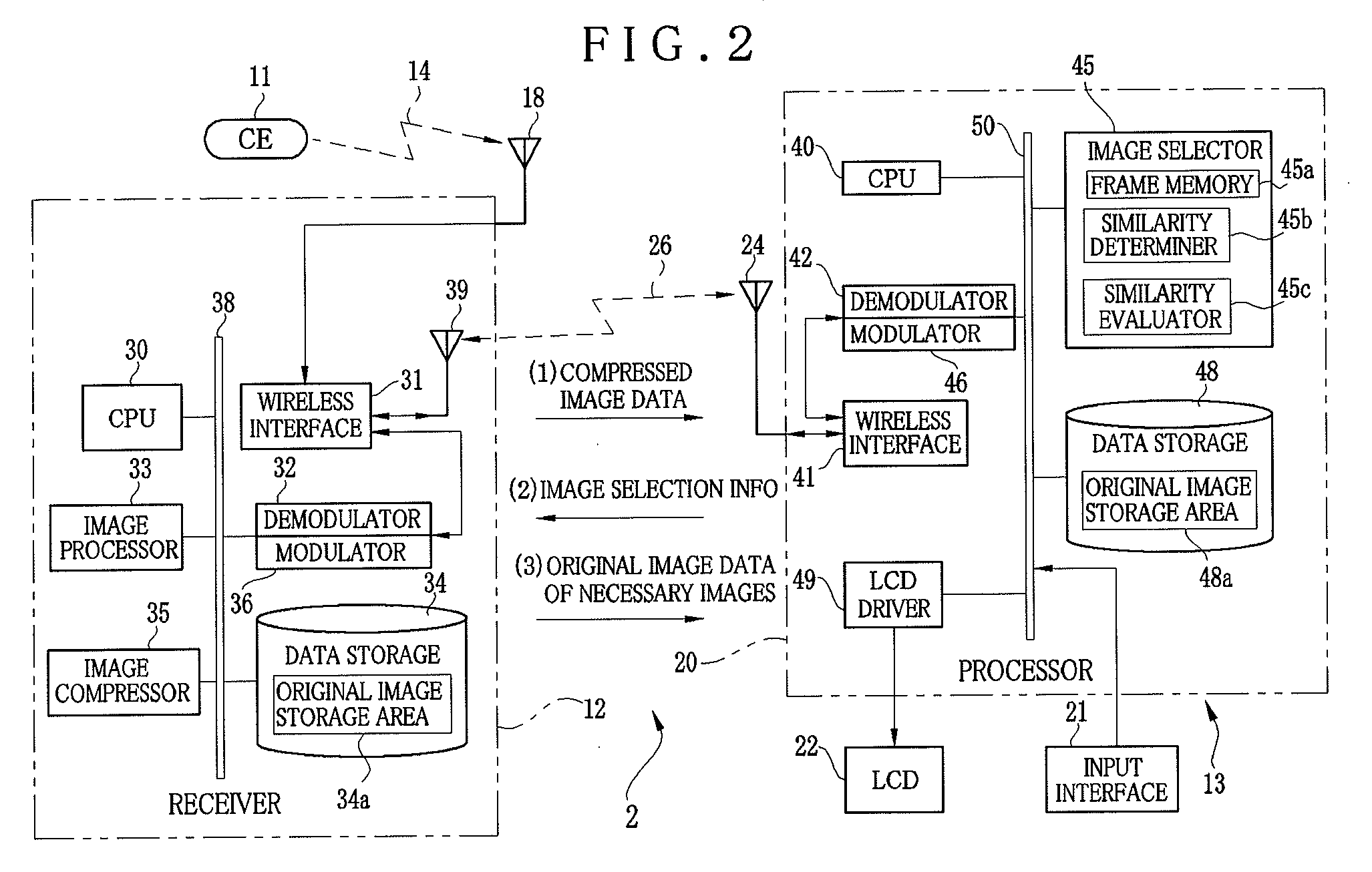
[0091]Another preferred embodiment of the invention is described now. In the first embodiment, the difference square sum of the pixel values of the images is utilized to evaluate similarity between the images. In contrast, in the present embodiment, similarity between preceding and succeeding frame images is evaluated according to detection of a motion vector from the preceding frame image to the succeeding frame image. To this end, the similarity determiner 45b in the image selector 45 determines the motion vector, according to which the similarity evaluator 45c evaluates similarity between the frame images. See FIG. 2.

[0092]The similarity determiner 45b determines a motion vector according to a feature point matching method. According to this, one pixel included in those in a sampling area of a preceding image is determined as a representative pixel before motion. A pixel value of the representative pixel before motion is detected. Also, pixel values of pixels in a sampling area o...

third embodiment

[0135]In the third embodiment, the image feature value extracted from the compressed image data is compared with the sample image feature value extracted previously from the sample image data, to evaluate similarity between the compressed image data and the sample image data. However, other methods of evaluating similarity may be used in the invention.

[0136]In the first, second and third embodiments, only original image data of necessary frame images are wirelessly transmitted from the receiver 12 to the workstation. However, the invention is not limited to those embodiments. For example, compressed image data may be data after lossless compression (compression encoding) of original image data in the receiver 12 or the image compressor 35. For this data, a decoder can be incorporated in the workstation for converting compressed image data of necessary frame images into original image data. It is unnecessary to send the original image data from the receiver 12 to the workstation, rem...


PUM


Abstract

A capsule endoscope system includes a capsule endoscope, swallowable in a body, for forming an image. A receiver is positioned on the body, for wirelessly receiving the image from the capsule endoscope, to store the image. A workstation as information manager operates for image filing of the image from the receiver. A first wireless interface is incorporated in the receiver, for wirelessly transmitting the image during imaging with the capsule endoscope. A second wireless interface is positioned on the workstation, for wirelessly receiving the image from the first wireless interface. Furthermore, an image selector selects the image for image filing in the workstation among plural images received by the receiver. The first wireless interface transmits the selected image to the workstation. The receiver includes an image compressor for reducing a data size of the image received from the capsule endoscope before transmission in the first wireless interface.

Description

BACKGROUND OF THE INVENTION[0001]1. Field of the Invention[0002]The present invention relates to a capsule endoscope system and endoscopic image filing method. More particularly, the present invention relates to a capsule endoscope system and endoscopic image filing method in which image reading of endoscopic images can be carried out with high efficiency.[0003]2. Description Related to the Prior Art[0004]A capsule endoscope has been recently developed for the purpose of medical examination of a patient's body. The capsule endoscope includes a capsule body of a very small size, and an image pickup device and a light source incorporated in the capsule body. For the examination, at first a patient orally swallows the capsule endoscope. The light source illuminates a body part in a gastrointestinal tract of his or her human body. The image pickup device creates an image of the body part. Image data are obtained. A receiver wirelessly receives a radio wave of the image data, and writes ...

Claims


Application Information

Patent Timeline
Patent Type & Authority Applications(United States)
IPC IPC(8): A61B1/04G16H30/20G16H40/63
CPCA61B1/00016H04N7/185G06F19/321A61B1/041G16H30/20G16H40/63
Inventor SHIMIZU, KUNIMASAKINJO, NAOTOKOIKE, KAZUMI
Owner FUJIFILM CORP
Why Eureka
Try Eureka