Magnetic resonance imaging apparatus and magnetic resonance imaging method

a magnetic resonance imaging and magnetic resonance imaging technology, applied in the direction of magnetic measurements, instruments, measurement devices, etc., can solve the problems of deteriorating image quality, inappropriate phase encode pulse application, image distortion, etc., and achieve the effect of reducing distortion

Active Publication Date: 2009-08-27
TOSHIBA MEDICAL SYST CORP
View PDF8 Cites 22 Cited by
  • Summary
  • Abstract
  • Description
  • Claims
  • Application Information

AI Technical Summary

Benefits of technology

[0023]The magnetic resonance imaging apparatus and the magnetic resonance imaging method according to the present invention as described above make it possible to reduce a distortion, of which amount varies depending on a spatial position of an imaging section, in an image occurring due to eddy currents.

Problems solved by technology

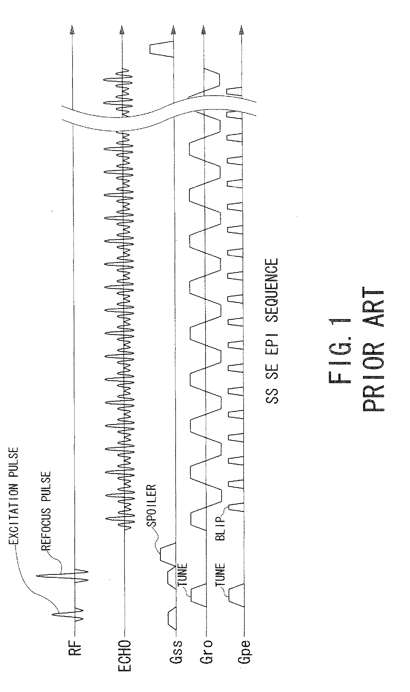
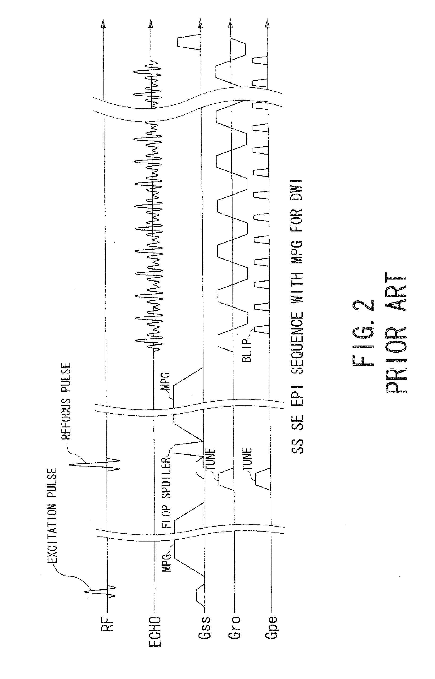
However, in the conventional SS EPI, there is a problem that an image may be distorted under the influence of eddy currents at some imaging positions depending on characteristics of the hardware.
That is, eddy currents having spatial distributions separately to an apparatus are generated due to factors such as a type of magnet, manufacturing error or how to wind a gradient magnetic field coil.
For this reason, the influence of an eddy current is different between imaging sections of which positions are mutually different spatially, and therefore, inappropriate phase encode pulses may be applied on some imaging positions.
In addition, since s special distribution of an eddy current changes depending on an imaging position and an application pattern of a gradient magnetic field, there is a problem that deterioration of an image quality becomes prominent at an imaging position in case of calculating an isotropic image using plural application patterns of gradient magnetic fields with applications of particularly high-intensity MPG pulses like imaging of a DWI especially.

Method used


Image

  • Magnetic resonance imaging apparatus and magnetic resonance imaging method
  • Magnetic resonance imaging apparatus and magnetic resonance imaging method
  • Magnetic resonance imaging apparatus and magnetic resonance imaging method

Examples


Embodiment Construction

[0038]A magnetic resonance imaging apparatus and a magnetic resonance imaging method according to embodiments of the present invention will be described with reference to the accompanying drawings.

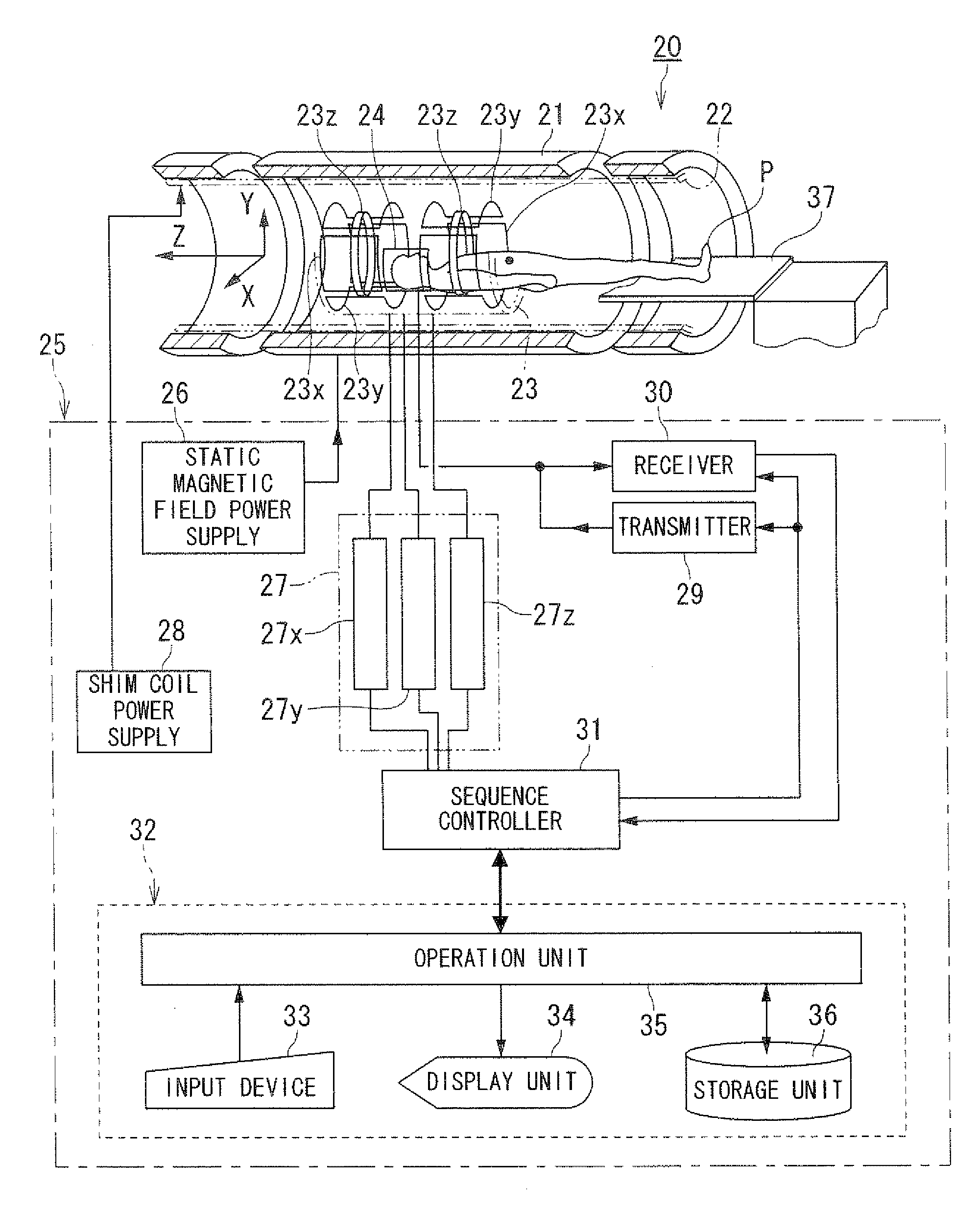
[0039]FIG. 4 is a block diagram showing a magnetic resonance imaging apparatus according to an embodiment of the present invention.

[0040]A magnetic resonance imaging apparatus 20 includes a static field magnet 21 for generating a static magnetic field, a shim coil 22 arranged inside the static field magnet 21 which is cylinder-shaped, a gradient coil 23 and a RF coil 24. The static fueled magnet 21, the shim coil 22, the gradient coil 23 and the RF coil 24 are built in a gantry (not shown) the magnetic resonance imaging apparatus 20 also includes a control system 25. The control system 25 includes a static magnetic field power supply 26, a gradient power supply 27, a shim coil power supply 28, a transmitter 29, a receiver 30, a sequence controller 31 and a computer 32. The gradient power s...


PUM


Abstract

A magnetic resonance imaging apparatus includes a data acquisition unit and an image generating unit. The data acquisition unit acquires echo signals by echo planar imaging which generates the echo signals with correcting and applying plural gradient magnetic fields for phase encode and with continuously inverting a gradient magnetic field for readout after one nuclear magnetic excitation. Each of the plural gradient magnetic fields for phase encode has an intensity set so as to compensate an influence of an eddy current distributing spatially adaptively to each of imaging positions. The image generating unit generates image data based on the echo signals.

Description

BACKGROUND OF THE INVENTION[0001]1. Field of the Invention[0002]The present invention relates to a MRI (magnetic resonance imaging) apparatus and a magnetic resonance imaging method which excite nuclear spin of an object magnetically with a RF (radio frequency) signal having the Larmor frequency and reconstruct an image based on NMR (nuclear magnetic resonance) signals generated due to the excitation, and more particularly, to a magnetic resonance imaging apparatus and a magnetic resonance imaging method which generate image data by single shot echo planar imaging.[0003]2. Description of the Related Art[0004]Magnetic Resonance Imaging is an imaging method which excite nuclear spin of an object set in a static magnetic field with a RF signal having the Larmor frequency magnetically and reconstruct an image based on MR signals generated due to the excitation.[0005]In the field of magnetic resonance imaging, there is an imaging method called echo planar imaging (EPI) (see, for example,...

Claims


Application Information

Patent Timeline
Patent Type & Authority Applications(United States)
IPC IPC(8): G01V3/00
CPCG01R33/5615G01R33/56518G01R33/5616
Inventor OOKAWA, MASASHI
Owner TOSHIBA MEDICAL SYST CORP
Why Eureka
Try Eureka