Method of revealing a biological process using a fret measurement

a biological process and fret technology, applied in the field of cell biology, can solve the problems of not being able to detect a fret, not enabling the interaction between biological entities to be detected, and measurement not making it possible to distinguish between a decrease in fluorescen

Active Publication Date: 2009-09-03
CIS BIO INT
View PDF18 Cites 13 Cited by
  • Summary
  • Abstract
  • Description
  • Claims
  • Application Information

AI Technical Summary

Benefits of technology

[0019]It has now been found that it is possible to reveal a biological process in a cell system by means of a FRET measurement using, as the donor fluorophore, a fluorophore which has

Problems solved by technology

On the other hand, it has not been possible to detect a FRET when one of the fluorophores is on the intracellular part and the other fluorophore is on the extracellular part of the receptor, i.e. when the fluorophores are on either side of the plasma membrane (cf. FIG. 4 of said document).
Therefore, these FRET systems do not enable the interactions between the biological entities to be detected when they are on either side of the plasma membrane.
This method of measurement does not make it possible to distinguish between a decrease in fluorescence due to the energy transfer between the donor and t

Method used


Image

  • Method of revealing a biological process using a fret measurement
  • Method of revealing a biological process using a fret measurement
  • Method of revealing a biological process using a fret measurement

Examples


example 1

Molecular Constructs

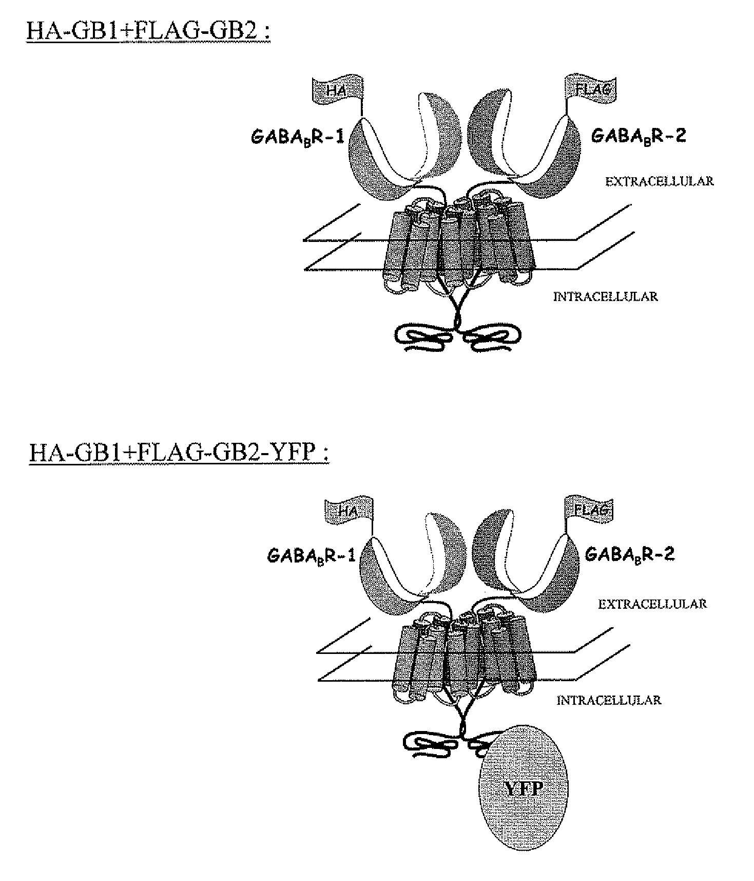
[0095]Two constructs, which are illustrated diagrammatically in FIG. 1, were used:[0096]Construct 1: HA-GABAB R-1 (“HA-GB1”)+FLAG-GABAB R-2 (FLAG-GB2)[0097]Construct 2: HA-GABAB R-1 (“HA-GB1”)+FLAG-GABAB R-2 YFP (FLAG-GB2-YFP)

example 2

Reagents

[0098]The following reagents are used in the experiments described below:[0099]an anti-FLAG antibody labeled with the following terbium crypate:

[0100]an anti-HA antibody labeled with Alexa 647: the anti-HA antibody was labeled with Alexa 647 succinimidyl ester (Molecular Probes, ref. A-2006). The labeling reaction was carried out in a 0.1 M carbonate buffer, pH 9, for 30 minutes at room temperature, with a molar excess of 4 Alexa 647 per antibody. The excess fluorescent probe which has not reacted with the antibody is removed by exclusion chromatography (Pharmacia Biotech G-25 super fine gel). The final labeling rate of the antibody, determined by means of the absorption spectrum of the conjugate, is 2.6 Alexa 647 per antibody.

example 3

Measurement of TR-FRET

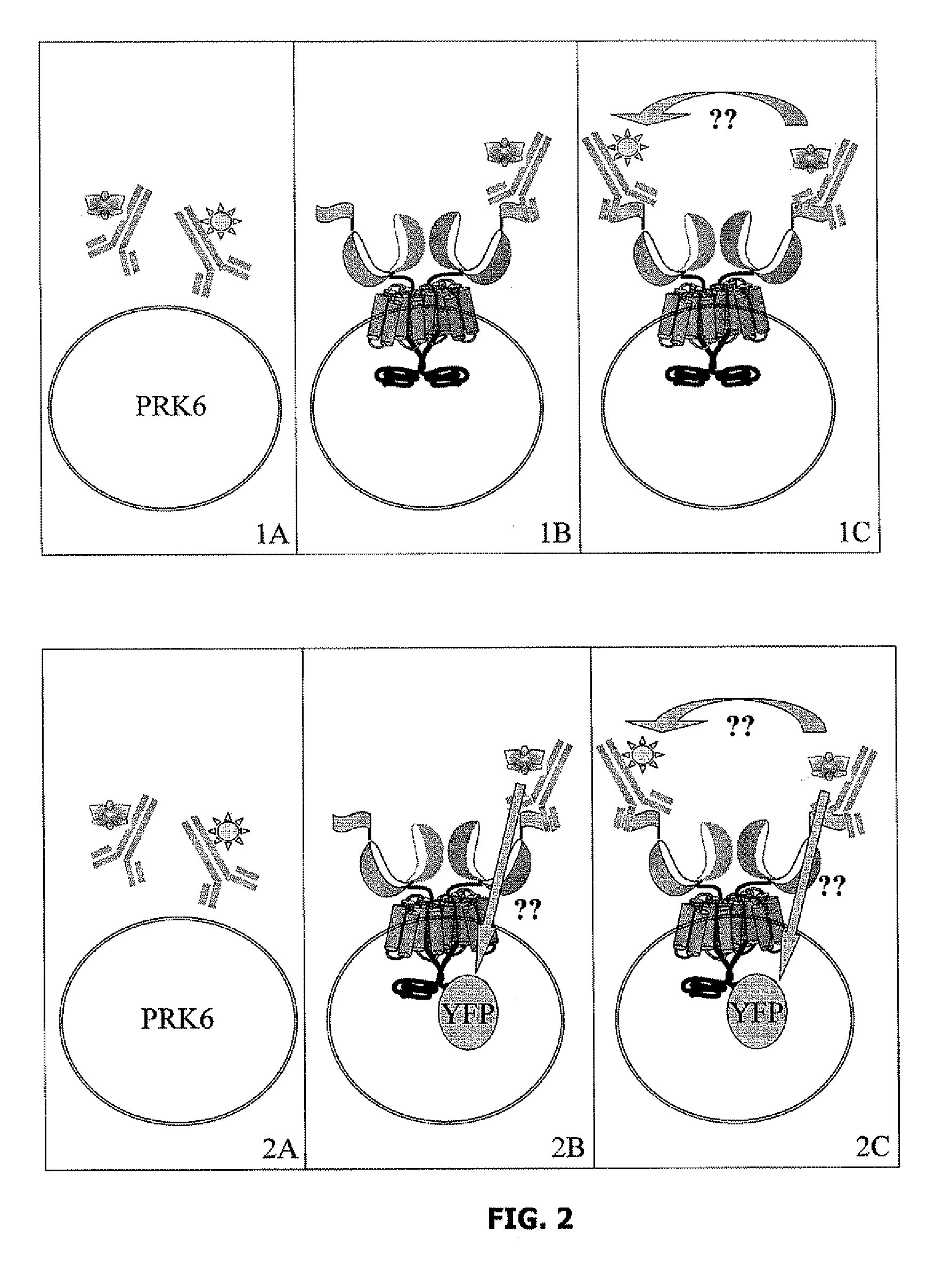
[0101]The TR-FRET signal is measured in wells each containing 50,000 or 100,000 cells, said cells containing the molecular constructs described in Example 1 in a final volume of 100 μl, to which the following reagents are added (FIG. 2):

1A: PRK6 cells+anti-FLAG antibody coupled with a terbium cryptate (hereafter “anti-FLAG / terbium cryptate”) 1 nM final+anti-HA antibody coupled with Alexa 647 (hereafter “anti-HA / A647”) 3 nM final;

1B: PRK6 cells expressing HA-GB1 and FLAG-GB2+anti-FLAG / terbium cryptate 1 nM final;

1C: PRK6 cells expressing HA-GB1 and FLAG-GB2+anti-FLAG / terbium cryptate 1 nM final+anti-HA / A647 3 nM final;

2A: PRK6 cells+anti-FLAG / terbium cryptate 1 nM final+anti-HA / A647 3 nM final;

2B: PRK6 cells expressing HA-GB1 and FLAG-GB2-YFP+anti-FLAG / terbium cryptate 1 nM final;

2C. PRK6 cells expressing HA-GB1 and FLAG-GB2-YFP+anti-FLAG / terbium cryptate 1 nM final+anti-HA / A647 3 nM final.

[0102]For each experiment the wells were incubated for 20 h at 4° C. prio...


PUM

PropertyMeasurementUnit
Timeaaaaaaaaaa
Timeaaaaaaaaaa
Timeaaaaaaaaaa

Abstract

The present invention relates to a method of revealing a biological process using a FRET measurement, which comprises the following steps:
    • incorporating, into a measurement medium containing a lipid membrane, a biological entity X coupled with a first member of a pair of FRET partners and a second biological entity Y coupled with the second member of the pair of FRET partners, the energy-donating member of the pair of FRET partners having a long lifetime and the members of said pair of FRET partners being located on either side of the lipid membrane;
    • exciting the measurement medium at the excitation wavelength of the energy-donating member; and
    • measuring the FRET signal or the variations in said signal emitted in said culture medium.

Description

FIELD OF THE INVENTION[0001]The present invention relates to the field of cell biology and more particularly to the study of the interactions between biological entities or between the different constituent parts of these entities, optionally in the presence of potential ligands, for the purpose of elucidating the biological or pathological processes and screening new drugs.[0002]Attempts are increasingly being made to study the biological or pathological processes within membrane preparations or live cells, and this is frequently done using fluorescence measurement tests, particularly fluorescence tests which measure the non-radiative resonance energy transfer between a donor fluorophore and an acceptor fluorophore.[0003]After luminous excitation at the excitation wavelength of the donor fluorophore, an energy transfer takes place between the donor fluorophore and the acceptor fluorophore if they are close to one another; this energy transfer is characterized by an emission of ligh...

Claims


Application Information

Patent Timeline
IPC IPC(8): G01N33/53C12Q1/02
CPCG01N33/6872G01N33/542Y10S435/968
Inventor TRINQUET, ERICANSANAY, HERVEMATHIS, GERARD
Owner CIS BIO INT
Why Eureka
Try Eureka