Methods for Treating Autoimmune Diseases in a Subject and In Vitro Diagnostic Assays

a technology for autoimmune diseases and in vitro diagnosis, applied in the direction of antibody medical ingredients, liposomal delivery, peptide/protein ingredients, etc., can solve the problems of devastating and crippling diseases of autoimmune diseases

Inactive Publication Date: 2009-10-22
BANCHEREAU JACQUES F +2
View PDF0 Cites 32 Cited by
  • Summary
  • Abstract
  • Description
  • Claims
  • Application Information

AI Technical Summary

Problems solved by technology

Autoimmune diseases are devastating and crippling diseases, and occur when a patient's own immune system turns against itself by attacking the patient's own body or tissues.

Method used


Image

  • Methods for Treating Autoimmune Diseases in a Subject and In Vitro Diagnostic Assays
  • Methods for Treating Autoimmune Diseases in a Subject and In Vitro Diagnostic Assays
  • Methods for Treating Autoimmune Diseases in a Subject and In Vitro Diagnostic Assays

Examples


example 1

SLE Sera Induces Monocytes to Differentiate into Cells with Properties Like Dendritic Cells

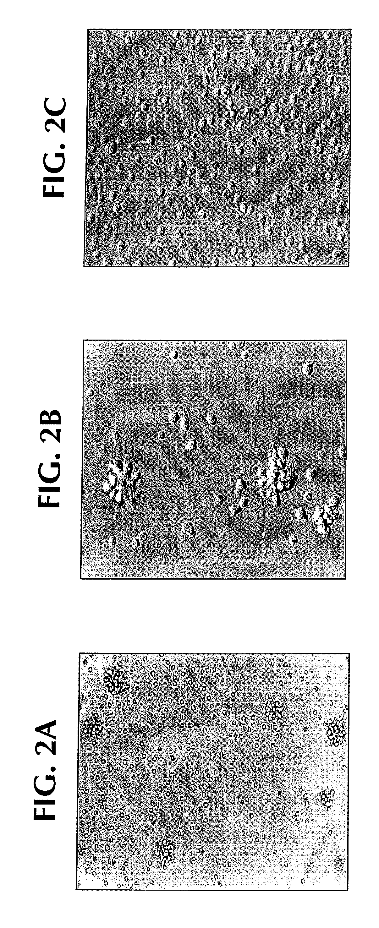
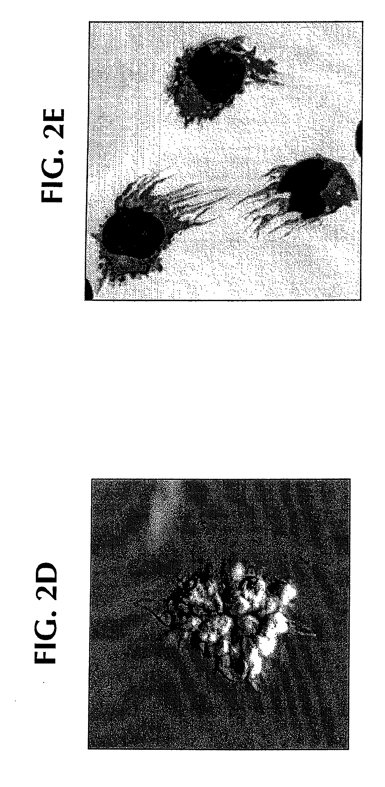
[0117]Normal monocytes were exposed to SLE sera in vitro: within 12-24 hours, clustering of monocytes was noted; and within 24-48 hours, clustered cells displayed fine cytoplasmic projections reminiscent of DC cultures (FIGS. 2A and 2B). Only SLE serum induced monocytes to cluster with 12-24 hours and acquire veiled cell morphology (FIGS. 2A-2B compared to AS in FIG. 2C).

[0118]SLE serum: After informed consent, blood was obtained from the patient who satisfied diagnostic criteria of American College of Rheumatology (ACR) for SLE. Whole blood was collected into tubes containing EDTA or heparin, and was separated immediately by centrifugation at 100×g at 4° C. The plasma was harvested, treated with thrombin (Jones Pharma Incorporated, MO) and stored at −80° C. until used. Disease activity was assessed by using the SLUE Disease Activity Index (SLEDAI) score (Lahita, R. G. 1999. Systemic Lupus Ery...

example 2

SLE-DCs Present Antigens from Captured Apoptotic Cells

[0127]The non-normal and inappropriate processing of apoptotic cells by the immune system is considered as one of the pathogenic events in SLE. Thus, it was next determined whether SLE-DCs can present antigens from captured apoptotic cells. To this end, SLE-DCs were shown to be able to capture apoptotic cell fragments in culture (FIGS. 7A-7B).

[0128]SLE-DCs could also capture DNA containing apoptotic bodies derived from melanoma cells. FIGS. 8A and 8B show the capture of allogeneic apoptotic cells and presentation of their antigens to autologous CD4+ T cells. SLE-DCs captured DNA containing apoptotic bodies (FIG. 8A) and presented their antigens to autologous CD4+ T cells as indicated by the induction of CD4+ T cell proliferation (FIG. 8B). HLA-DR+ monocytes induced by AS serum SLE serum and GM-CSF / IL-4 captured 7AAD labeled DNA-bodies (melanoma cell line killed by gamma-irradiation (150 Gy)). To allow capture of apoptotic bodies,...

example 3

Only IFN-α Containing Active SLE Sera Induce Monocytes to become SLE-DCs

[0130]Experiments were performed to determine whether all SLE sera were able to direct monocyte differentiation into DCs. Monocytes were cultured with 19 different SLE sera to be tested for their ability to stimulate a mixed leukocyte response or mixed lymphocyte reaction (MLR). As shown in Table 1, monocytes cultured with autologous serum were only able to induce very low T cell proliferation (negative control), and monocytes cultured with GM-CSF / IL-4 to produce DCs elicited a 100% MLR (positive control), the mean proliferation was 7.5% (±6.4% Y, n=5). When monocytes cultured with SLE sera were evaluated in the same way, considering 20% proliferation as a cut-off point (mean+2SD of autologous serum cultured monocytes) 11 of the 19 sera induced monocyte-s to become allostimulatory DCs with mean proliferation of 41%±15%. As sera from patients with dermatomyositis were unable to reproduce this skewing and since th...


PUM

PropertyMeasurementUnit
concentrationaaaaaaaaaa

Abstract

The invention provides a method for treating an autoimmune disease in a subject by administering an interferon antagonist and a Flt3 ligand (Flt3L) antagonist. The invention also provides compositions containing one or more interferon antagonists, and one or more Flt3L antagonists, an in vitro assay for determining a subject's risk for developing an autoimmune disease, and kits for use, inter alia, with the assay.

Description

CROSS-REFERENCES TO RELATED APPLICATIONS[0001]This application claims the priority to U.S. Provisional Patent Application Ser. No. 60 / 260,541, filed Jan. 9, 2001, the entire contents of which are hereby incorporated by reference.FIELD OF THE INVENTION[0002]This invention relates to treating autoimmune diseases and diagnostic assays related to autoimmune diseases.BACKGROUND OF THE INVENTION[0003]Autoimmune diseases are devastating and crippling diseases, and occur when a patient's own immune system turns against itself by attacking the patient's own body or tissues. One example of an autoimmune disease is systemic lupus erythematosus (SLE), which is characterized by multi-organ involvement and immunological abnormalities that include the presence of autoreactive T cells and B cells. Autoantibodies against the nucleosome appear to be a hallmark characteristic of SLE and suggest that inappropriate handling of dying (apoptotic) cells may represent a key pathogenic event in the developme...

Claims


Application Information

Patent Timeline
Patent Type & Authority Applications(United States)
IPC IPC(8): A61K9/127A61K39/395G01N33/53A61K38/00A61K31/7088A61K38/17A61K38/19A61K38/21A61K45/06A61P3/10A61P7/00A61P7/06A61P17/02A61P17/06A61P19/00A61P19/02A61P19/08A61P21/04A61P25/00A61P29/00A61P31/00A61P31/18A61P37/06C07K16/22C07K16/24C07K16/40G01N21/76G01N21/78G01N33/564G01N33/577G01N33/58G01N33/68
CPCA61K38/191A61K2039/505C07K16/22C07K16/24C07K16/249A61K38/1793G01N33/564G01N33/6863G01N33/6866G01N2800/24A61K38/179C07K2317/73A61P17/02A61P17/06A61P19/00A61P19/02A61P19/08A61P21/04A61P25/00A61P29/00A61P31/00A61P31/18A61P37/02A61P37/06A61P43/00A61P5/14A61P7/00A61P7/06A61P3/10A61K39/395
Inventor BANCHEREAU, JACQUES F.PALUCKA, ANNA KAROLINABLANCO, PATRICK
Owner BANCHEREAU JACQUES F
Why Eureka
Try Eureka