Methods and compositions for the production and use of magnetosomes

a technology of magnetosomes and compositions, applied in the field of molecular biology and medical imaging, can solve the problems of lack of widespread biomedical molecular application

Inactive Publication Date: 2009-12-17
EMORY UNIVERSITY +1
View PDF0 Cites 21 Cited by
  • Summary
  • Abstract
  • Description
  • Claims
  • Application Information

AI Technical Summary

Problems solved by technology

However, due to their relatively large size and clearance by the reticuloendothel...

Method used


Image

  • Methods and compositions for the production and use of magnetosomes
  • Methods and compositions for the production and use of magnetosomes
  • Methods and compositions for the production and use of magnetosomes

Examples


example 1

[0067]MagA is known to be involved in the production of magnetosomes in the species Magnetospirillum magneticum, (Nakamura et al. (1995) J. Biol. Chem., 270(47):28392-28396; Nakamura et al. (1995) J. Biochem. (Tokyo) 118(1):23-7; Bazylinski and Frankel (2004) Nat. Rev. Microbiol., 2(3):217-230). Nakamura and co-workers (Nakamura et al. (1995) J. Biol. Chem., 270(47):28392-28396 and Nakamura et al. (1995) J. Biochem. (Tokyo) 118(1):23-7) found that MagA encodes a protein with significant sequence homology to the cation-efflux proteins, KefC, a K+-translocating protein in Escherichia coli, and NapA, a putative Na+ / H+ antiporter from Enterococcus hirae. The MagA protein is present in both the cytoplasmic and magnetosome membranes of M. magneticum strain AMB-1. MagA was expressed in E. coli and inverted membrane vesicles prepared from these cells were shown to transport Fe(II) in an energy-dependent manner, leading to accumulation of Fe(II) in the vesicle, which indicates that MagA func...

example 2

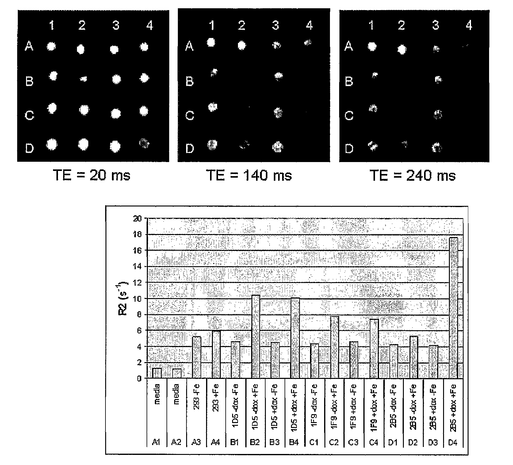
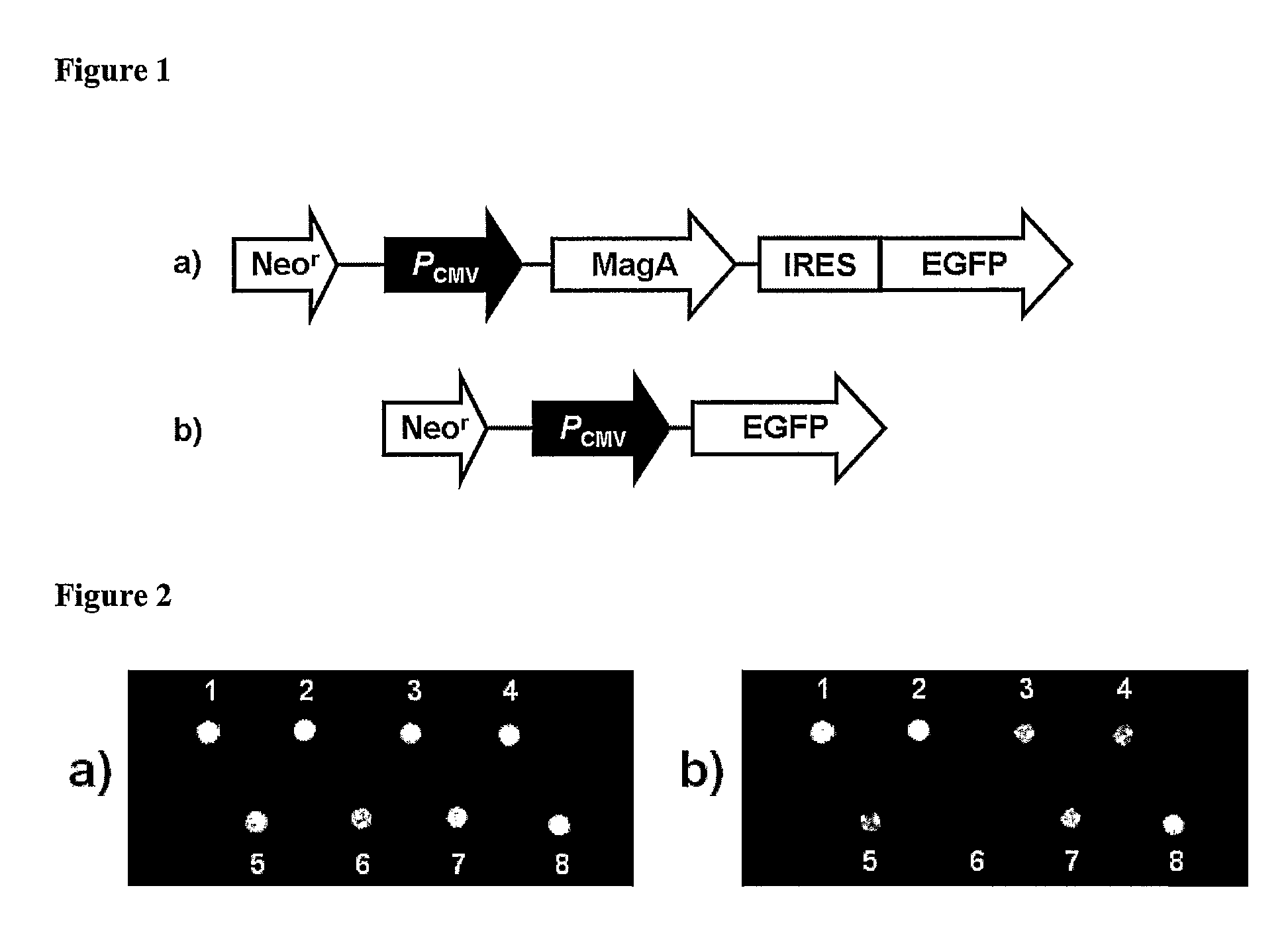
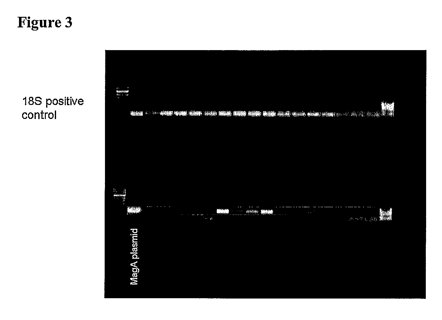
[0073]In this example, cell lines producing magnetosomes were created. As described below, these magnetosomes could be seen by electron micrography and they affected the MR signal as expected. A major advantage of these methods is that the magnetosome production can continue, even after the cells divide, since the genes have been incorporated into the cellular DNA. In this way, long term identification of cells can be made, even after many cell divisions. Long-term studies can be conducted since the magnetosomes can be regenerated and MR imaging itself is non-invasive and does not destroy the sample being imaged. Furthermore, by developing a controllable system, long-term studies can be conducted in which cells are allowed to grow, migrate, divide and develop over a period of time in the absence of magnetosomes. This minimizes the chances of the magnetosomes to interfere with normal cellular processes. Expression of magnetosomes can then be induced and magnetosomes allowed to form, ...


PUM

PropertyMeasurementUnit
Fractionaaaaaaaaaa
Fractionaaaaaaaaaa
Fractionaaaaaaaaaa

Abstract

Methods and compositions for using magnetosomes as cellular contrast agents and markers for magnetic resonance imaging are provided. Certain methods involve synthesizing magnetosomes in a cell as directed by a nucleotide construct comprising an exogenous polynucleotide sequence, wherein the magnetosome serves as a contrast agent or marker for magnetic resonance imaging. Methods of synthesizing and isolating magnetosomes for introduction into immune-matched cells within a tissue or subject for use as a contrast agent or marker for magnetic resonance imaging are also provided. Also provided are methods for stably transfecting cells to express a polypeptide that drives or modulates magnetosome production in the cell, cells produced by such methods and methods for their isolation, transgenic animals comprising at least one eukaryotic cell produced by such methods, and vectors and delivery systems for the transfection of such cells. Further provided are methods for non-invasively generating a visible image of a tissue or subject containing at least one cell such transfected cell, as well as use of such methods to monitor the location, migration, or proliferation of cells in a tissue or subject or to detect or monitor gene expression in a tissue or subject.

Description

FIELD OF THE INVENTION[0001]The invention relates to the fields of molecular biology and medical imaging, more particularly to the genetic manipulation of cells to produce magnetosomes that serve as intracellular contrast agents and markers for magnetic resonance imaging and other purposes.BACKGROUND OF THE INVENTION[0002]Currently, magnetic resonance imaging (MRI) is most often used to construct an image based on the intrinsic contrast provided from the relaxation of spin of hydrogen atoms. These images provide an accurate anatomical picture that has greatly advanced health care today. Clinically, the images produced by MRI are invaluable, but the full potential of using MRI in acquiring functional, physiological, and molecular information is only beginning to be realized.[0003]Contrast agents are important in achieving such goals. Superparamagnetic iron oxide nanoparticles are regularly used for applications such as high-density magnetic storage, catalytic and separation processes...

Claims


Application Information

Patent Timeline
IPC IPC(8): A61K49/00C12N15/87C12N5/06A01K67/027C12N15/00A61K31/7088C12N13/00
CPCC07K14/195A61K49/1818
Inventor HU, XIAOPING PHILIPCHAN, ANTHONY WING SANGZURKIYA, OMAR
Owner EMORY UNIVERSITY
Why Eureka
Try Eureka