Methods for detection and characterization of atypical vessels in cervical imagery

a technology of cervical imaging and atypical vessels, applied in the field of medical imaging, can solve the problems of random distribution or non-uniform distribution of atypical vessels, and achieve the effects of smoothing the image, suppressing high intensity components, and enhancing image contras

Inactive Publication Date: 2010-02-04
STI MEDICAL SYST
View PDF15 Cites 27 Cited by
  • Summary
  • Abstract
  • Description
  • Claims
  • Application Information

AI Technical Summary

Benefits of technology

[0016]The presently preferred embodiment of the invention discloses a method for the detection of atypical vessels by applying a pre-processing stage for contrast enhancement, a segmentation stage to identify regions of interest, and a post-processing stage to determine the final region of interest. The pre-processing stage smoothes the image and suppresses high intensity components using a top hat filter; enhances the contrast of the image using a top hat and bottom hat filter; enhances the red component of the image; and then enhances the contrast of the red component of the image. The segmentation stage identifies regions of interest for atypical vessels by locating regions of maximum texture and maximum gradient information in the image and calculating the intersection of that information. The post-processing stage identifies and removes features that are unrelated to atypical vessels and further refines the image using known color, shape and size information on atypical vessels to create an output image with a final region of interest. This automated method of atypical vessel detection is especially useful for diagnostic purposes such as cancer detection, namely, cervical cancer detection.

Problems solved by technology

In addition, atypical vessels are distributed randomly or non-uniformly.

Method used


Image

  • Methods for detection and characterization of atypical vessels in cervical imagery
  • Methods for detection and characterization of atypical vessels in cervical imagery
  • Methods for detection and characterization of atypical vessels in cervical imagery

Examples


Embodiment Construction

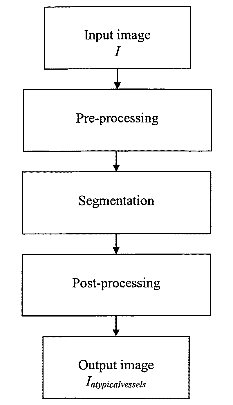
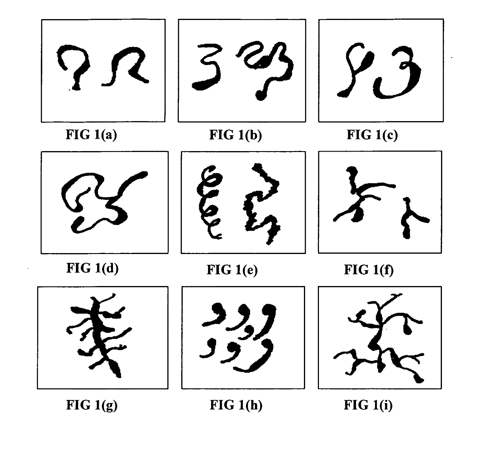
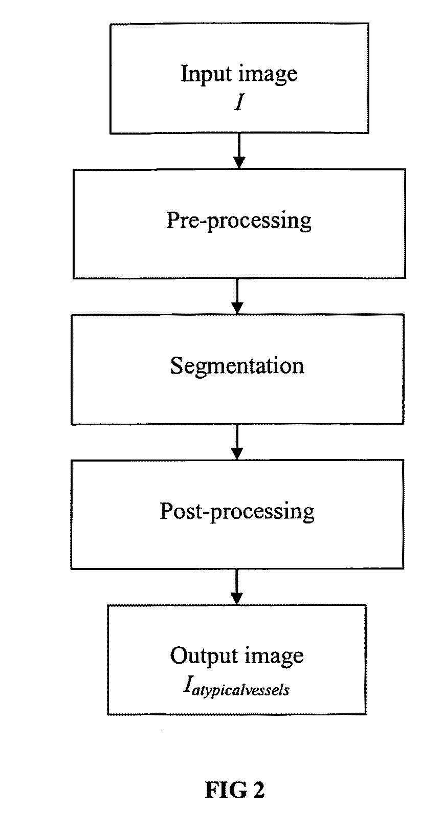
[0026]The presently preferred embodiment of the invention provides a systematic framework for the automated detection of atypical blood vessels in cervical imagery. A flowchart of the preferred embodiment of the invention is shown in FIG. 2. Due to the large variation of shapes, diameters, spacing and branching patterns of atypical vessels, a three-stage algorithm process is preferably implemented.

[0027]The atypical vessels detection of the presently preferred embodiment of the invention starts by collecting high-quality image data (digital images) of an organ (such as the cervix) in real-time during examination with a colposcope. A pre-processing stage is applied to the image data, which provides contrast enhancement of the original cervical image. The pre-processing stage conditions the image data for the segmentation stage by enhancing visualization of the vasculature in the image compared to the surrounding tissue. The second stage is the segmentation stage in which regions of i...


PUM


Abstract

The present invention discloses a method for the detection of atypical vessels in digital cervical imagery. A pre-processing stage is applied to enhance the contrast of blood vessel features compared to the surrounding tissue. Next, a segmentation stage is applied to identify regions of interest for atypical vessels using texture and gradient information. Finally, a post-processing stage (1) identifies other clinically relevant features in the cervical imager, and removes these features from the region of interest; and (2) uses color, size, and shape information to further refine the region of interest to eliminate false positives and determine a final region of interest. This automated method of atypical vessel detection is especially useful for diagnostic purposes such as cervical cancer detection.

Description

TECHNICAL FIELD[0001]This invention relates to medical imaging and, more specifically, to the computer aided detection and diagnosis of uterine cervical cancer and pre-cancerous lesions. The invention provides an automated method to detect atypical blood vessels in cervical imagery for the diagnosis of cancer.BACKGROUND ART[0002]Although this invention is being disclosed in connection with cervical cancer, it is applicable to many other areas of medicine. Uterine cervical cancer is the second most common cancer in women worldwide, with nearly 500,000 new cases and over 270,000 deaths annually (IARC, “Globocan 2002 database,” International agency for research in cancer, 2002, incorporated herein by reference). Because invasive disease is preceded by pre-malignant Cervical Intraepithelial Neoplasia (CIN), if detected early and treated adequately, cervical cancer can be universally prevented (D. G. Ferris, J. T. Cox, D. M. O'Connor, V. C. Wright, and J. Foerster, Modern Colposcopy. Tex...

Claims


Application Information

Patent Timeline
Patent Type & Authority Applications(United States)
IPC IPC(8): A61B5/00
CPCA61B1/00009A61B5/0084A61B5/02007G06T2207/30101G06T7/0081G06T2207/30096G06T7/0012G06T7/11A61B1/000094
Inventor VENKATARAMAN, SANKARLI, WENJINGGUSTAFSSON, ULF PETER
Owner STI MEDICAL SYST
Why Eureka
Try Eureka