In vivo spectral micro-imaging of tissue

a tissue and spectral micro-imaging technology, applied in the field of medical diagnostics for tissue investigation, can solve the problems of poor prognosis, difficulty in early detection, and onset is often asymptomatic, and achieve the effects of less time, high contrast, and higher image contras

Active Publication Date: 2012-11-27
LAWRENCE LIVERMORE NAT SECURITY LLC +1
View PDF16 Cites 51 Cited by
  • Summary
  • Abstract
  • Description
  • Claims
  • Application Information

AI Technical Summary

Benefits of technology

[0015]It is an object of the present invention to provide in vivo endoscopic methods for implementation of AF microscopy, without the use of exogenous agents, to effectively (with resolution sufficient to image nuclei) visualize and categorize various abnormal esophageal tissue forms.
[0016]Another object is to provide apparatuses capable of in vivo endoscopic methods for implementation of AF (without exogenous agents) microscopy to effectively visualize and categorize various abnormal esophageal tissue forms.
[0019]A microscope system described in US20070160279(A1), incorporated herein by reference, has been used to determine the most effective excitation-emission combinations for imaging changes of structural and biochemical characteristics in esophagus tissue [1]. Ultra violet (UV) wavelengths were implemented in this platform as the excitation sources for ex vivo human esophagus tissue biopsies. The present inventors have made the discovery that imaging of the superficial tissue microstructure can be achieved using conventional wide field autofluorescence microscopy under excitation with UV light of less than 400 nm. Although the exact mechanism is still not completely clear, they hypothesize that this becomes possible via two main mechanisms. The first mechanism now discussed is that the excitation UV light only penetrates tissue to about 100 μm or less and as a result, a sufficient amount of the fluorescence signal produced in the superficial tissue layer can be contained within the thickness of the image plane of the microscope, thus the out of focus signal (background) is sufficiently reduced to allow the formation of high contrast images of tissue microstructure using the native fluorescence (autofluorescence) of the tissue cells and intracellular components. There is no need for a pinhole because substantially all of the AF is produced within this superficial tissue layer that has thickness comparable to the thickness of the image plane. This hypothesis is supported by additional experimental results that the contrast of AF images obtained with excitation wavelengths longer that about 300 nm is lower to that when the excitation wavelength in shorter than about 300 nm, suggesting that the photon penetration depth for shorter UV wavelength is also shorter giving rise to a higher image contrast. The second mechanism now discussed is that there is sufficient variability in the concentration of chromophores (that can be photo-excited with UV light) contained within subcellular and intracellular components so that the microscopic autofluorescence image of the tissue reveals the microstructure and organization of the superficial layer in a similar way to that provided by H&E staining.
[0020]Further, since the present invention provides much more light for imaging than the confocal microscope, it is able to produce an autofluorescence image in much less time (˜10 microseconds or less). This reduced time required to collect enough light to produce an adequate image enables use of autofluorescence microscopy in vivo. The limit on the duration of the image acquisition window is that the image must be collected in an exposure time in which the target tissue (such as esophagus) and / or instrument (such as micro-endoscope) moves no more that the resolution of the optical system. Thus, if the desired resolution of the optical system is 1 micrometer, then the time it takes the target tissue to move 1 micrometer is the limit of the duration of the image acquisition window. The resolution S is equal to the speed, in not all, that the tissue is moving times the limit on the exposure time. Thus, if S=1 micrometer and the combined speed of motion is on the order of V=5 millimeter / second, then the time duration limit is 200 μsec. This technology does not require exogenous agents and is non-destructive to the sample. The present invention provides a minimally (or non) invasive method to visualize and categorize various abnormal esophageal tissue forms such as Barrett's esophagus (BE), low-grade dysplasia (LGD), high-grade dysplasia (HGD), and adenocarcinoma (ADC) in vivo and in real time. Endoscopic implementations are provided.
[0021]The invention herein utilizes the AF optical signal and provides resolution sufficient to image nuclei. This technology thus bypasses the complications of contrast agents while still obtaining diagnostic information sufficient to meet the current Hematoxylin and Eosin (HE) Staining Protocol, which is sometimes referred to in the field as the H&E gold standard. Embodiments of the present invention have been successfully implemented into endoscopes. A prototype zoom video endoscope probe was originally designed to operate in contact mode and is equipped with a white light guide, objective lens, and CCD encased at the tip of the endoscope probe. The probe distal end outer diameter is 3.2 mm and has a 300×300 μm field of view. The design has previously been shown to image cell nuclei with methylene blue contrast enhancement [34-36]. The present invention is able to acquire and categorize AF images of tissue without the use of contrast agents or hardware modification.

Problems solved by technology

A significant factor of the poor prognosis is the difficulty of early detection when disease initiates in the epithelial tissues before becoming invasive.
Additionally, onset is often asymptomatic until the disease has advanced to the point where the curative therapy may be unavailable.
This asymptomatic, and potentially heterogeneous nature of diseased esophageal mucosa makes early detection of abnormal progression almost impossible to distinguish using current endoscopic surveillance of the esophagus [8].
Often times, dysplastic progression is invisible to SVE due to low resolution, and missed during random sampling.
This random sampling is of great concern due to the inherent sampling error and delayed turn-around time for results [12].
While spectroscopy is very sensitive in detecting these changes, the point measurements are averaged over an area and do not provide a comprehensive image.
Investigators acknowledged the difficulties in fitting multiple exponentials to resulting data, as well as achieving the required resolution with these techniques.
While these features allow visibility of normal squamous mucosa and specialized intestinal metaplasia in the esophagus, OCT has not yet been shown to adequately differentiate between dysplastic and intramucosal carcinoma [21].
However, as the image plane of a conventional microscope has a thickness on the order of 10-50 μm, nearly all the signal collected by the microscope's optics is out of focus, thus not contributing to the formation of an image but act as background signal and is equivalent to a background noise.
This problem is further enhanced by multiple scattering of the photons arriving from deeper tissue layers resulting in a loss of the image information.
However, due to the fact that much of the light from sample fluorescence is blocked at the pinhole, this increased resolution is at the cost of decreased signal intensity, and is more time intensive as it requires image formation via point-by-point scanning.
In addition, the ability of an operator to keep a microscope system to a still (no motion) position is also limited and practically impossible when microscopic resolution is needed.
Furthermore, for in-vivo application, the scanning speed for image acquisition directly determines how large an area within the region of interest (such as esophagus) can be examined with the microscope because the time available in the OR is limited and very expensive.
Additionally, it was noted that encasement of the individual white light and confocal optics at the distal end of the endoscope limited the ability of the white light image to guide placement of the confocal collection point [26].
While histopathologically comparable images were obtained from the sectioned and stained samples, the destructive nature in preparing the sample eliminates this approach from in vivo application.
The second prototype used fluorescein to obtain in vivo images of blood vessels, but suffered from leakage over time and did not provide cellular images.
In addition to requiring complex nonlinear instrumentation, these methods focus on the esophageal stroma rather than the nuclei.

Method used


Image

  • In vivo spectral micro-imaging of tissue
  • In vivo spectral micro-imaging of tissue
  • In vivo spectral micro-imaging of tissue

Examples


Embodiment Construction

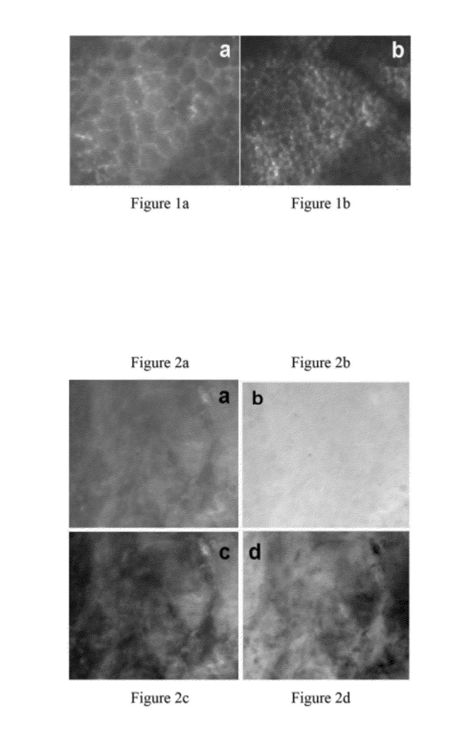
[0040]FIG. 1 exemplifies the different 245 μm×215 images acquired, using a prototype microscope, of a single leaf sample under the two specific excitation wavelengths, 266 nm and 355 nm respectively. The prototype microscope system has been built to test the designing principles of this invention. These autofluorescence images demonstrate the different microstructures visible within the leaf under different excitation.

[0041]FIGS. 2a-d show 245 μm×215 μm images of one high-grade dysplastic human esophagus biopsy under excitation from light at a wavelength of 266 nm, 355 nm, 266 nm / 355 nm and 355 nm / 266 nm, respectively. Clearly, the abnormal tissue autofluorescence appears different under 266 nm and 355 nm excitation. Image processing was used in the images of FIGS. 2c and 2d to further enhance contrast.

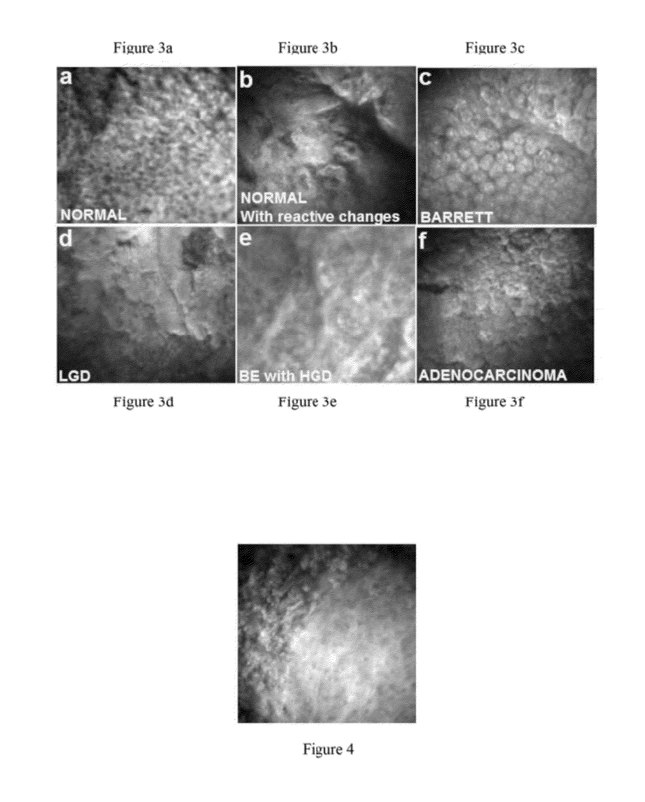
[0042]Images under 266 nm excitation were seen to provide spatial resolution and contrast sufficient to visualize nuclei. FIG. 3 shows examples of human esophagus optical biopsies, co...


PUM


Abstract

In vivo endoscopic methods an apparatuses for implementation of fluorescence and autofluorescence microscopy, with and without the use of exogenous agents, effectively (with resolution sufficient to image nuclei) visualize and categorize various abnormal tissue forms.

Description

[0001]This application claims priority to U.S. Provisional No. 61 / 112,509, filed Nov. 7, 2008. This is a continuation-in-part of U.S. application No. 11 / 292,406, filed Nov. 30, 2005 now U.S. Pat. No. 7,945,077, incorporated herein by reference.[0002]The United States Government has rights in this invention pursuant to Contract No. DE-AC52-07NA27344 between the United States Department of Energy and Lawrence Livermore National Security, LLC.BACKGROUND OF THE INVENTION[0003]1. Field of the Invention[0004]The present invention relates to medical diagnostics for tissue investigation and more particularly, it relates instruments for real-time monitoring of tissues in vivo.[0005]2. Description of Related Art[0006]Esophageal adenocarcinoma will claim approximately 89.6% of all cases within 5 years of diagnosis [2,3]. This rare but fatal disease has been increasing in the United States at the alarming rate of approximately 2% every year, and has been linked to conditions such as obesity [3-...

Claims


Application Information

Patent Timeline
Patent Type & Authority Patents(United States)
IPC IPC(8): G06K9/00
CPCA61B5/0071A61B5/0084A61B5/411A61B5/417G01N21/6458G01N21/6486G02B21/082G02B21/16G02B23/24A61B5/0088A61B5/0091G01N2021/4792
Inventor DEMOS, STAVROS G.URAYAMA, SHIROLIN, BEVINSAROUFEEM, RAMEZ MOUSSA GHOBRIAL
Owner LAWRENCE LIVERMORE NAT SECURITY LLC
Why Eureka
Try Eureka