Endoscope system

a technology of endoscope and endoscope, which is applied in the field of endoscope system, can solve the problems of difficult to improve the diagnostic performance of identifying cancer cells with a single type of fluorescent probe, and achieve the effect of improving the diagnostic performance of cancer cells

Inactive Publication Date: 2010-02-11
OLYMPUS CORP
View PDF21 Cites 14 Cited by
  • Summary
  • Abstract
  • Description
  • Claims
  • Application Information

AI Technical Summary

Benefits of technology

[0013]The present invention provides advantages in that the acquisition of a fluorescence distribution image for each fluorescent agent from a fluorescence image acquired

Problems solved by technology

Since molecules that are overexpressed in cancer cells are often overexpressed in inflamed areas/benign tumors etc., it

Method used


Image

  • Endoscope system
  • Endoscope system
  • Endoscope system

Examples


Embodiment Construction

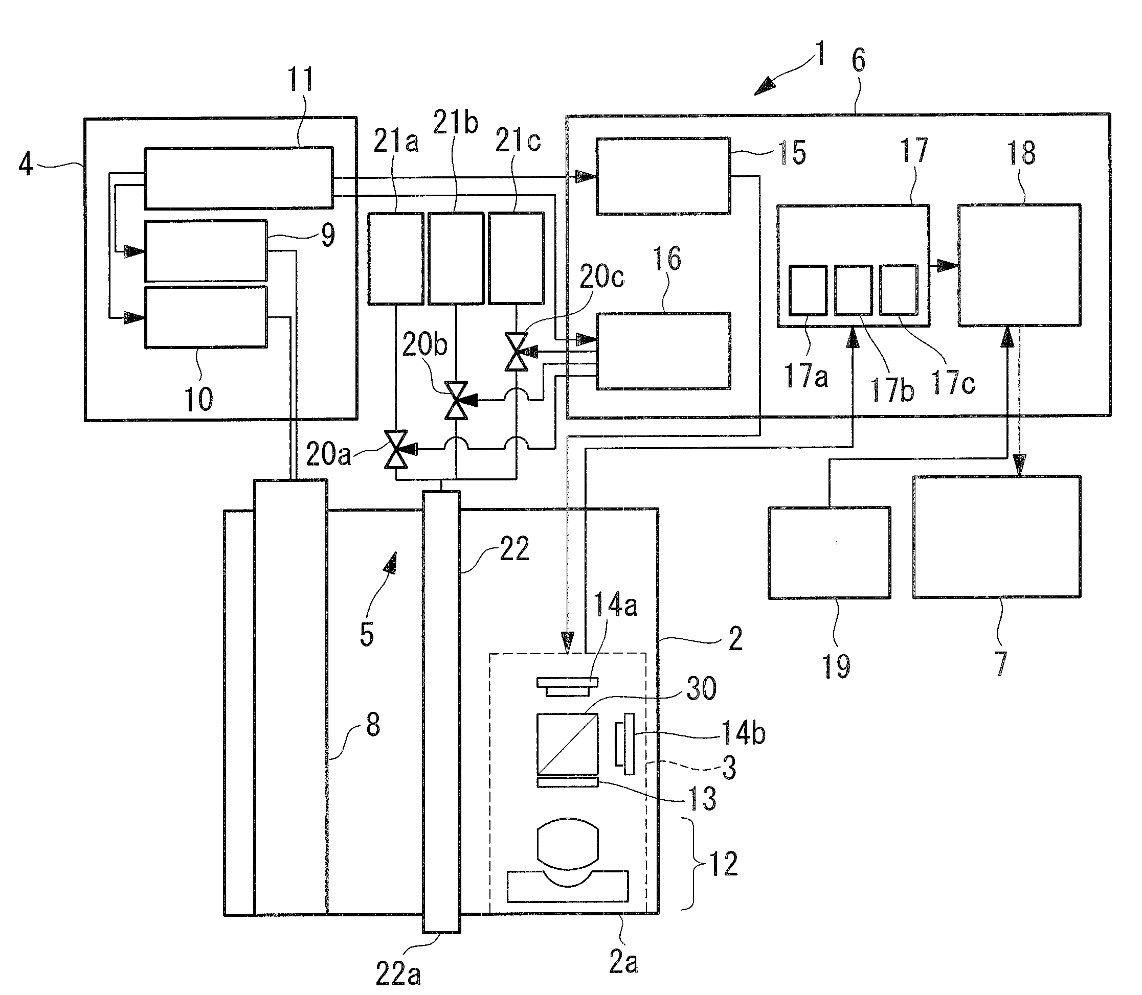
[0028]An endoscope system 1 according to a first embodiment of the present invention will be described below with reference to FIGS. 1 to 4.

[0029]As shown in FIG. 1, the endoscope system 1 according to this embodiment includes an insertion portion 2 that is inserted into a body cavity of a living body, an image-acquisition unit 3 that is disposed inside the insertion portion 2, a light source unit 4 that generates excitation light and illumination light for normal optical examination, a fluid supplying unit 5 that supplies fluid to be discharged from a tip 2a of the insertion portion 2, a control unit 6 that controls the image-acquisition unit 3, the light source unit 4, and the fluid supplying unit 5, and a display unit (display) 7 that displays an image acquired by the image-acquisition unit 3.

[0030]The insertion portion 2 has an extremely thin external shape that allows it to be inserted into the body cavity of a living body and accommodates the image-acquisition unit 3 and a lig...


PUM


Abstract

It is possible to acquire a fluorescence distribution image for each fluorescent agent from a fluorescence image acquired in a mixed state, thus improving the diagnostic performance of cancer cells. There is provided an endoscope system (1) configured to insert at least a part into a body cavity of a living body and to acquire an image of an image-acquisition subject in the body cavity, the endoscope system including a light source unit (10) configured to emit excitation light for exciting two or more different types of fluorescent agents having different optical characteristics; two or more image-acquisition units (14a, 14b) provided in a section inserted in the body cavity and configured to simultaneously capture fluorescence emitted from the image-acquisition subject as fluorescence in two or more different wavelength bands; a storage unit configured to store information associated with the relative relationship between the intensity of fluorescence generated when excited by the excitation light and the concentrations of the fluorescent agents; and a concentration-information calculating unit (18) configured to calculate and output concentration information of the fluorescent agents on the basis of fluorescence intensity of images in two or more wavelength bands captured by the image-acquisition units and the information associated with the relative relationship stored in the storage unit.

Description

TECHNICAL FIELD[0001]The present invention relates to an endoscope system.BACKGROUND ART[0002]Conventionally, diagnosis and treatment in which a fluorescent material having affinity to a disease, such as cancer, is injected in advance into the body of a test subject and excitation light that excites the fluorescent material is emitted to detect fluorescence from the fluorescent material accumulated in the diseased site have been attracting attention. According to this diagnosis method, since intense fluorescence is radiated from the diseased site, the presence of a lesion can be determined from the brightness of a fluorescence image.[0003]Patent Document 1 discloses an endoscope apparatus that diagnoses cancer cells using such a method.[0004]Patent Document 1:[0005]Japanese Unexamined Patent Application, Publication No. HEI-10-201707DISCLOSURE OF INVENTION[0006]Since molecules that are overexpressed in cancer cells are often overexpressed in inflamed areas / benign tumors etc., it is ...

Claims


Application Information

Patent Timeline
IPC IPC(8): A61B1/06
CPCA61B5/0084A61B5/0071
Inventor NAKAOKA, MASAYAMORISHITA, KOKI
Owner OLYMPUS CORP
Why Eureka
Try Eureka