
Multi-imager compatible robot for image-guided interventions and fully automated brachytherapy seed

Active Publication Date: 2010-02-18
THE JOHN HOPKINS UNIV SCHOOL OF MEDICINE
View PDF13 Cites 90 Cited by
  • Summary
  • Abstract
  • Description
  • Claims
  • Application Information

AI Technical Summary

Benefits of technology

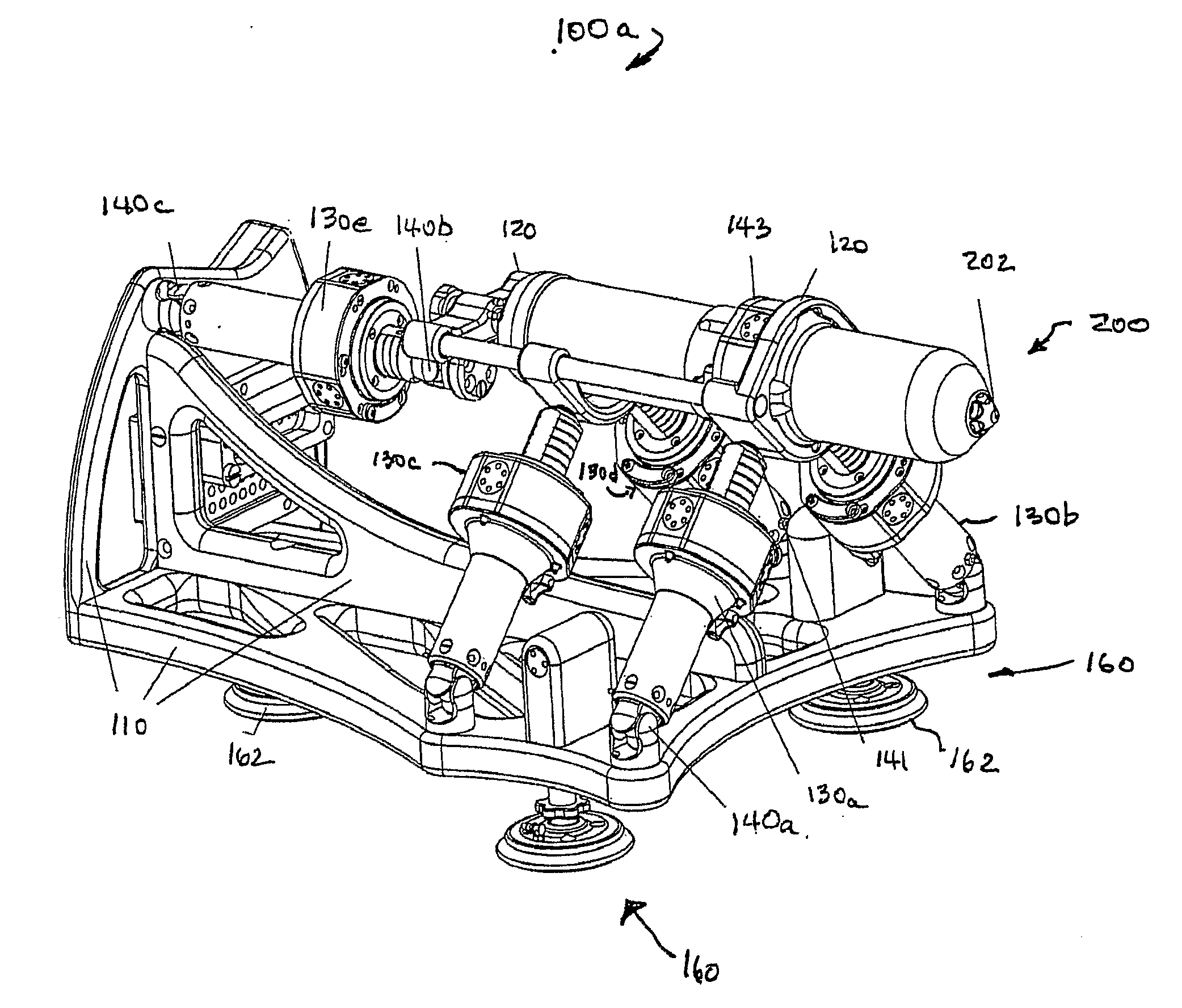
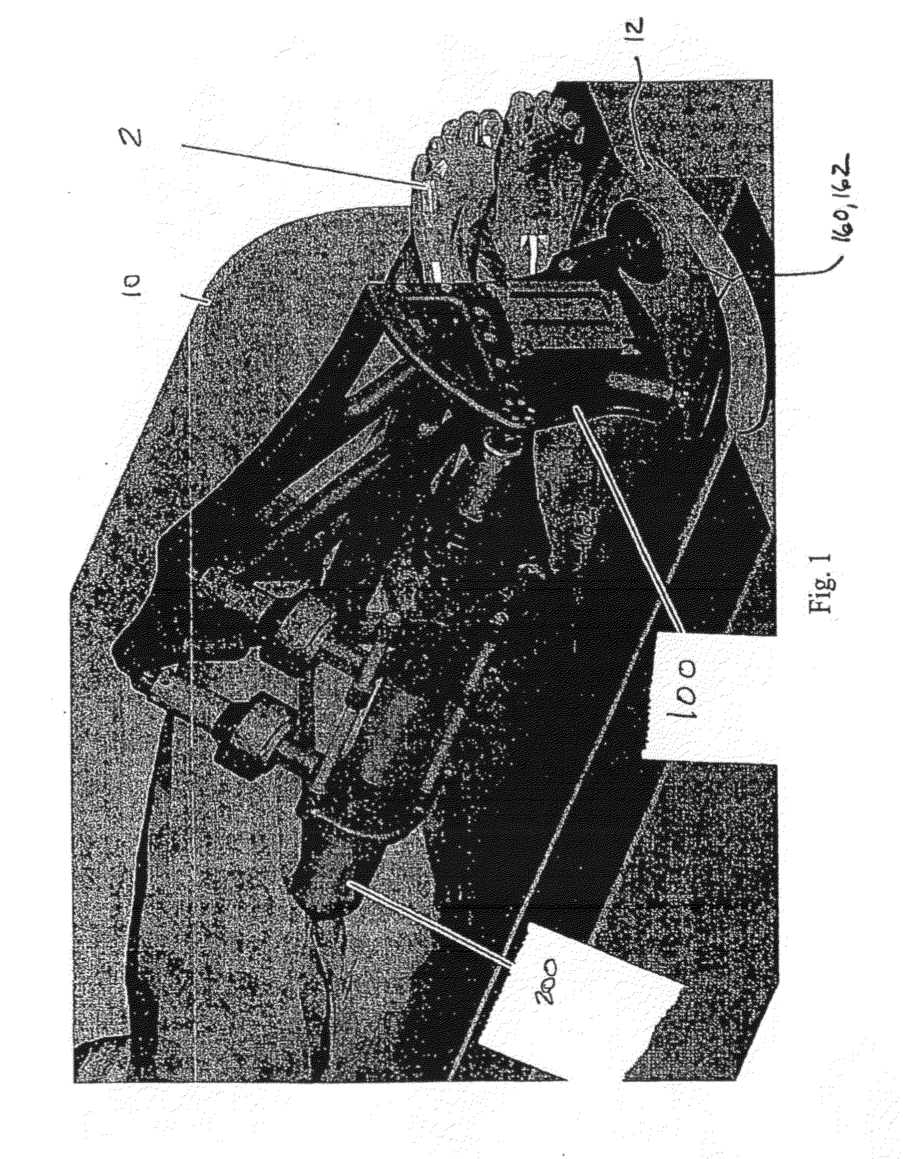
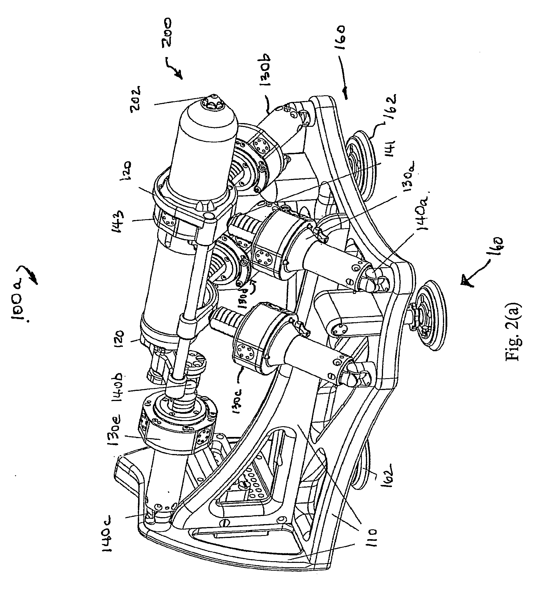
[0021]Moreover, because the size of the bore of a closed MRI scanner is on the order of about 500 mm in diameter, the space in such closed bore MRI scanners along the patient is very limited. Previous clinical trials revealed that perineal access legroom may be gained if the patient is positioned in the MRI head first in the left lateral decubitus position (i.e., on his side). As such, the robot of the present invention is configured and arranged so that the patient and the robot can be placed within the bore of an MRI scanner. Since other imager types provide larger access, the arrangement of the robot is acceptable for multi-imager accessibility, but other robot mounting orientations provide versatility of use.
[0025]The actuators of such a robot includes a fiber optic encoder for sensing motor operation and a fiber optic limit switch that provides a zero reference for linear movement and the robot further includes fiber optic cables as are known in the art. The fiber optic cables optically interconnect the fiber optic encoder and limit switch to electro-optical interface elements that detects light beam interruptions caused by designated moving elements in the actuator and provide electrical output signals therefrom. The optical cables have a length so that the electro-optical interface elements are located at least a predetermined distance from an imaging device so operation of the electro-optical interference elements at least substantially minimizes, if not avoids, the potential for creating signal interference during the imaging process.
[0036]The first interior member chamber and the needle piston are configured so that when pressurized fluid (e.g., gas) is admitted in one variable end portion of the first interior chamber, that is opposite to the needle piston, the needle moves in a first direction. Also, when fluid is removed from the one variable end portion of the first interior chamber so as to create a negative pressure therein, the needle moves in a second direction opposite to the first direction. The length of the first interior member chamber is set so a first end of the first interior member chamber defines a stop for the needle piston so as to thereby limit movement of the needle in the first direction.

Problems solved by technology

Medical imaging data, in particular, gives robots abilities unattainable to humans, because, unlike humans, robots and imagers are digital devices.
The design and construction of M compatible robots is a very challenging engineering task because most of the components commonly used in robotics can not be used in close proximity of the imager.
The use of electricity also can cause interference which can create image artifacts and / or robot signal distortions.
The problem becomes more intricate for robot actuation, where the ubiquitous electromagnetic motors are incompatible by principle when using MRI.
The major limitation of pneumatic actuators, however, has been their reduced precision in controlled motion [H. S. Choi, C. S. Han, K. Y. Lee, and S. H. Lee, “Development of hybrid robot for construction works with pneumatic actuator,”Automation in Construction, vol.
While research on MRI compatible robotics has been quantitatively limited, there is a commercial IGI system.
On the clinical application side, for needle access of the prostate gland under MRI guidance, manual procedures have been experimentally performed in a very few clinical trials, due to the complexity of the MR-IGI and the lack of proper instrumentation.

Method used


Image

  • Multi-imager compatible robot for image-guided interventions and fully automated brachytherapy seed
  • Multi-imager compatible robot for image-guided interventions and fully automated brachytherapy seed
  • Multi-imager compatible robot for image-guided interventions and fully automated brachytherapy seed

Examples


Embodiment Construction

[0068]The present invention features a robot, a robotic system, needle delivery apparatus that can be used with such a robot as well as methods and devices related thereto. In more particular aspects, such a robot generally includes a modular system structure that can be adapted for use with any of number of intervention specific medical devices such as intervention specific needle injectors. Such medical devices include, but are not limited to various end-effectors for different percutaneous interventions such as biopsy, serum injections, or brachytherapy. As also described herein, in particular embodiments such an end-effector is a fully-automated low dose radiation seed brachytherapy injector. Additionally, the present invention includes an automated seed magazine for delivering seeds to such a seed brachytherapy injector. Thus, in an all inclusive aspect, a robotic system of the present invention includes all of the foregoing features.

[0069]Such a robot also is particularly adap...


PUM


Abstract

Featured is a robot and a needle delivery apparatus. Such a robot comprises a plurality of actuators coupled to control locating any of number of intervention specific medical devices such as intervention specific needle injectors. Such a robot is usable with image guided interventions using any of a number of types of medical imaging devices or apparatuses including MRI. The end-effector can include an automated low needle delivery apparatus that is configured for dose radiation seed brachytherapy injection. Also featured is an automated seed magazine for delivering seeds to such an needle delivery apparatus adapted for brachytherapy seed injection.

Description

[0001]This application claims the benefit of U.S. Provisional Application Ser. No. 60 / 742,170 filed Dec. 2, 2005, the teaching of which are incorporated herein by reference.STATEMENT REGARDING FEDERALLY-SPONSORED RESEARCH[0002]The present invention was supported by grants from the National Institute of Health (NIH-NCI), grant number CA88232. The U.S. Government may have certain rights to the present invention.FIELD OF INVENTION[0003]The present invention relates to a robot for medical use, brachytherapy and includes methods, devices and apparatus related thereto and more particularly to such a robot that can be used with any of a number of imaging modalities and / or for image guided interventions, apparatus for automated brachytherapy and including methods, devices and apparatus related thereto.BACKGROUND OF THE INVENTION[0004]A count of robotics publications in the medical literature reveals that the impact of medical robotics has exponentially grown since its inception in late 1980...

Claims


Application Information

Patent Timeline
IPC IPC(8): A61M36/00G01D5/30A61B19/00
CPCA61N5/1007A61N2005/101A61N2005/1011A61B10/0233A61B2019/2226A61B2017/00544A61B2019/462A61B17/3468A61B19/2203A61B34/30A61B2034/304A61B2090/062
Inventor STOIANOVICI, DANPATRICIU, ALEXANDRUMAZILU, DUMITRUPETRISOR, DORUKAVOUSSI, LOUIS R.
Owner THE JOHN HOPKINS UNIV SCHOOL OF MEDICINE
Learn More