
Surgical Device For Minimal Access Surgery

Inactive Publication Date: 2010-04-01
ENDOROBOTICS
View PDF8 Cites 274 Cited by
  • Summary
  • Abstract
  • Description
  • Claims
  • Application Information

AI Technical Summary

Benefits of technology

[0012]The present invention also provides a system and single or multi-functional element insertable instrument that can be inserted and temporarily placed or implanted into a structure having a lumen or hollow space, such as a subject's abdominal cavity to provide access to the site of interest in connection with minimally invasive surgical procedures. The insertable instrument may be configured such that the functional elements have various degrees of freedom of movement with respect to orienting the functional elements or elements to provide access to the site from multiple and different orientations / perspectives as the procedure dictates, e.g., to provide multiple selectable views of the site, and may provide a stereoscopic view of the site of interest.

Problems solved by technology

However, even though multiple incisions are small, each is still often a source of pain for a patient, each may still result in scarring, and each still has a small but finite likelihood of leading to complications such as a wound infection or incisional hernia.
The greater the number of incisions, the incrementally greater likelihood of complications.
In the past, reducing the number of incisions has been attempted rarely, and with limited success.
However, these procedures require a different level of skill on the part of the surgeon, as well as different instruments than typically used when multiple incisions are made.
However, the surgeon must still use a laparoscope, and the laparoscope generally either occupies space in the large port that could otherwise be used for instruments for the operation, or the laparoscope requires its own port placed through the larger skin incision.
In each case, the laparoscope has the tendency to get in the way of the other instruments or otherwise complicate the surgery, further diminishing the surgeon's ability to manipulate the instruments and visualize the site of the surgery.

Method used


Image

  • Surgical Device For Minimal Access Surgery
  • Surgical Device For Minimal Access Surgery
  • Surgical Device For Minimal Access Surgery

Examples


example 1

Prototype I

[0144]The prototype created is a fully insertable, micro robotic imaging platform for minimally invasive surgery as a replacement for conventional laparoscopes and endoscopes. The product offers multiple degrees-of-freedom (pan, tilt and zoom movement) with picture-in-picture imaging, automatic instrument tracking and alternative joystick control.

(a) Dimensions:Length of 110 mm and width of 11 mm insertable through standard12 mm trocar(b) Housing (Shell) Material:Stainless steel(c) Components:+ Camera head - ¼″color video with active pixels 752(H) ×582 (V) at PAL System - 450 TV lines (H) × 420 (V)+ Camera head 6.5 mm in diameter+ Lens - miniature pin-hole lens machined to 6.5 mm o / s dia.(focal length 5.0 mm and F number 4.0)+ Pan / tilt mechanism− Brushless DC motor− 625:1 Planetary gear head− Dimensions: 27 mm × 5.8 mm diameter+ Focusing range: 40-100 mm.+ Zoom: Digital via software+ Worm gear --- 16:1 reduction ratio (machined to 125:1)+ Motor driver board+ Integrated LE...

example ii

Prototype II

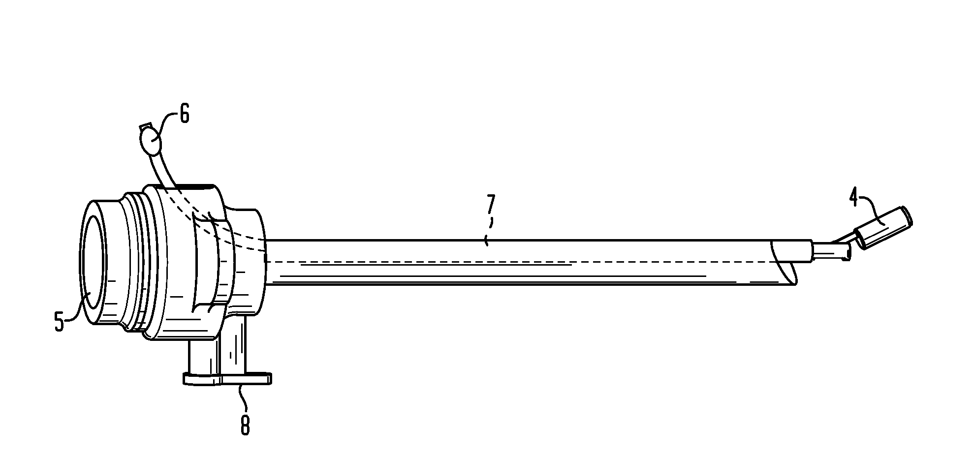
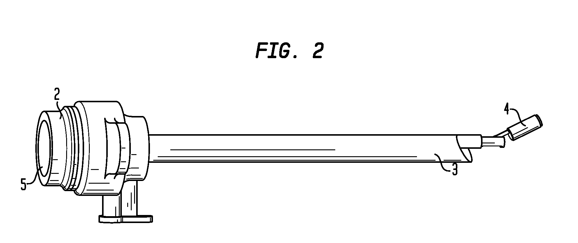
[0145]Another insertable instrument, as depicted in FIG. 8, available for attachment to a trocar within the scope of the present invention, is an imaging device whose outer shell is a tube 22 mm in diameter, 19 cm long, and has cabling emerging from its proximal end. The device contains a first motor that controls the tilting motion of the cameras, and is parallel with the central axis of the shell and near the proximal end of the device. The motor rotates in an inner shell that contains both cameras and additional motors. A 5.8 cm long section of the outer tube is cut away at the distal end to allow the cameras to tilt 180° when they are extracted. Visual serving experiments with Prototype II demonstrate that the system is capable of keeping a moving target pattern within its field of view. Image-based visual serving was used to track the target automatically.

[0146]In suturing experiments with a laparoscopic training box, the insertable instrument can provide an image t...

example iii

Prototype III

[0147]Another insertable instrument is available for attachment to a trocar within the scope of the present invention. The insertable instrument is smaller than that of Example II, and it includes an integrated light source. The surgical device has a two motor design with pan and tilt axes for a single camera module. The total length of the device is 110 mm, and the diameter is 11 mm, and it can be inserted into a 12 mm trocar. For the integrated light source, an LED array fits around the camera module, and provides lighting with low power requirements.

[0148]This insertable instrument has only a single camera, but an alternative insertable instrument includes 2 cameras side by side in a single camera module, thus sharing the pan / tilt axes but providing stereo 3D imaging. Including 2 cameras in a side-by-side design increases the device's diameter to 15 mm. Modular design was used advantageously to make the device components interchangeable and extendable.

[0149]After the...


PUM


Abstract

The present invention is directed to devices and methods for performing minimal access surgery; in various embodiments, to a surgical device comprising an insertable instrument attached to a trocar, as well as methods of performing a surgical procedure comprising inserting in insertable instrument / trocar combination into the body of a patient through an incision.

Description

CROSS REFERENCE TO RELATED APPLICATIONS[0001]The present application claims benefit of provisional Application No. 61 / 218,316 filed Jun. 18, 2009. This application is also a continuation-in-part of application Ser. No. 11 / 475,737, filed on Jun. 26, 2006, which is a continuation of application Ser. No. 11 / 226,665, filed on Sep. 13, 2005, now abandoned, which is a continuation-in-part of application Ser. No. 10 / 620,298, filed on Jul. 15, 2003, now U.S. Pat. No. 7,066,879; all of which are hereby incorporated by reference in their entireties.FIELD OF THE INVENTION[0002]This invention relates to the field of devices, in particular, devices that are useful in procedures such as minimal access or minimally invasive surgery, and that can be inserted and temporarily placed or implanted into a structure having a lumen or hollow space, such as a patient's abdominal cavity or elsewhere within the body of a patient, to provide access to a site of interest in connection with such minimal access ...

Claims


Application Information

Patent Timeline
IPC IPC(8): A61B17/34A61B1/00
CPCA61B1/00149A61B2019/2276A61B1/00188A61B1/00193A61B1/041A61B1/313A61B1/3137A61B5/0059A61B5/01A61B5/024A61B5/0816A61B5/145A61B5/14539A61B5/6882A61B8/00A61B17/00234A61B17/0469A61B17/320068A61B17/3421A61B17/3476A61B18/20A61B19/2203A61B19/5202A61B19/5212A61B2017/00221A61B2017/00398A61B2017/00734A61B2017/3447A61B2019/2207A61B2019/2211A61B2019/2242A61B2019/2249A61B1/00183A61B34/32A61B2034/742A61B34/71A61B2034/301A61B90/30A61B90/361A61B34/30A61B34/72A61B2017/320069A61B2017/32007
Inventor FOWLER, DENNISALLEN, PETER
Owner ENDOROBOTICS
Learn More