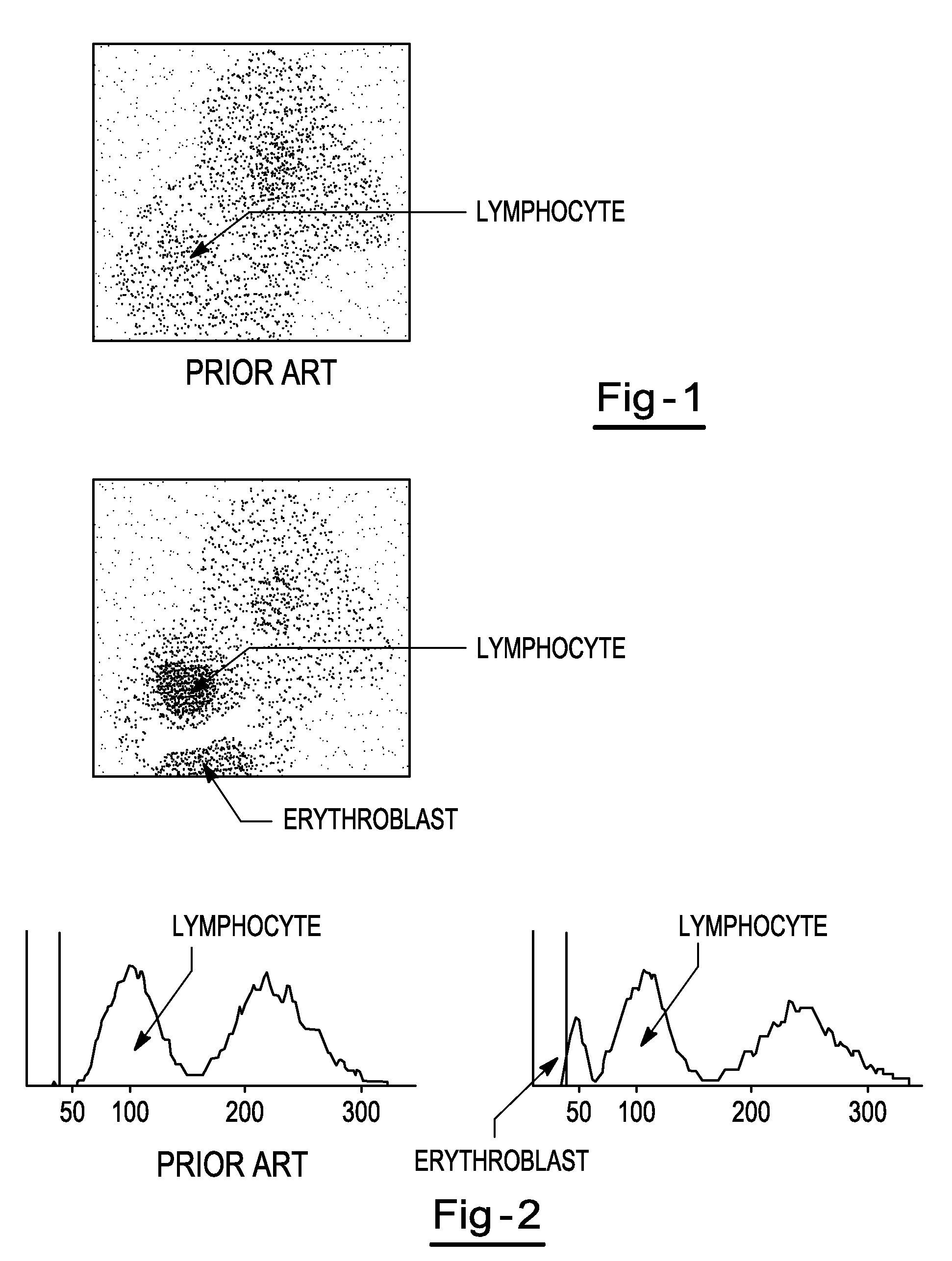
Hematology controls and methods
a hematology and control technology, applied in the field of simulated blood components, can solve the problems of difficult to achieve reproducibility across all desired subpopulations, the stability of the resulting product and/or the performance of individual instruments is not necessarily predictable, etc., and achieves the effects of stable hematology control, relative ease of handling, and cost-effective manufacturing
- Summary
- Abstract
- Description
- Claims
- Application Information
AI Technical Summary
Benefits of technology
Problems solved by technology
Method used
Image
Examples
example 1
[0060]Chicken red blood cells are selected and run on an automated hematology analyzer. The cells are centrifuged for 10 minutes at 1000 rpm. The supernatant is aspirated and the cells are re-suspended in Solution D of Table 1 or 2. The cells are again centrifuged for 10 minutes at 1000 rpm followed by re-suspension of the cells in Solution D of Table 1 or 2. The centrifugation / re-suspension process is repeated for a third time. The red blood cell count is then adjusted to 200,000 / μl using an automated hematology analyzer. A hypotonic fixative reagent is prepared, including a water solution containing 0.04% glutaraldehyde (percentage of stock glutaraldehyde which is 25%). The red blood cells are then added to the fixative reagent. The preparation is then incubated for 18 to 24 hours in a 40° C. water bath. The cells are then re-suspended and centrifuged for 10 minutes at 1000 rpm. Supernatant is aspirated and the cells are re-suspended in approximately 300 ml of a solution of phosph...
example 2
[0061]Turkey red blood cells are selected and run on an automated hematology analyzer. The cells are centrifuged for 10 minutes at 1000 rpm. The supernatant is aspirated and the cells are re-suspended in Solution D of Table 1 or 2. The cells are again centrifuged for 10 minutes at 1000 rpm followed by re-suspension of the cells in Solution D of Table 1 or 2. The centrifugation / re-suspension process is repeated for a third time. The red blood cell count is adjusted to 200,000 / μl using an automated hematology analyzer. A hypotonic fixative solution is prepared, by diluting glutaraldehyde to 0.3% in water (percentage of stock glutaraldehyde which is 25%). The fix ratio is 1:100. 1 ml of the cells is then added to 100 ml the fixative solution. The preparation is then incubated for 18 to 24 hours in a 40° C. water bath. The cells are then re-suspended and centrifuged for 10 minutes at 1000 rpm and the supernatant is aspirated. The cells are then re-suspended in a solution of phosphate bu...
example 3
[0062]Human blood cells are contacted with 150 ml of Solution E of Table 1 or 2 plus 0.11% formaldehyde for about 70 minutes at refrigerated temperature to stabilize the white blood cells. The cells are then centrifuged for 10 minutes at 1000 rpm and all but 100 ml of the supernatant is removed. The red blood cells are lysed with ammonium chloride tris solution for 25-35 minutes at refrigerated temperature. All but 100 ml of the supernatant is removed and the sample is again contacted with ammonium chloride tris solution for 60-70 minutes at refrigerated temperature. The sample is then washed with Solution F of Table 1 or 2 and the sample is centrifuged at a refrigerated temperature for 6 minutes at 600 rpm. The supernatant is carefully removed as the lymphocytes are located in the upper portion. The washing / centrifugation steps are repeated until the lymphocyte percentage is 10-15%. The white blood cells are then contacted with an expansion solution including Solution D of Table 1 ...
PUM
 Abstract
Description
Claims
Application Information
 - R&D
- Intellectual Property
- Life Sciences
- Materials
- Tech Scout
- Unparalleled Data Quality
- Higher Quality Content
- 60% Fewer Hallucinations
Browse by: Latest US Patents, China's latest patents, Technical Efficacy Thesaurus, Application Domain, Technology Topic, Popular Technical Reports.
© 2025 PatSnap. All rights reserved.Legal|Privacy policy|Modern Slavery Act Transparency Statement|Sitemap|About US| Contact US: help@patsnap.com

