Method for automatic boundary segmentation of object in 2d and/or 3D image

a technology of object boundary segmentation and image, applied in the field of medical imaging systems, can solve the problems of pain in ejaculation, difficulty in erectile contraction, and difficulty in starting a urinary tract infection, so as to improve the effectiveness and utility of trus, reduce inter and intra-operator variability, and minimize the effect of tim

Active Publication Date: 2010-06-03
THE UNIV OF WESTERN ONTARIO ROBARTS RES INST
View PDF13 Cites 72 Cited by
  • Summary
  • Abstract
  • Description
  • Claims
  • Application Information

AI Technical Summary

Benefits of technology

[0033]Shown in this present invention is that the fully-automatic version of the algorithm performs well. Thus this method for automatic prostate boundary segmentation eliminates user initialization for segmenting the prostate from 2D TRUS images, minimizes the time required for complete prostate segmentation, reduces inter and intra-operator variability and improves the effectiveness and utility of TRUS in diagnosis and treatment of prostate cancer. Although shown here is the use of this method in prostate boundary segmentation, this method is also applicable to segmentation of any object including, tumors, kidneys, and other organs. Also shown in this present invention is the use of 2D TRUS images for initialization, however, it is also applicable to other 2D or 3D US, CT and MRI images.

Problems solved by technology

Some of these symptoms include frequent and burning urination, difficulty starting a urinary stream, difficulty in having an erection, and painful ejaculation.
Although this procedure is low-risk, complication may occur.
1996), it is time consuming and is prone to user variability.
This method gave good results but it could generate erroneous edges due to artifacts in the image (Shao et al.
Its insensitivity to edges parallel to the radial direction from the centre of the prostate was the weakness of this method (Chiu et al.
In addition, this method failed if the image centre and the prostate boundary centroid were far from each other.
One of the drawbacks of this method was that the resulting prostate may be represented by a set of disconnected regions (Chiu et al.
Their method had the disadvantage of needing extensive training data.
This algorithm is very sensitive to the initial curve and only works well when this initial curve is reasonably close to the prostate boundary (Shao et al.
However, this method requires careful manual initialization of the contour and further user interaction to edit the detected boundary, which introduces complexity and user variability.

Method used


Image

  • Method for automatic boundary segmentation of object in 2d and/or 3D image
  • Method for automatic boundary segmentation of object in 2d and/or 3D image
  • Method for automatic boundary segmentation of object in 2d and/or 3D image

Examples


Embodiment Construction

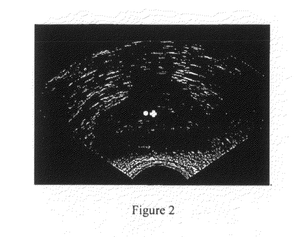
[0060]In accordance with one aspect of the present invention, as an input, the algorithm requires the approximate prostate centre. For prostate brachytherapy, the prostate gland is typically centred at the approximate centre of the image. In this case, the user does not need to select the prostate centre; as a result, the algorithm of the present invention would be fully-automatic. In cases when the prostate is not centred at the centre of the image, the user must select the approximate centre; as a result, our algorithm would be semi-automatic. FIG. 2 shows a 2D prostate TRUS image with the location of the user-selected centre shown by a dot and the centre of the image shown as a cross.

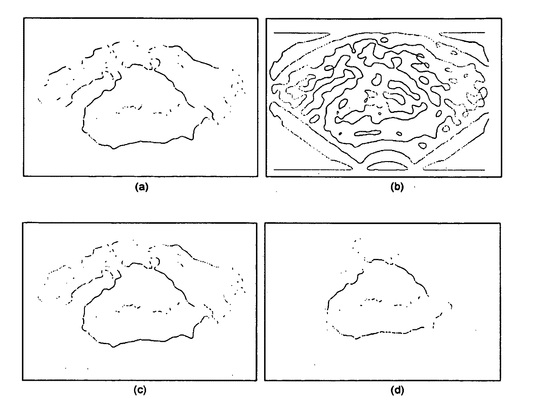
[0061]The Gaussian filter is used to blur the image and remove some detail and noise by convolving it with the 2D Gaussian distribution h(x, y):

h(x,y)=12πσ2-x2+y22σ2(1)

where σ is the standard deviation that determines the degree of smoothing. A digital approximation to the Gaussian function with σ=1 ...


PUM


Abstract

Segmenting the prostate boundary is essential in determining the dose plan needed for a successful bracytherapy procedure—an effective and commonly used treatment for prostate cancer. However, manual segmentation is time consuming and can introduce inter and intra-operator variability. This present invention describes an algorithm for segmenting the prostate from two dimensional ultrasound (2D US) images, which can be full-automatic, with some assumptions of image acquisition. Segmentation begins with the user assuming the center of the prostate to be at the center of the image for the fully-automatic version. The image is then filtered to identify prostate edge candidates. The next step removes most of the false edges and keeps as many true edges as possible. Then, domain knowledge is used to remove any prostate boundary candidates that are probably false edge pixels. The image is then scanned along radial lines and only the first-detected boundary candidates are kept the final step includes the removal of some remaining false edge pixels by fitting a polynomial to the image points and removing the point with the maximum distance from the fit. The resulting candidate edges form an initial model that is then deformed using the Discrete Dynamic Contour (DDC) model to obtain a closed contour of the prostate boundary.

Description

CLAIM OF PRIORITY[0001]The present application claims priority to the provisional application 60 / 939,443 filed to the United States Patent and Trademark Office on May 22, 2007, the content of which is hereby incorporated by reference into the present application.FIELD OF THE INVENTION[0002]The present invention relates in general to medical imaging systems, and more specifically, to a method for generating an automatic segmentation of the prostate boundary from 2D and 3D images.BACKGROUND OF THE INVENTION[0003]The prostate gland is an essential part of the male reproductive system. Located in the pelvis behind the pubic bone and right in front of the rectum and below the neck of the bladder, the prostate completely surrounds the urethra, which is the passageway that carries urine from the bladder through the penis and out of the body. In a healthy adult male, the prostate is approximately the size and shape of a walnut, weighing about 20 grams and measuring about 3 cm in width and 2...

Claims


Application Information

Patent Timeline
Patent Type & Authority Applications(United States)
IPC IPC(8): G09G5/00
CPCG06T7/0012G06T7/0083G06T7/0089G06T2207/30081G06T2207/20101G06T2207/20168G06T2207/10132G06T7/12G06T7/149
Inventor SAIKALY, MANALEFENSTER, AARONLADAK, HANIF
Owner THE UNIV OF WESTERN ONTARIO ROBARTS RES INST
Why Eureka
Try Eureka