
Apparatus and method for processing microscopic single cell biological specimens with a single microtool

a single microtool and microtool technology, applied in the field of new oocyte, embryo and specimen positioning apparatus and a method for inspection, observation, biopsy and micromanipulation, can solve problems such as not being designed to accommodate, and achieve the effect of improving efficiency, improving efficiency and efficiency

Inactive Publication Date: 2010-07-08
COOPERSURGICAL INC
View PDF8 Cites 4 Cited by
  • Summary
  • Abstract
  • Description
  • Claims
  • Application Information

AI Technical Summary

Benefits of technology

[0017]The oocyte, cell or embryo can be removed from the restricting narrow areas of the compartment after the microprocedure by using a flow of fluid or culture medium released from a standard pipette or by gently probing the oocyte or embryo towards the tip of the narrowest end of the lane. A small alcove in each of the compartments may allow deposition and holding of biopsied cells or fragments from a biopsy micropipette after biopsy of the oocyte, cell or embryo. The biopsied cells or fragments can be pipetted safely using a narrow bored pipette. The dish may also contain separate wells for prepared samples of spermatozoa to be used for microinjection of one or more spermatozoa when the dish is used for intra-cytoplasmic sperm injection (ICSI). The dish may also contain other wells for washing oocytes or embryos before and after micromanipulation.
[0018]The apparatus of this invention may allow users to better and more effectively and efficiently perform the following procedures:1) Intra-cytoplasmic sperm injection (ICSI) and other forms of assisted fertilization;2) Polar body biopsy;3) Assisted hatching, which is assisting the hatching process of a three day old embryo, or a blastocyst embryo, from the zona pellucida with a microtool, when the embryo is unable to hatch on its own;4) Blastomere biopsy;5) Blastocyst biopsy;6) Manipulation of the zona pellucida (chemical, enzymatic or by laser);7) Injecting solutions into the oocytes, blastomeres or blastocysts for transferring genes or other bio-molecules; and8) Removing and injecting cytoplasm or organelles into oocytes or embryos.

Problems solved by technology

Most current dishes are flat and are not designed to accommodate any particular procedure.

Method used


Image

  • Apparatus and method for processing microscopic single cell biological specimens with a single microtool
  • Apparatus and method for processing microscopic single cell biological specimens with a single microtool
  • Apparatus and method for processing microscopic single cell biological specimens with a single microtool

Examples


Embodiment Construction

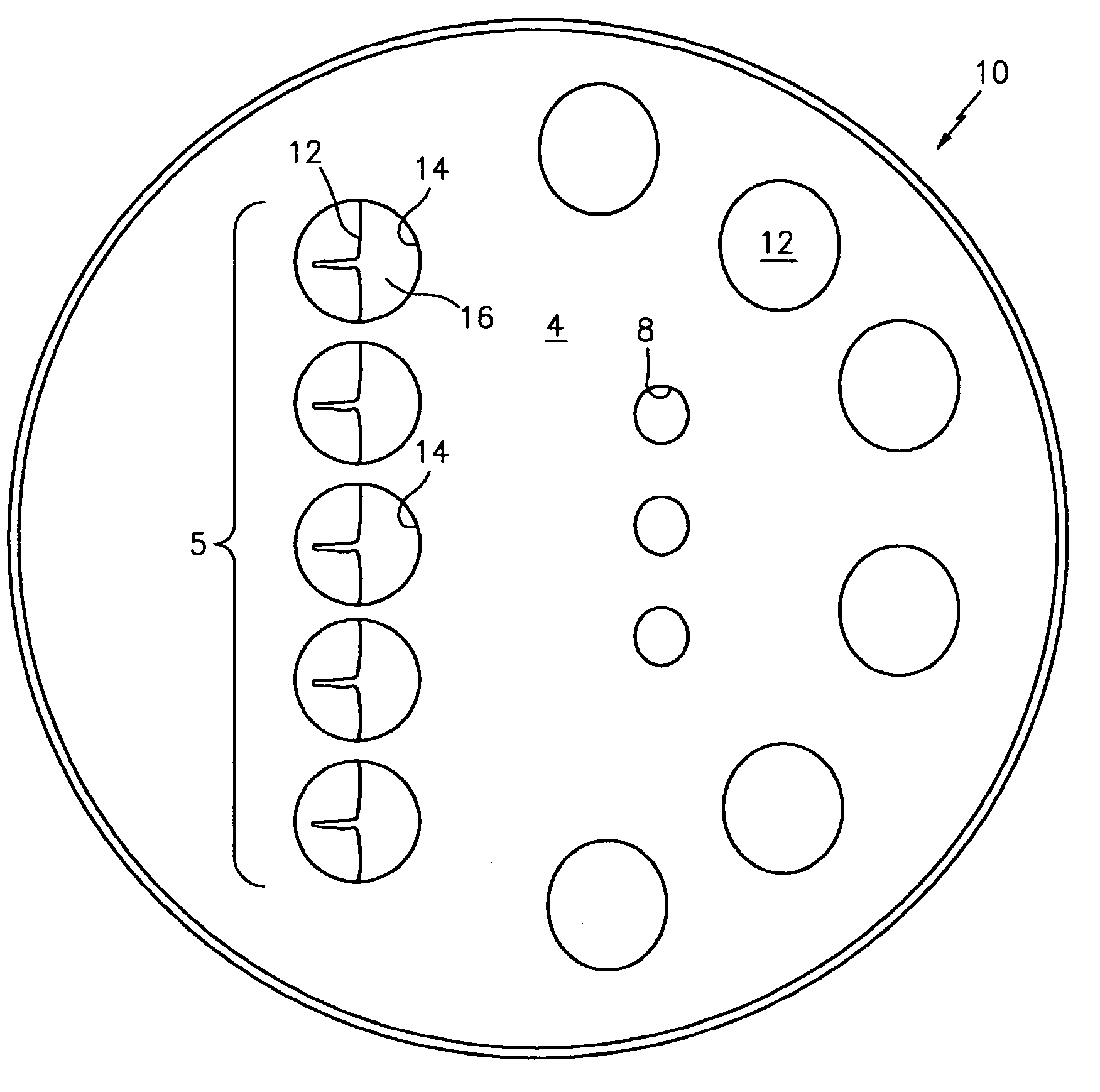
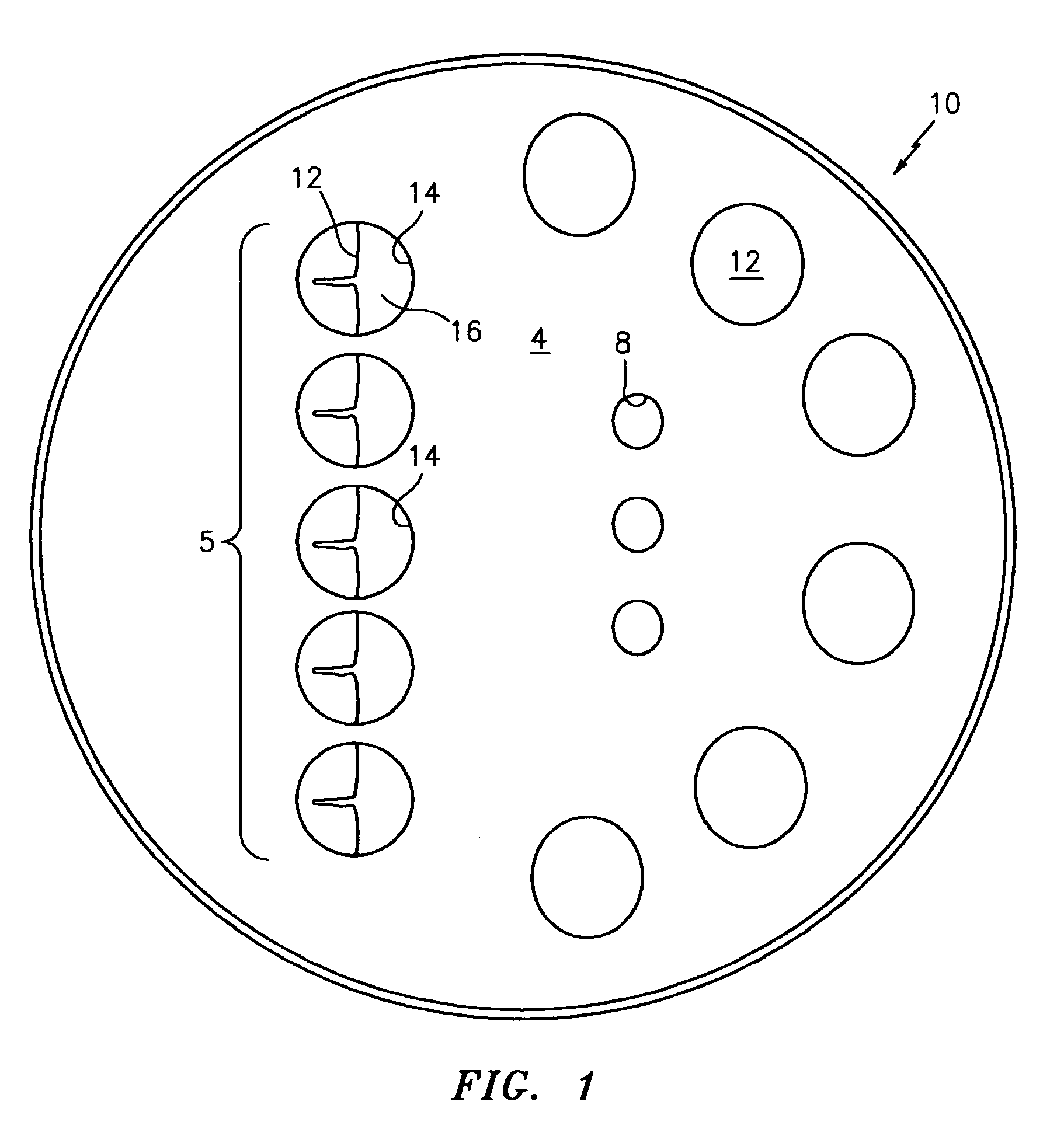
[0031]Referring now to FIG. 1 there is shown a plan view of the apparatus of this invention 12 inserted into a Petrie dish 10. The apparatus 12 is surrounded by walls 14 creating a small well 16. The walls 14 of the well 16 are taller than the apparatus 12. The walls 14 allow the user to place culture media and oils into the well area 16 to fully submerge the apparatus 12. There are five wells 16 in this embodiment of the invention. The number of wells 16 can be five, ten, or more, depending on relationship of the sizes of the wells 16 and the size of the dish 10, and the needs of the users will determine the exact number. Within the dish 10 are additional wells 8 which may be used to hold reagents, media, stem cells, embryos, sperm or other materials related to each procedure.

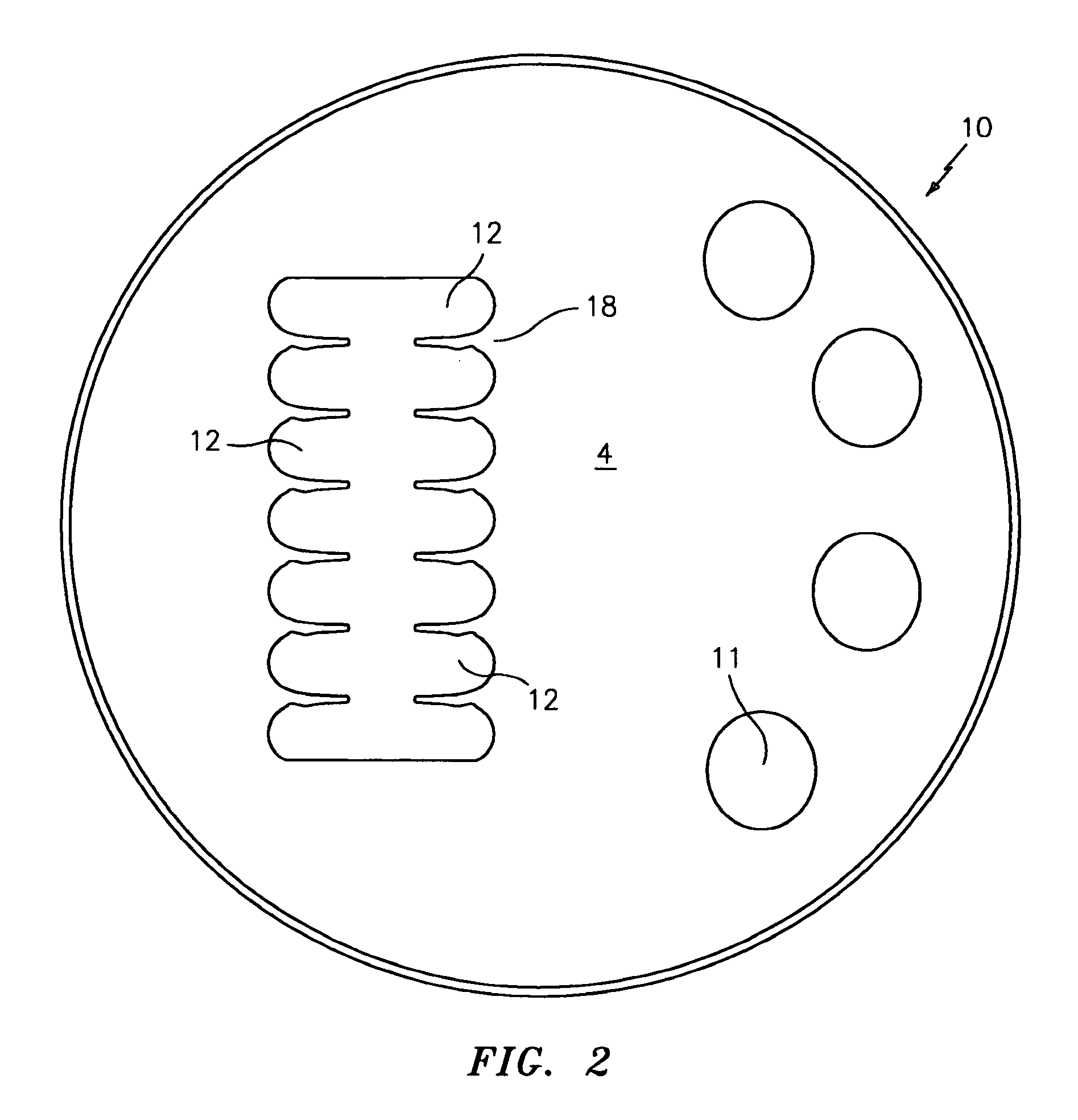
[0032]FIG. 2 shows a configuration of the apparatus 12 in a Petrie dish 10. In this example, the apparatus 12 consists of rows 18 of cell capture compartments, in a back to back fashion. This configuration ill...


PUM


Abstract

An oocyte and embryo-positioning apparatus enables a plurality of oocytes, embryos or other cell specimens to be held and positioned for injection therein of fluid molecular reagents such as DNA, RNA, protein, cells, spermatozoa and cells; and removal of cells, fragments of cells, cytoplasm, nuclei and cellular organelles. The oocytes and embryos are kept separate from each other in open marked or unmarked well-compartments that are located in the culture container. Each well-compartment comprises one or more finger like structures that can position one oocyte or one embryo. The lanes between the digits of the fingers may be sloped allowing the oocytes or embryo to position at the narrowest and lowest point of the compartment. The method and oocytes and embryo-positioning apparatus of this invention includes a culturing container, such as a Petrie dish, in which the oocytes and embryos are held for micromanipulation. The Petrie dish preferably contains a plurality of the oocyte or embryo-holding finger like-structures which can be positioned in the Petrie dish in a predetermined pattern. For example, the well-compartments can be positioned in the Petrie dish as a strip of well-compartments between the 12 o'clock and 6 o'clock positions, or in any other planned deployment. Marked well-compartments can be identified by letters or numbers or symbols.

Description

[0001]This application claims the benefit of U.S. Ser. No. 61 / 200,462, filed Dec. 1, 2008.TECHNICAL FIELD[0002]This invention relates to a novel oocyte, embryo and specimen positioning apparatus and a method for inspection, observation, biopsy and the micromanipulation of oocytes, embryos and cells without the use of a second device, such as a holding pipette. The invention relates to the use of a series of structures within the apparatus that will allow the specimen to be placed into the apparatus and into the structures whereby the specimen is held in place may be easily located, and may be biopsied, manipulated or such without the use of a holding pipette. The apparatus allows the use of a single microtool, such as a micro injection pipette for the injection of materials, or a pipette to biopsy the specimen without a holding pipette to hold and stabilize the specimen during the procedure. The apparatus can be used for a wide range of specimens of animal and human cells, tissues, ...

Claims


Application Information

Patent Timeline
Patent Type & Authority Applications(United States)
IPC IPC(8): A61B17/43A61B17/435
CPCC12M21/06C12M33/04C12M23/34C12M23/10
Inventor COHEN, JACQUESSCHIMMEL, TIMCECCHI, MICHAEL D.
Owner COOPERSURGICAL INC
Learn More