
Dual Modality Imaging Of Tissue Using A Radionuclide

Inactive Publication Date: 2010-07-15
MAYO FOUND FOR MEDICAL EDUCATION & RES
View PDF4 Cites 4 Cited by
  • Summary
  • Abstract
  • Description
  • Claims
  • Application Information

AI Technical Summary

Benefits of technology

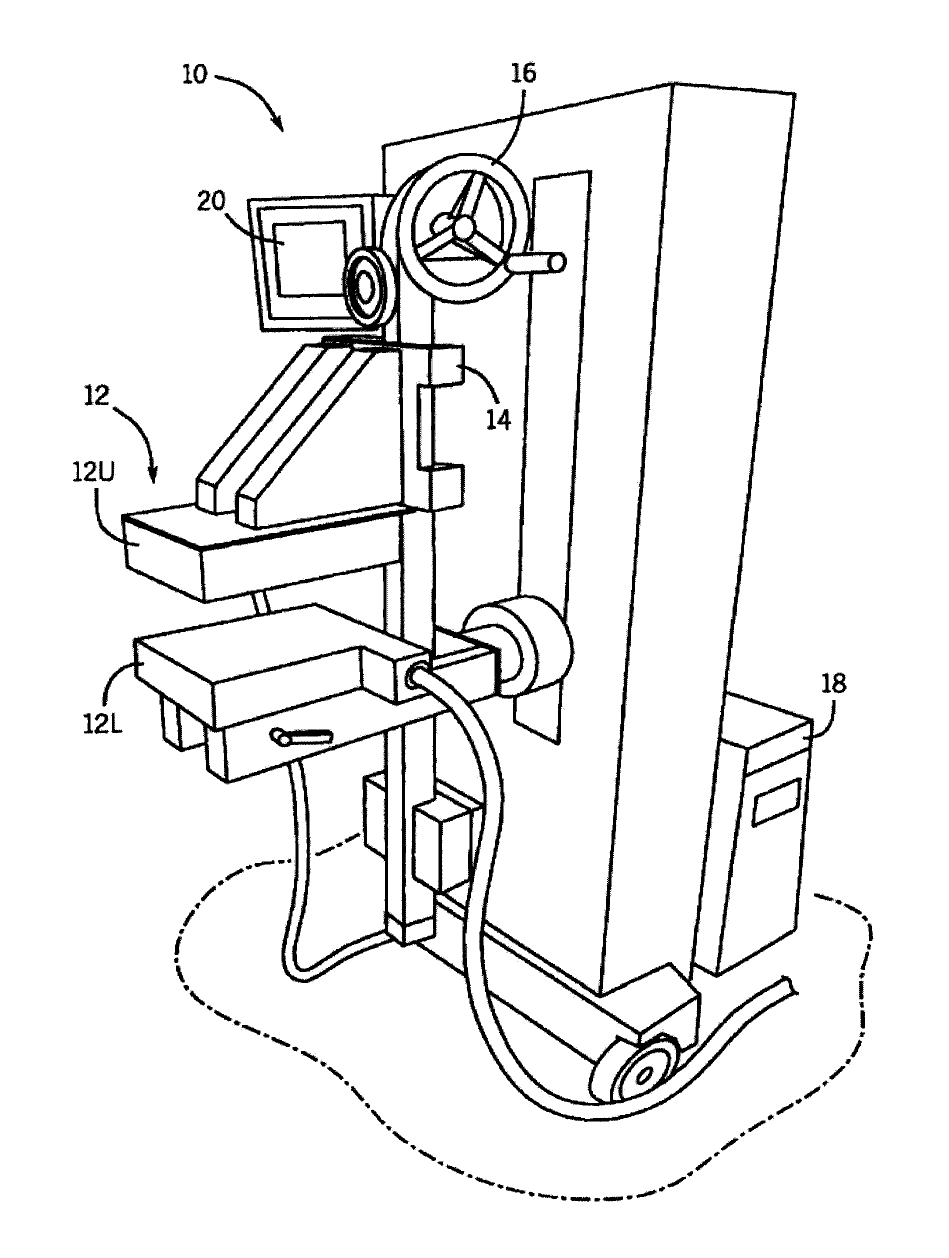
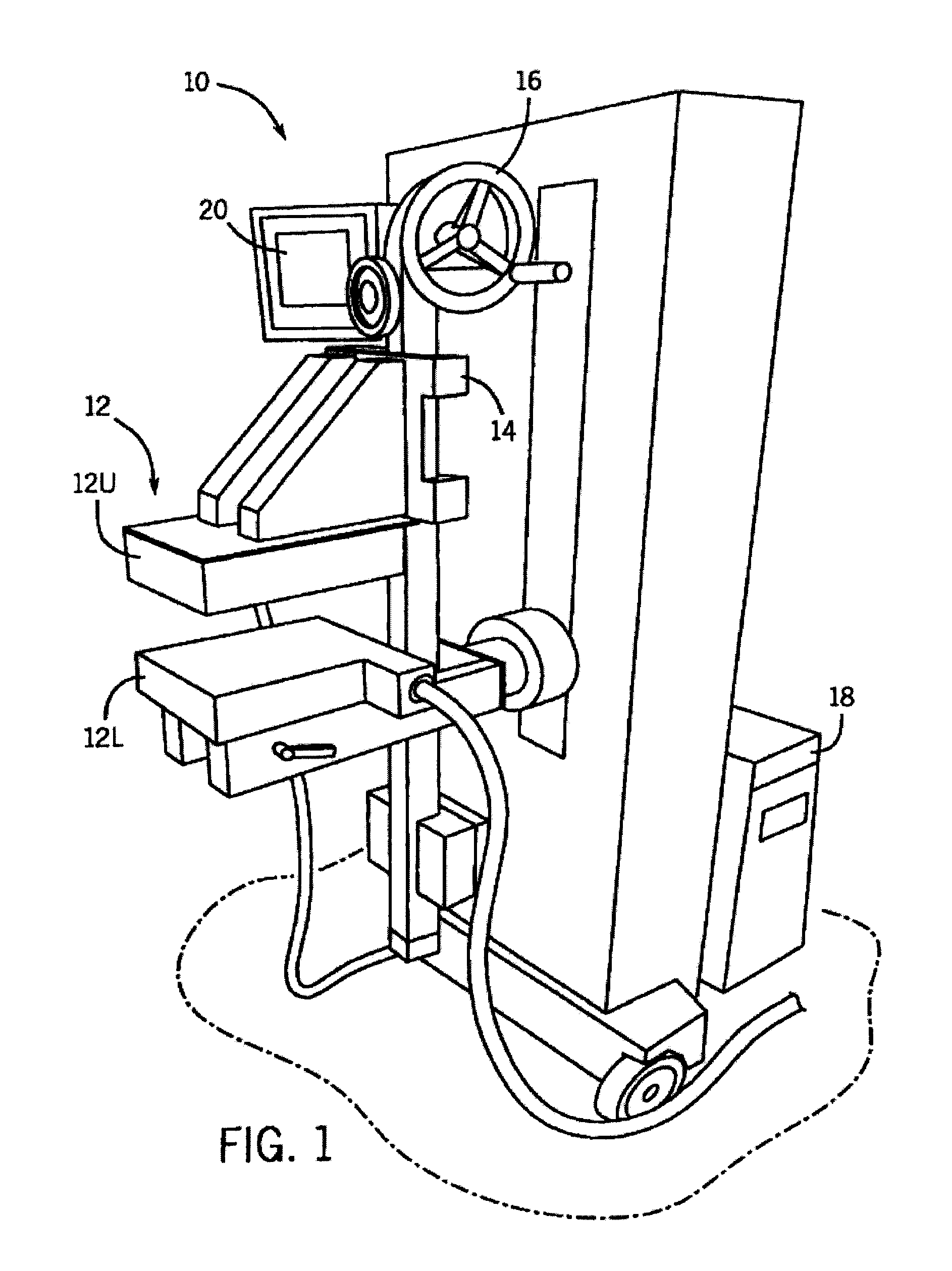
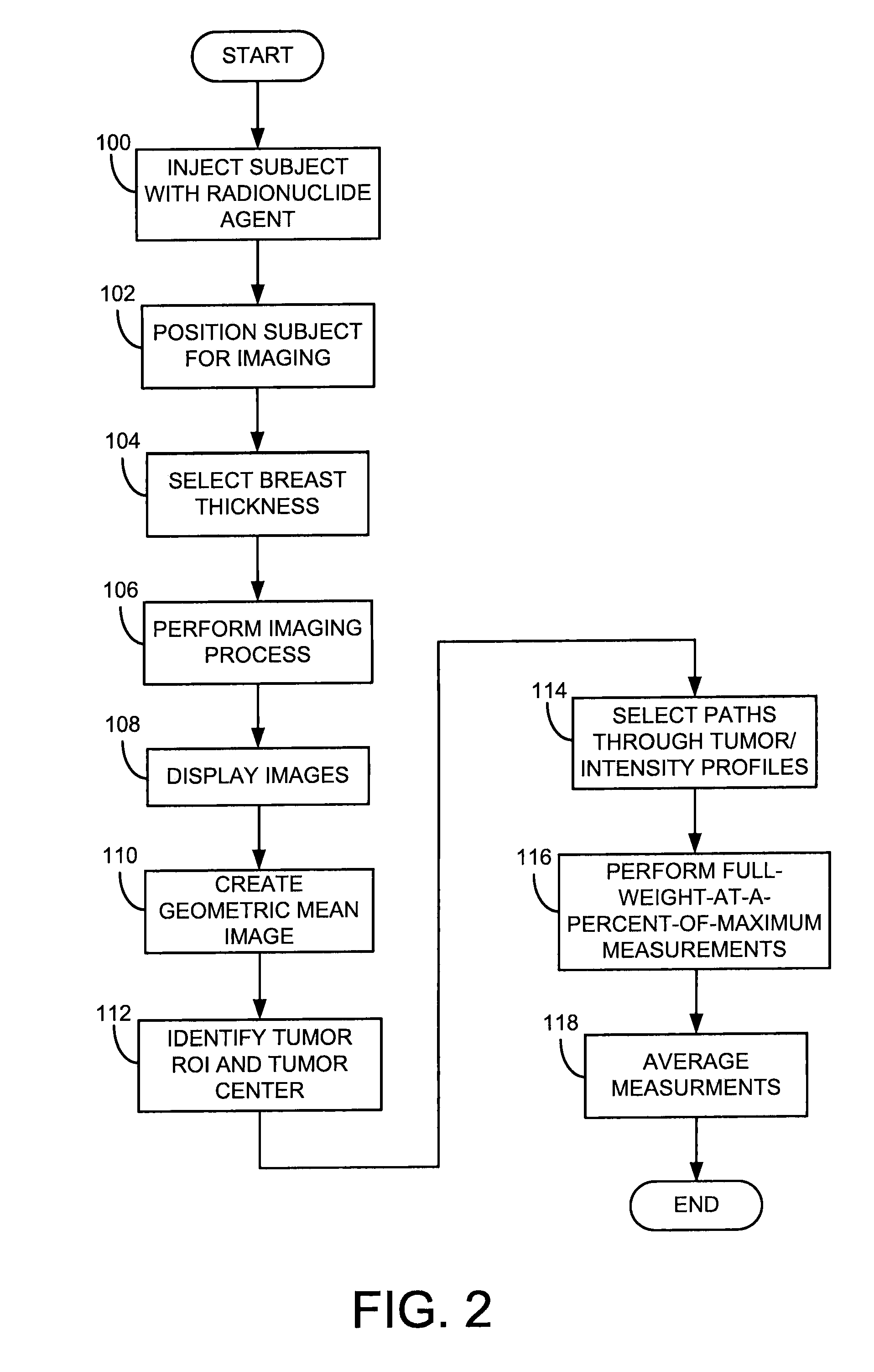
[0016]The present invention overcomes the aforementioned drawbacks by providing a system and method for performing quantitative tumor analysis using information acquired with a dual-headed, high resolution molecular breast imaging (MBI) system at a regiment that can be coordinated with other nuclear imaging, such as with myocardial perfusion imaging. The MBI process can accommodate for an injection dosage that is higher than 20 mCi Tc-99m sestamibi or Tc-99m tetrofosmin, such as the dosages used during MPI. By recognizing that MBI is not impacted by uptake of these radiopharmaceuticals in other organs, such as the myocardium or the liver, the present invention permits breast imaging to commence shortly after the injection of the radiopharmaceutical, such as 5 minutes post-injection, and thereby, temporally couple an MBI procedure with other nuclear imaging processes to perform a breast analysis without the patient being subjected to any additional radiation dose.

Problems solved by technology

In fact, women suffer a higher risk of mortality resulting from cardiovascular disease than from cancer.
For example, if the diminished radionuclide concentration is worse when the radionuclide is administered during stress as compared to when the radionuclide is administered during rest, the zone of decreased concentration during stress is likely due to ischemia.
On the other hand, if the radionuclide concentration remains the same after an injection of the radionuclide at rest, the zone of decreased concentration is likely due to scar tissue.
One factor in the failure of mammography to detect breast cancer is radiographic breast density.
However, since bilateral breast MRI is currently approximately 20 times more expensive than mammography, it is not in widespread use as a screening technique.
However, the currently available radiotracer, F-18 Fluoro deoxyglucose, requires that a patient fast overnight; the patient have low blood levels (this is often a problem for diabetics); and the patient should wait 1-2 hours after the injection for optimum uptake of F-18FDG in the tumor.
The high cost of these PET procedures coupled with the long patient preparation time reduces the usefulness of this procedure and makes it difficult to employ for routine breast evaluation.
However, these results only hold for large tumors and several studies have shown that the sensitivity falls significantly with tumor size.
This limitation is particularly relevant in light of the finding that up to a third of breast cancers detected by screening mammography are smaller than 10 mm.
The failure of conventional scintimammography to meet this limit led to its abandonment as a useful technique in the United States.
However, when larger amounts are given, it may cause adverse effects on the body may ensue.

Method used


Image

  • Dual Modality Imaging Of Tissue Using A Radionuclide
  • Dual Modality Imaging Of Tissue Using A Radionuclide
  • Dual Modality Imaging Of Tissue Using A Radionuclide

Examples


Embodiment Construction

[0026]In nuclear medicine, both breast cancer and myocardial diseases can be connected through the fortuitous biological behavior of radiopharmaceuticals such as Tc-99mm sestabmibi and Tc-99m tetrofosimin. These radiopharmaceuticals were originally developed in the 1980s for myocardial perfusion imaging and have become the standard radiopharmaceuticals used in the United States in the evaluation of myocardial function. In the late 1980′s it was noticed that these compounds also localized in a variety of cancers and showed strong uptake in breast cancer. This finding lead to the development of a technique known as scintimammography. However, technical limitations significantly reduced the sensitivity of the technique for the detection of small breast tumors (sensitivity>50% for sub 10 mm lesions), and the technology never gained widespread clinical use. Recent studies have overcome some of these limitations. For example, a co-pending patent application with an Application Ser. No. WO...


PUM


Abstract

A method for imaging a subject includes injecting the subject with a single dose of a radionuclide and acquiring, with a molecular breast imaging (MBI) system, a first set of medical image data of a breast of the subject after injection of the single dose of a radionuclide. The method also includes acquiring, with a myocardial perfusion imaging (MPI) system, a second set of medical image data of a portion of a cardiovascular system of the subject after the single dose of a radionuclide and reconstructing the first set of medical image data into a medical image of the breast of the subject and reconstructing the second set of medical image data into a medical image of the portion of the cardiovascular system of the subject.

Description

REFERENCE TO RELATED APPLICATIONS[0001]This application is based on provisional application Ser. No. 61 / 145,053, filed Jan. 15, 2009, and entitled “DUAL MODALITY IMAGING OF TISSUE AFTER ADMINISTRATION OF A RADIONUCLIDE,” and claims the benefit thereof.FIELD OF THE INVENTION[0002]The present invention related to systems and methods for imaging of organic tissue, and more particularly, to a system and method for using a radionuclide as an agent imaging, such as in imaging breast and myocardial tissue.BACKGROUND OF THE INVENTION[0003]Cardiovascular disease and cancer remain the leading causes of death in America. In fact, women suffer a higher risk of mortality resulting from cardiovascular disease than from cancer. In 2007 alone, almost twice as many women died of cardiovascular disease (both heart and stroke) than from all cancers combined. For example, studies have shown that one in three American women will die of heart disease, while approximately one in seven American women will ...

Claims


Application Information

Patent Timeline
IPC IPC(8): A61B6/00
CPCA61B5/4312A61B6/504A61B6/502A61B6/037
Inventor O'CONNOR, MICHAEL K.
Owner MAYO FOUND FOR MEDICAL EDUCATION & RES
Learn More