

Method for acquiring 3-dimensional images of coronary vessels, particularly of coronary veins
a technology of coronary veins and 3D images, applied in image enhancement, angiography, instruments, etc., can solve the problems of restricting the available space around the patient, reconstructed 3-dimensional models may only yield a rough representation of the coronary veins, and cannot be calculated on 3D reconstruction or models, so as to improve the quality of the acquired x-ray images, improve the filter quality, and achieve the effect of precise resulting centerline models
- Summary
- Abstract
- Description
- Claims
- Application Information
AI Technical Summary
Benefits of technology
Problems solved by technology
Method used
Image

Examples
Embodiment Construction
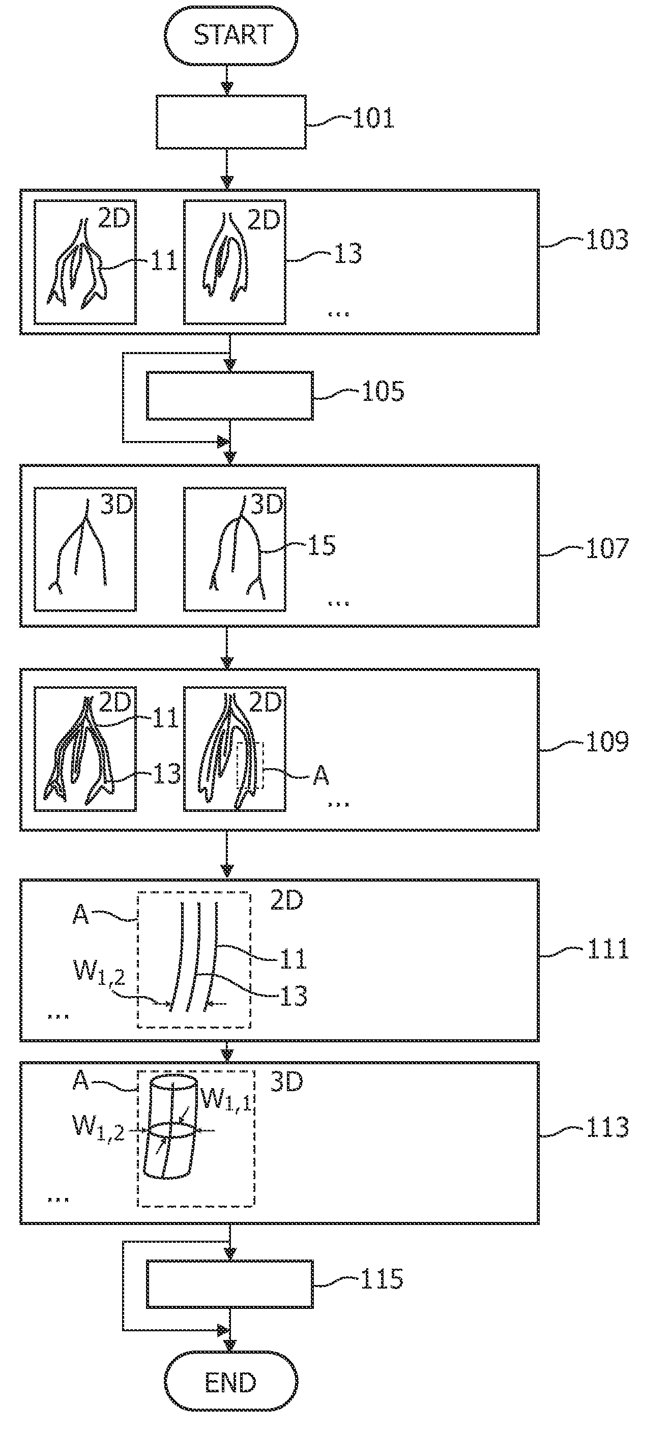
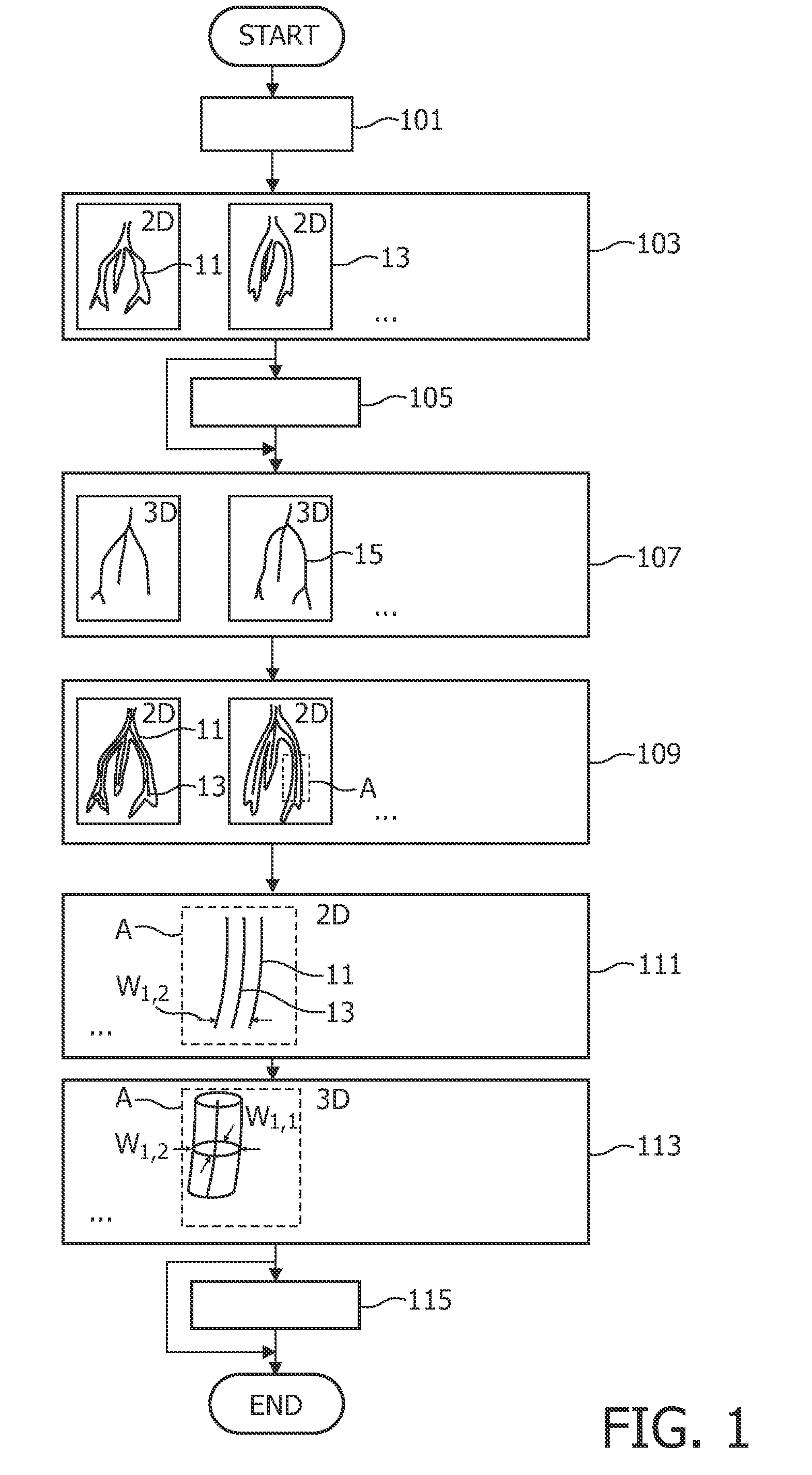
[0035]FIG. 1 can be used to explain the basic steps of a method for acquiring a 3-dimensional image of a coronary vein according to an embodiment of the present invention.
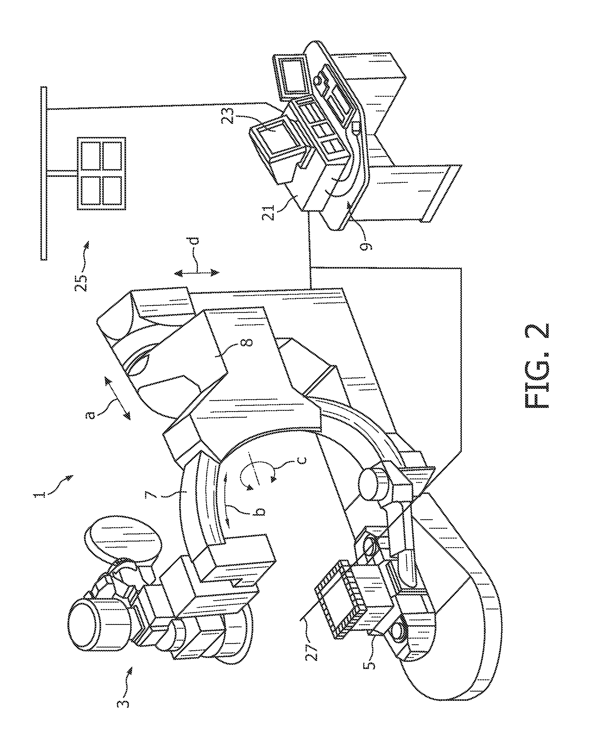
[0036]After locating a patient in a suitable apparatus such as a C-arm X-ray apparatus, contrast medium is injected into a coronary vein to be imaged using a catheter (step 101).
[0037]Then, a plurality of 2-dimensional X-ray images of an observation region including the veins 11 is acquired under different projection angles while rotating the C-arm around the patient's corpus (step 103) (only two images 13 shown exemplary).
[0038]Optionally, the acquired 2D images may be downsampled and / or filtered using a high-pass filter and / or a vessel enhancement filter (step 105) thereby improving the image quality with respect to the veins to be imaged.
[0039]From a specific number of 2D images acquired for a same motion phase such as the end-diastolic phase where there is minimum cardiac motion, a 3D centerline model 15 of the...
PUM
 Abstract
Description
Claims
Application Information