
System and method for descemet's stripping automated endothelial keratoplasty (DSAEK) surgery

Inactive Publication Date: 2010-08-19
WESTON PHILIP DOUGLAS +2
View PDF20 Cites 26 Cited by
  • Summary
  • Abstract
  • Description
  • Claims
  • Application Information

AI Technical Summary

Benefits of technology

[0009]Accordingly, the broad objects of the invention are to provide an apparatus and method for inserting a corneal implant (also referred to variously as a “donor,” a “donor cornea,” a “corneal lenticule,” a “lenticule,” and a “donor corneal lenticule”) into the eye of a recipient without inducing significant endothelial damage.
[0010]A more specific object of the invention is to provide such an apparatus and method wherein and whereby the donor corneal lenticule is optimally temporarily deformed for effective insertion, while providing enhanced protection against significant endothelial damage before and during surgical implantation.
[0011]Additional objects of the invention are to provide an apparatus having the foregoing features and advantages which, in addition, is readily employed and is highly efficient and effective for its intended purposes; and to provide such a method which is readily carried out and is highly efficient and effective.

Problems solved by technology

One of the most challenging aspects of this procedure is the insertion of the donor posterior lenticule into the anterior chamber (AC) through a small incision, without inducing significant endothelial damage.
This traumatic handling of the donor has been criticized because of its propensity for damaging endothelial cells, with primary graft failure rates due to intraoperative endothelial cell loss and damage ranging from 6% to 45% in the current literature with this folding technique (Mearza A A, Qureshi M A, Rostron C K: “Experience and 12-month results of Descemet-stripping endothelial keratoplasty (DSEK) with a small-incision technique”; Cornea 2007 Apr.
Damage to endothelial cells may occur as a consequence of mechanical folding of the donor, compression with holding forceps, and may also occur during intraocular manipulations to unfold the donor within the AC without the presence of an ophthalmic visco-surgical device (OVD).
Our own extensive in-vitro work has confirmed that significant endothelial damage occurs with the conventional folding technique, despite the use of commercially available ‘non-compression’ forceps (Goosey forceps, model no.
Our scanning electron microscope (SEM) studies confirm that significant reduction in endothelial loss occurs with this technique, with a mean cell loss of 9%, mostly occurring at the peripheral rim, which may be due to contact of the donor edges with the plastic sheets glide, despite the use of OVD, and some damage must still occur when the donor is dragged through the lips of the wound, as the donor endothelial surface is still potentially in contact with the inferior lip of the scleral wound.
Our only primary graft failure occurred during our first case using this technique and can be attributed to the use of an excessively thick donor lenticule (400 μm) which resulted in Descemet's detachment.

Method used


Image

  • System and method for descemet's stripping automated endothelial keratoplasty (DSAEK) surgery
  • System and method for descemet's stripping automated endothelial keratoplasty (DSAEK) surgery
  • System and method for descemet's stripping automated endothelial keratoplasty (DSAEK) surgery

Examples


Embodiment Construction

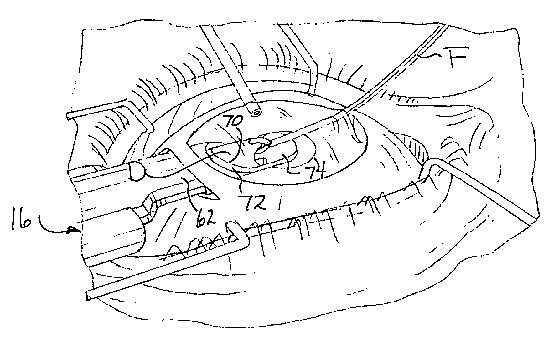
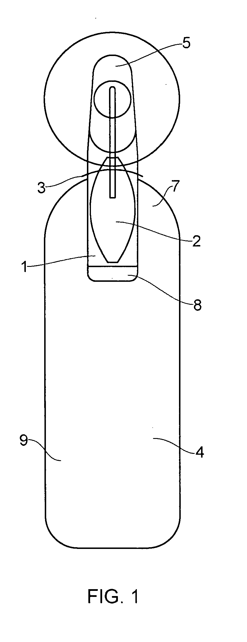
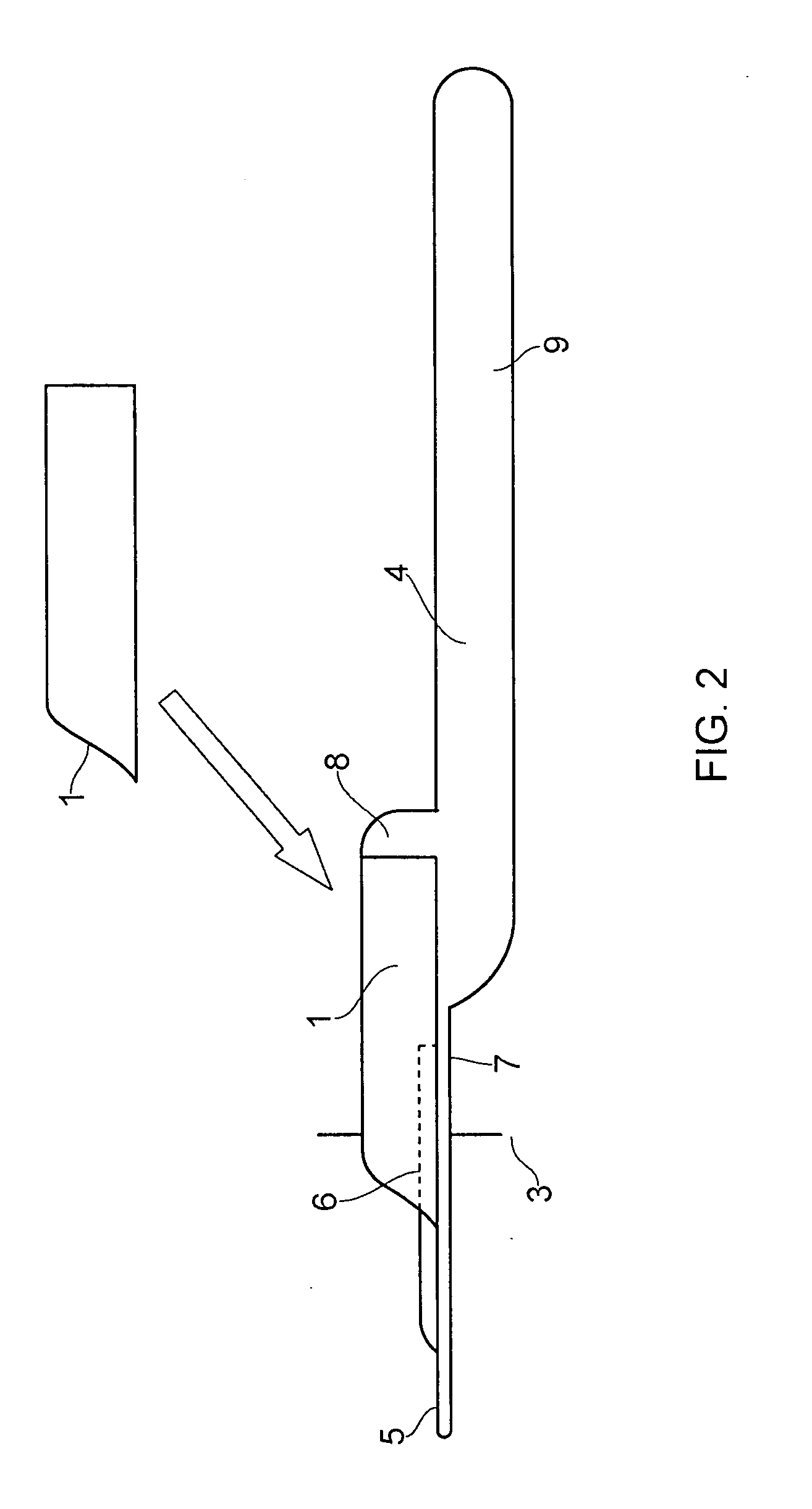
[0081]Turning in detail initially to FIGS. 1-4 and 7-9 of the drawings, the donor chamber 1 of a first embodiment is composed of clear plastic, and consists of a curved chamber, open at both ends. The donor lenticule 2 is dragged into the chamber and coiled up in a specific proprietary configuration, with the stromal surface snugly aligned to the inner surface of the chamber 1. The front end of the chamber 1 is bevelled in a cut-away design to allow ease of insertion through a scleral wound opening 3. The outer chamber diameter is designed to fit exactly through the scleral wound 3 with a semi-elliptical opening. The superior aspect of the anterior edge has a cut-away anterior surface to allow space for a pair of glide forceps (not shown) to enter the chamber 1 and grasp the leading stromal edge of the coiled donor 2. The posterior margins of the chamber 1 are straight-edged and grooved, to allow sliding of a glide platform 4 to close the chamber 1 inferiorly, with a glide member 5 ...


PUM


Abstract

The system for donor cornea implantation includes a preparation base having a well for receiving a posterior lamellar donor corneal lenticule, a cartridge disengageably mounted on the base adjacent the well, and a handle for disengageable attachment to a posterior end portion of the cartridge. In drawing the donor lenticule from the well into and through a bore or chamber of the cartridge, from the posterior end, the lenticule is caused to assume a double coil configuration. After attachment of the handle, removal of the assembly from the preparation base, and insertion of blade and adjacent body portions of the cartridge through an incision in the cornea, the coiled donor lenticule is pulled from the cartridge bore through its forward end, to uncoil automatically within the anterior chamber of the recipient's eye.

Description

CROSS-REFERENCE TO RELATED APPLICATION[0001]This is a continuation-in-part of International Application No. PCT / GB2008 / 050943 entitled “Ophthalmic Surgical Device for Endothelial Keratoplasty for Descemet's Stripping Automated Endothelial Keratoplasty (DSAEK) Surgery,” filed 16 Oct. 2008 and published under International Publication No. WO 2009 / 050511, the entire specification of which is incorporated hereinto by reference thereto.BACKGROUND OF THE INVENTION[0002]A paradigm shift in the approach to corneal transplantation is occurring, with new forms of anterior and posterior lamellar keratoplasty now enabling targeted replacement of only diseased layers of the cornea. These forms of lamellar corneal surgery are gradually replacing conventional full thickness penetrating keratoplasty (Tan D T, Mehta JS: “Future Directions in Lamellar Corneal Transplantation”; Cornea; in press).[0003]Descemet's stripping automated endothelial keratoplasty (DSAEK) is a form of small incision and essen...

Claims


Application Information

Patent Timeline
IPC IPC(8): A61F9/007
CPCA61F2/143A61F2009/00872A61F9/0017A61F2/148
Inventor WESTON, PHILIP DOUGLASTANMEHTA
Owner WESTON PHILIP DOUGLAS
Learn More