Apparatus and methods for forming and securing gastrointestinal tissue folds

a technology of gastrointestinal tract and folds, applied in the direction of oesophagoscope, surgical staples, surgical forceps, etc., can solve the problems of atypical diarrhea, inconvenient operation, and inability to perform morbid procedures, and achieve the effect of improving flexibility

Active Publication Date: 2010-08-19
USGI MEDICAL
View PDF99 Cites 39 Cited by
  • Summary
  • Abstract
  • Description
  • Claims
  • Application Information

AI Technical Summary

Benefits of technology

[0014]It is another object of the present invention to provide methods and apparatus for forming gastrointestinal tissue folds using anchors that can be reconfigured from a reduced delivery profile to an expanded deployed profile.
[0016]It is a further object of the present invention to provide methods and apparatus for forming gastrointestinal tissue folds, wherein the anchor assembly is deployed in a manner that reduces a possibility of injuring neighboring organs.
[0020]Optionally, a third tissue contact point may be established at another location initially proximal of, or in line with, the first tissue contact point. Upon movement of the tissue engaged by the tissue grabbing assembly to a position proximal of both the second and third tissue contact points, a tissue fold is formed with the second and third contact points on opposing sides of the fold. The third contact point may provide backside stabilization upon delivery of the anchor assembly across the tissue fold from a vicinity of the second tissue contact point.
[0022]More preferably, the distal region of the catheter includes a bendable section that permits the first tissue contact point to be positioned relative to the second tissue contact point so that the tissue fold is oriented substantially perpendicular to the anchor delivery system. In this manner, the anchor delivery system, when deployed, pierces the tissue fold and exits into the interior of the GI lumen, rather than the exterior of the tissue wall, thereby reducing a risk of injury to adjacent organs.

Problems solved by technology

These procedures are difficult to perform in morbidly obese patients because it is often difficult to gain access to the digestive organs.
In particular, the layers of fat encountered in morbidly obese patients make difficult direct exposure of the digestive organs with a wound retractor, and standard laparoscopic trocars may be of inadequate length.
In addition, previously known open surgical procedures may present numerous life-threatening post-operative complications, and may cause atypical diarrhea, electrolytic imbalance, unpredictable weight loss and reflux of nutritious chyme proximal to the site of the anastomosis.
Further, the sutures or staples that are often used in these surgical procedures may require extensive training by the clinician to achieve competent use, and may concentrate significant force over a small surface area of the tissue, thereby potentially causing the suture or staple to tear through the tissue.
One problem with conventional gastrointestinal reduction systems is that the anchors (or staples) must engage at least the muscularis tissue layer in order to provide a proper foundation.
In other words, the mucosa and connective tissue layers typically are not strong enough to sustain the tensile loads imposed by normal movement of the stomach wall during ingestion and processing of food.
This problem of capturing the muscularis or serosa layers becomes particularly acute where it is desired to place an anchor or other apparatus transesophageally rather than intraoperatively, since care must be taken in piercing the tough stomach wall not to inadvertently puncture adjacent tissue or organs.

Method used


Image

  • Apparatus and methods for forming and securing gastrointestinal tissue folds
  • Apparatus and methods for forming and securing gastrointestinal tissue folds
  • Apparatus and methods for forming and securing gastrointestinal tissue folds

Examples


Embodiment Construction

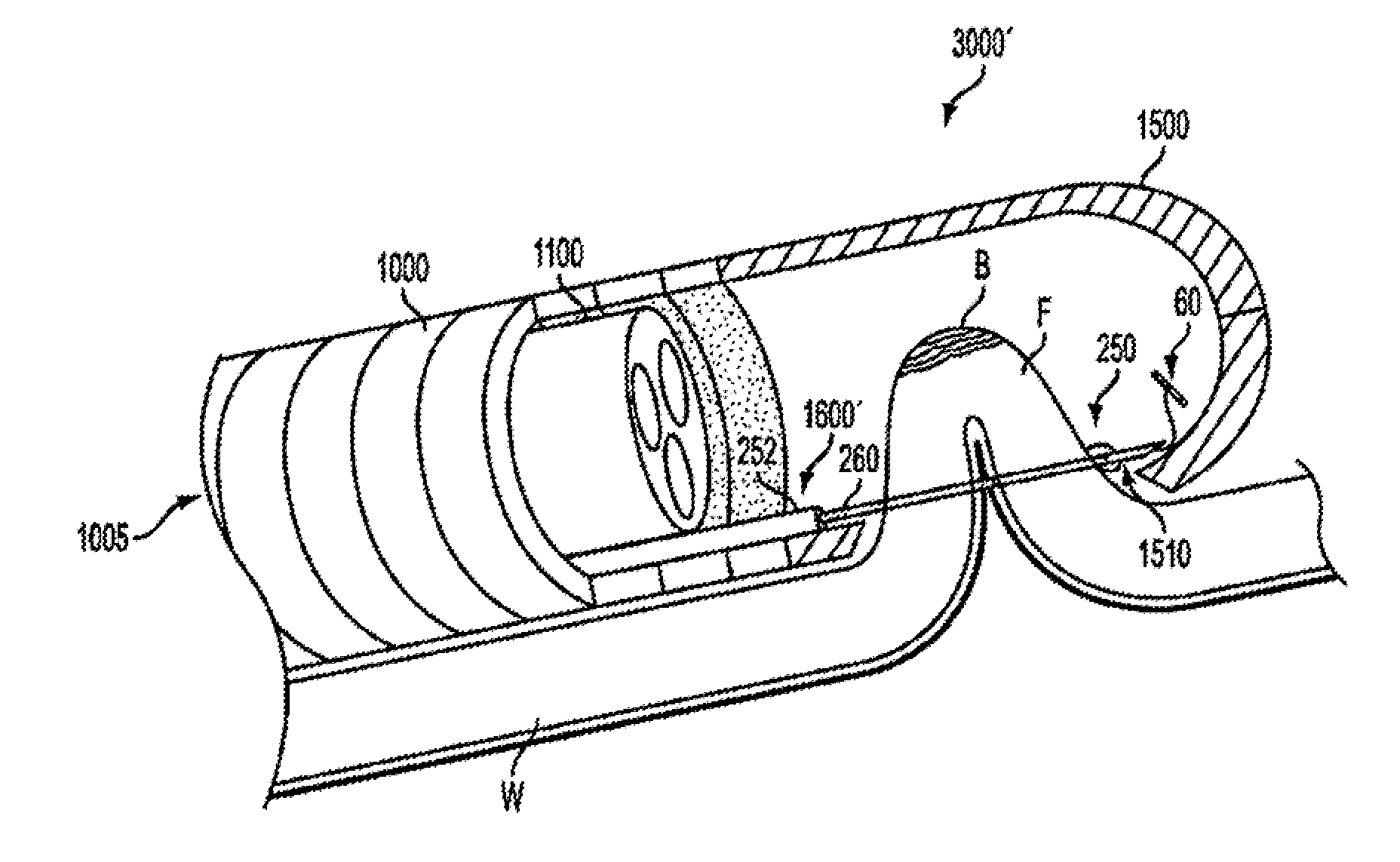
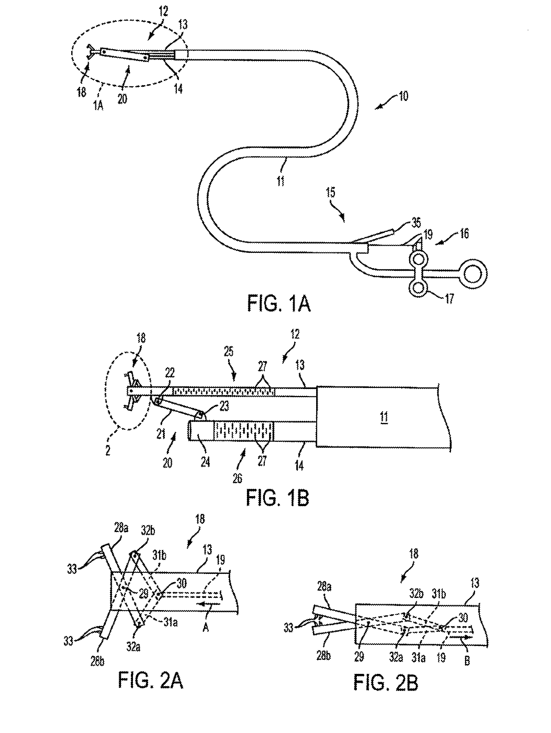
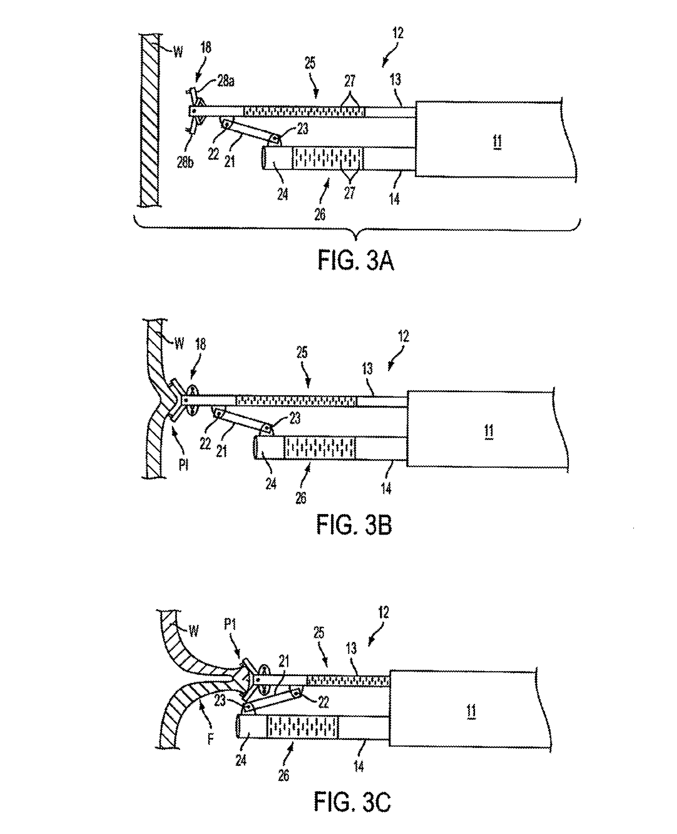
[0073]In accordance with the principles of the present invention, methods and apparatus are provided for intraluminally forming and securing gastrointestinal (“GI”) tissue folds, for example, to reduce the effective cross-sectional area of a GI lumen. These methods and apparatus may be used to treat obesity by approximating the walls of a gastrointestinal lumen to narrow the lumen, thus reducing the area for absorption in the stomach or intestines. More particularly, the present invention involves endoscopic apparatus that engages a tissue wall of the gastrointestinal lumen, creates one or more tissue folds and disposes one or more anchor assemblies through the tissue fold(s). Preferably, the anchor assemblies are disposed through the muscularis and / or serosa layers of the gastrointestinal lumen. In operation, a distal tip of the probe engages the tissue and then moves the engaged tissue to a proximal position relative to the catheter tip, thereby providing a substantially uniform p...


PUM


Abstract

Apparatus and methods are provided for forming a gastrointestinal tissue fold by engaging tissue at a first tissue contact point and moving the first tissue contact point from a position initially distal to, or in line with, a second tissue contact point to a position proximal of the second contact point, thereby forming the tissue fold, and extending an anchor assembly through the tissue fold from a vicinity of the second tissue contact point. Adjustable anchor assemblies; as well as anchor delivery systems, shape-lockable guides and methods for endoluminally performing medical procedures, such as gastric reduction, treatment of gastroesophageal reflux disease, resection of lesions, and treatment of bleeding sites; are also provided.

Description

CROSS-REFERENCE TO RELATED APPLICATIONS[0001]This application is a divisional of U.S. patent application Ser. No. 10 / 992,306, filed on Nov. 17, 2004, which is a continuation of U.S. patent application Ser. No. 10 / 734,547 filed Dec. 12, 2003. This application is related to, but does not claim the benefit of priority from, the following applications: U.S. patent application Ser. No. 10 / 672,375, filed Sep. 25, 2003, U.S. provisional patent application Ser. No. 60 / 500,627, filed Sep. 5, 2003; U.S. patent application Ser. No. 10 / 612,170, filed Jul. 1, 2003, and Ser. No. 10 / 639,162, filed Aug. 11, 2003; U.S. provisional patent application Ser. No. 60 / 433,065, filed Dec. 11, 2002; U.S. patent application Ser. No. 10 / 173,203, filed Jun. 13, 2002; U.S. patent application Ser. No. 10 / 458,060, filed Jun. 9, 2003, U.S. patent application Ser. No. 10 / 346,709, filed Jan. 15, 2003, U.S. provisional patent application Ser. No. 60 / 471,893, filed May 19, 2003;U.S. patent application Ser. No. 10 / 288,6...

Claims


Application Information

Patent Timeline
Patent Type & Authority Applications(United States)
IPC IPC(8): A61B17/11A61B1/00A61B1/005A61B1/018A61B1/273A61B1/31A61B17/00A61B17/02A61B17/04A61B17/06A61B17/08A61B17/10A61B17/28A61B19/00
CPCA61B1/00135A61F5/0086A61B1/018A61B1/2736A61B1/31A61B17/00234A61B17/0218A61B17/0401A61B17/0469A61B17/0482A61B17/0487A61B17/062A61B17/0625A61B17/0644A61B17/29A61B17/295A61B17/3421A61B17/3468A61B17/3478A61B2017/00269A61B2017/003A61B2017/00353A61B2017/00827A61B2017/0404A61B2017/0417A61B2017/0419A61B2017/0443A61B2017/0451A61B2017/0454A61B2017/0458A61B2017/0461A61B2017/0462A61B2017/0464A61B2017/0488A61B2017/0496A61B2017/06052A61B2017/061A61B2017/06176A61B2017/0649A61B2017/2905A61B2017/2927A61B2017/2943A61B2017/3445A61B2017/3488A61B2019/5217A61B1/0055A61B2090/3614A61B17/0483
Inventor EWERS, RICHSAADAT, VAHID C.MICHLITSCH, KENROTHE, CHRISBRENNEMAN, RODNEYLAM, CANGCHEN, EUGENE
Owner USGI MEDICAL
Why Eureka
Try Eureka