Mechanically deployable upper airway stent

a stent and upper airway technology, applied in the field of pharyngeal obstruction, can solve the problems of obstructive sleep apnea patients who receive sedation, analgesia or anesthesia for diagnostic or therapeutic procedures, increased risk of perioperative complications, and drop in blood oxygen level, so as to enhance positioning and removal of the stent. , the effect of preventing the collapse of pharyngeal tissues

Inactive Publication Date: 2010-08-26
OBERLE FAMILY TRUST
View PDF18 Cites 14 Cited by
  • Summary
  • Abstract
  • Description
  • Claims
  • Application Information

AI Technical Summary

Benefits of technology

[0016]To deploy the preferred embodiment stent, the linkage rod is moved axially in a distal direction within the housing of the central tube. The movement of the linkage rod causes the spokes to move through the openings of the central tube and project radially outward from the tube. In a preferred embodiment, each spoke is made of a sufficiently rigid material such that when the stent is deployed the spokes are sufficiently strong to prevent collapse of pharyngeal tissues. As a spoke moves radially outward, each rib (along with attached web members) moves outwardly away from the central tube. In the preferred embodiment the fully deployed spoke will extend outwardly at an angle approximately normal (70-110 degrees) to the central tube. When all spokes have been deployed and are radially extended, spaces between adjacent deployed spokes permit the easy passage of air along the length of the stent. The inter-spoke spaces of the preferred embodiment stent are wedge-shaped.
[0017]In use, the collapsed (un-deployed) stent of the present invention is inserted through a naris and into the nasal passageway. The stent is typically fully inserted when the distal end of the stent is proximal to the soft palate structures of the nasophraynx. In cases where the obstruction is due to lingual collapse, the stent may be inserted into position in the oropharynx where its distal end is proximal to and presses against the base of the tongue. In the full insertion mode a sufficient length of the proximal end of the stent extends out through the naris to permit manipulation and deployment of the stent. Once fully inserted, a portion of the stent is proximal to the anatomic structure or structures of the pharynx (nasopharynx and / or oropharynx) exhibiting undesirable inflammation, configuration, growth or motility. The stent can then be deployed and fixed into position. The proximal end of the central tube is provided with gripping means such as knurlments to enhance positioning and removal of the stent. In its deployed state, the perimeter ribs and web members of the stent press outwardly against the tissues of the pharynx prohibiting their collapse or incursion into the airway. By virtue of the inter-spoke spaces, air may freely pass through the nasal passageway into the pharynx.

Problems solved by technology

A reduction or cessation of airflow during sleep causes a resultant drop in blood oxygen level.
Consequently, when an apnea occurs, sleep is disrupted.
Obstructive sleep apnea occurs when the brain sends the signal to the muscles and the muscles make an effort to take a breath, but they are unsuccessful because the airway becomes obstructed and prevents the flow of air.
In addition, patients with obstructive sleep apnea who receive sedation, analgesia or anesthesia for diagnostic or therapeutic procedures are at increased risk for perioperative complications.
These events can obstruct the airway.
This tissue action can exacerbate the degree of obstruction because the harder the chest tries to pull air in, the greater the negative pressure, and the more the tissues of the airway are sealed together.
Surgical options for sleep apnea, however, may involve painful recoveries and extended time off from work to heal.
Because these surgeries carry risk and offer no guarantee of improvement, most patients with obstructive sleep apnea go through a regimen of non-surgical treatments before considering surgery.
This is often difficult because of the discomfort engendered by masks.
Critically, if the mask does not fit correctly, the efficacy of CPAP drops significantly.
Hence another disadvantage of CPAP is that it is not always easy to use.
The fact that the tube is constructed of such material and that the tube has a large surface area increases the possibility of irritating contact with body tissues.
Similarly, the large surface area of the tube can interfere with the natural secretory functions of surrounding tissues and sinuses of the nasal cavities.

Method used


Image

  • Mechanically deployable upper airway stent
  • Mechanically deployable upper airway stent
  • Mechanically deployable upper airway stent

Examples


Embodiment Construction

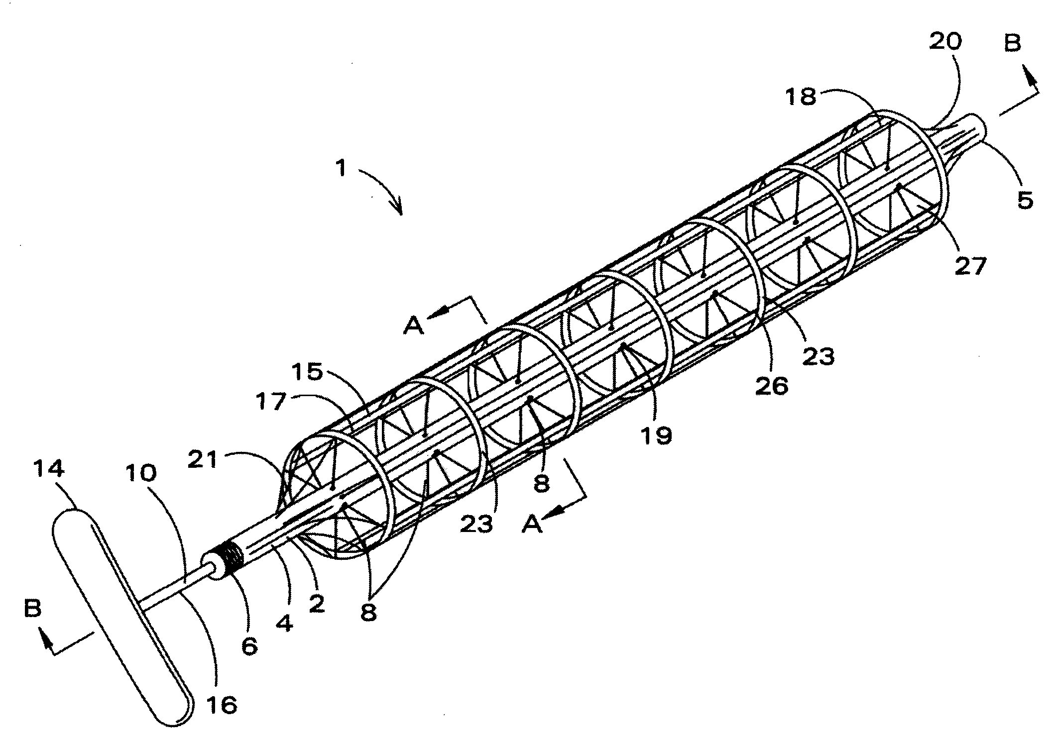
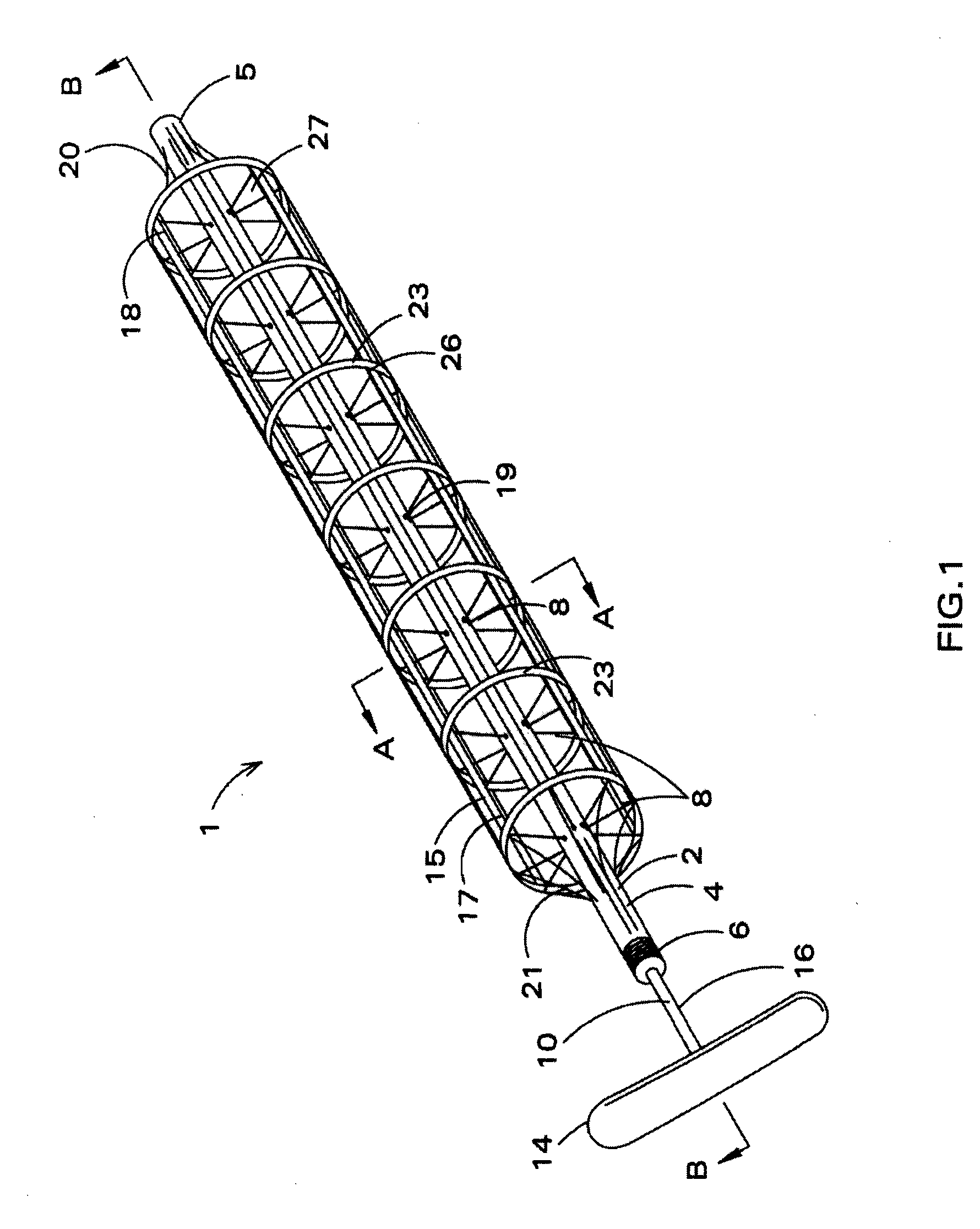
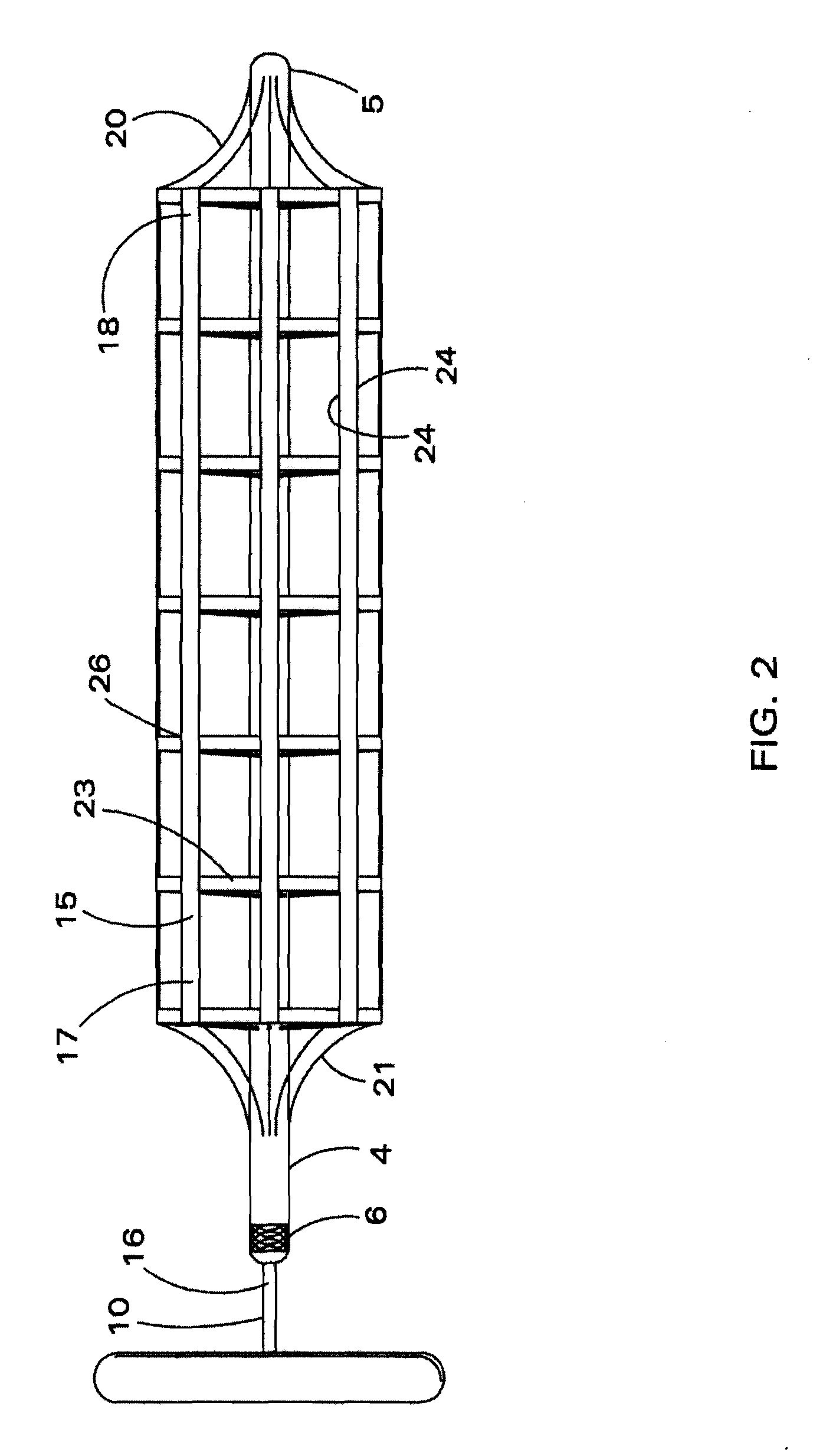
[0030]The present invention relates to a mechanically deployable pharyngeal stent and method of using same. In its un-deployed state, the stent has a reduced diametric profile and is insertable into the nasal passageway via one of the nares. Once fully inserted and positioned, the stent is deployed. Deploying the stent causes radially movable spokes bearing perimeter ribs to move outwardly from the stent such that the ribs and their optional adjoining web members press against the tissues of the pharyngeal cavity that define the patient airway. The pressing force from the stent restricts tissue swelling, incursion or motility and therefore prevents the structures of the nasopharynx or oropharynx from collapsing or intruding into the airway. Inter-spoke spaces allow the flow of air along the length of the stent and result in airway patency.

[0031]For purposes of description herein, the term “proximal” shall refer to that portion of the stent or component thereof that is situated close...


PUM


Abstract

A mechanically deployable upper airway stent is disclosed along with a method of using same. The stent comprises a central tube having an inner chamber and openings on its surface. A linkage rod is housed within the central tube and is provided with branched spokes that pass through the openings. In the un-deployed state the branched spokes are contained within the central tube. Deployed spokes are preferably aligned in groupings along the central tube. The outer ends of the spokes of each grouping connect to a rib. The un-deployed stent is inserted into the nasal passageway and positioned relative to occluding anatomic structures. Upon deployment, the ribs along with web members extending between the ribs move outwardly from the central tube and press upon the tissues of the pharyngeal cavity. Spaces between the extended spokes permit the passage of air along the stent and maintain airway patency.

Description

CROSS REFERENCE TO RELATED APPLICATION[0001]Not applicable.STATEMENT REGARDING FEDERALLY SPONSORED RESEARCH OR DEVELOPMENT[0002]Not applicable.SEQUENCE LISTING TABLE OR COMPUTER PROGRAM OR COMPACT DISC[0003]Not applicable.FIELD OF THE INVENTION[0004]This invention relates generally to methods and devices for treating pharyngeal obstructions and maintaining pharyngeal airway patency. More specifically, this invention relates to an improved method and apparatus for treating sleep apnea and breathing disorders caused by undesirable inflammation, configuration, growth or motility of the structures of the nasopharynx or oropharynx.BACKGROUND OF THE INVENTION[0005]An apnea is a period of time during which breathing stops or is markedly reduced. In simplified medical terms, an apnea occurs when a person stops breathing for 10 seconds or more. Apneas usually occur during sleep. Sleep apnea is a disorder characterized by a reduction or cessation of breathing airflow during sleep. A reduction...

Claims


Application Information

Patent Timeline
Patent Type & Authority Applications(United States)
IPC IPC(8): A61M29/00
CPCA61F5/56
Inventor OBERLE, PAUL
Owner OBERLE FAMILY TRUST
Why Eureka
Try Eureka