
Method of detecting infection with urogenital mycoplasmas in humans and a kit for diagnosing same

a technology for detecting human infection and mycoplasma, which is applied in the field of human infection detection of mycoplasma, can solve the problems of inability to detect infection, lack of diagnosis and appropriateness, and inability to be widely availabl

Inactive Publication Date: 2010-09-09
MOR RES APPL LTD
View PDF1 Cites 4 Cited by
  • Summary
  • Abstract
  • Description
  • Claims
  • Application Information

AI Technical Summary

Benefits of technology

[0009]The present invention provides a method of detecting infection with urogenital mycoplasma, by measuring the level of specific anti-mycoplasma antibodies in a biological sample taken from a patient to be diagnosed, comprising i) providing a plurality of biological specimens from a plurality of pathological states, in patients suffering from mycoplasmal urogenital infections; ii) preparing a whole cell preparation from said several mycoplasma strains obtained from patients; iii) providing a plurality of mycoplasmal antigenic determinants (from each species) to be used as the material for detection of specific antibodies in the patients' sample; iv) binding said mixed antigen on an immunoassay surface; v) binding of the patients' biological sample with said surface of step iv, thereby binding of anti-mycoplasma antibodies eventually present in said sample onto said surface; vi) contacting the surface with a reagent for detecting human antibodies. Thus, the determination of infection will be performed by measuring a wide spectrum (plurality) of specific antibodies to the same species. Said immunoassay surface means a protein-binding surface, used in the art, that usually comprises polymers having strong affinity for proteins, not releasing them even during washing steps; the polymers may comprise modified natural materials or synthetic materials. Said plurality of human strains preferably comprises at least six patients each infected with one or more mycoplasma species selected from the group consisting of Ureaplasma urealyticum, Ureaplasma parvum, Mycoplasma hominis, Mycoplasma fermentans, and Mycoplasma genitalium. In a preferred embodiment of the invention, said a concentrated pellet of mycoplasma cells, obtained by centrifugation from a mycoplasma culture (of each strain), is sonicated and the whole cell sonicate preparations from several strains are mixed (in equal amounts of proteins) to form the “mixed antigen” preparation. Essential is that each of the protein antigens comprising the mixture, is obtained from a strain isolated from a patient with a known pathology (i.e. urethritis, pregnancy complication, infertility, arthritis etc.) and that protein antigens are obtained from a plurality of pathologies, thereby providing the best possible representation of array of antigens expressed in disease. Said immunoassay surface may comprise an item selected from well plate and polymer sheet, or other means known in the art of the immunoassay design. Said reagents may comprise anti-human antibodies conjugated with an enzyme yielding a color or fluorescent reaction, and an enzyme substrate, or other means enabling the antibody visualization.

Problems solved by technology

There is an increasing amount of evidence accumulated in the literature regarding mycoplasmal pathogenic mechanisms, but well-established tests, that could significantly promote the analysis of urogenital mycoplasmal diseases, are not generally available.
The lack of reliable and rapid diagnostic tests makes the identification of urogenital mycoplasma as the cause of disease very difficult, resulting in underestimation of their role in disease and therefore lack of diagnosis and appropriate treatment
The role of urogenital mycoplasmas in the development of human diseases has been emerging only in the last decade, and determining mycoplasmal serology has been difficult, due to lack of standardized antigen being further complicated by unavailability of a practical test in the market.
In spite of various “home-made” techniques introduced in various countries, no efficient and reliable general diagnostic test has been available so far for detecting human urogenital mycoplasmas in clinical samples.
As known to a person skilled in the art, the techniques that are available suffer from various drawbacks, including limited sensitivity and limited specificity (appearance of false-positive or false negative results), to name some.

Method used


Image

  • Method of detecting infection with urogenital mycoplasmas in humans and a kit for diagnosing same
  • Method of detecting infection with urogenital mycoplasmas in humans and a kit for diagnosing same
  • Method of detecting infection with urogenital mycoplasmas in humans and a kit for diagnosing same

Examples


example 1

[0028]A plurality of samples were taken from a plurality of patients suffering from acute or chronic urogenital infection. A mycoplasma strain was cultured from each sample, verifying the presence of mycoplasma by PCR. Six of the samples, in which proved mycoplasma belonged to either of Ureaplasma urealyticum and Ureaplasma parvum by means of PCR, were cultured in a 10B broth, containing 7.5% FCS (fetal calf serum). The cells were centrifuged at 27,000×g for 40 min and washed twice with phosphate buffer saline. The washed pellet of cells was sonicated in the presence of 1 mM phenylmethyulsulfonylfluoride (a proteases inhibitor), and the sonicate, containing all components of the cell, namely un fractionated mixture of proteins, lipoproteins and nucleic acids was used as the mycoplasma antigen.

example 2

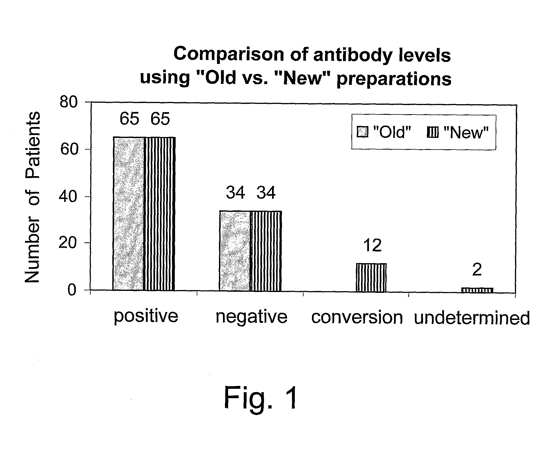
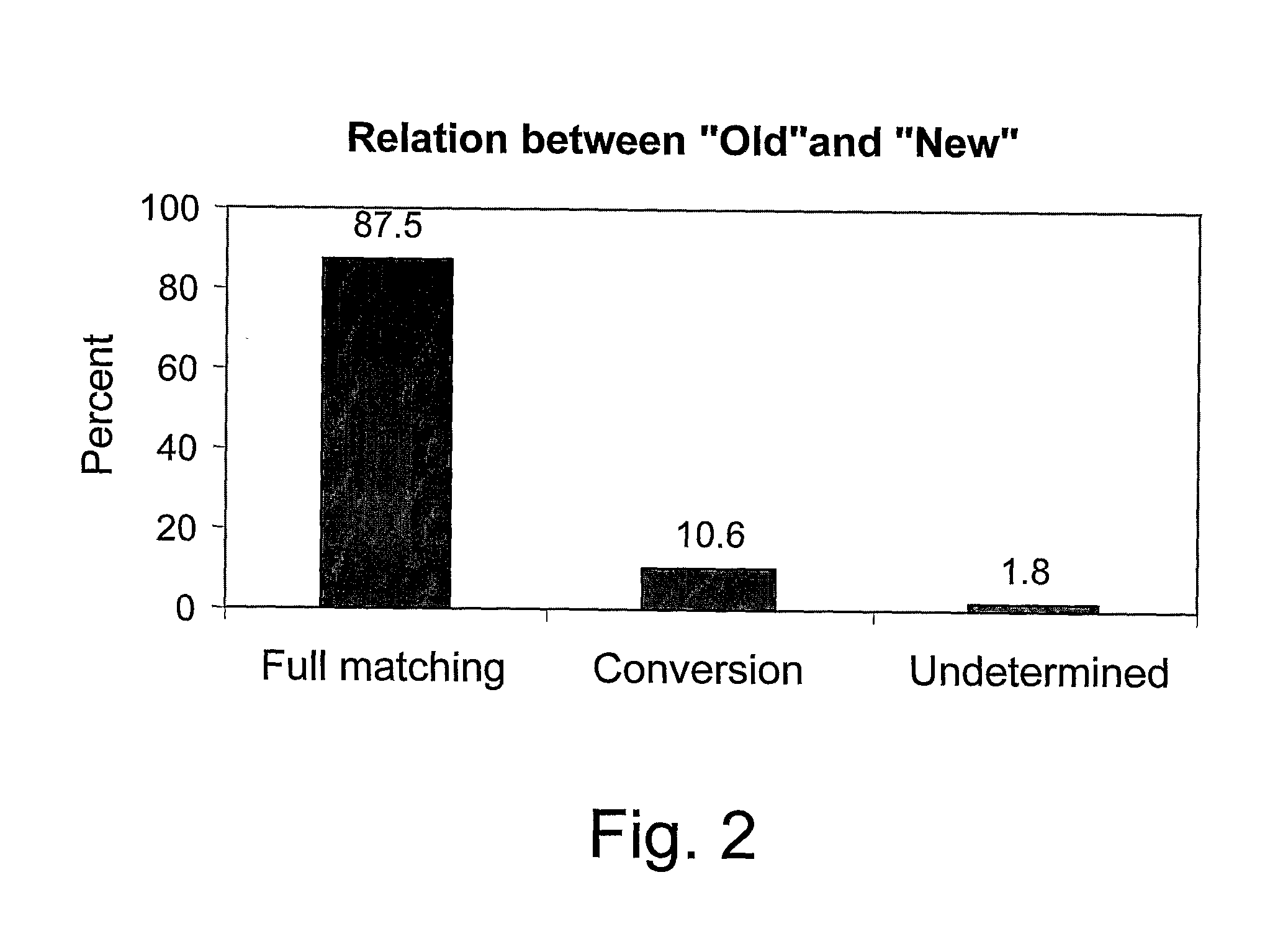
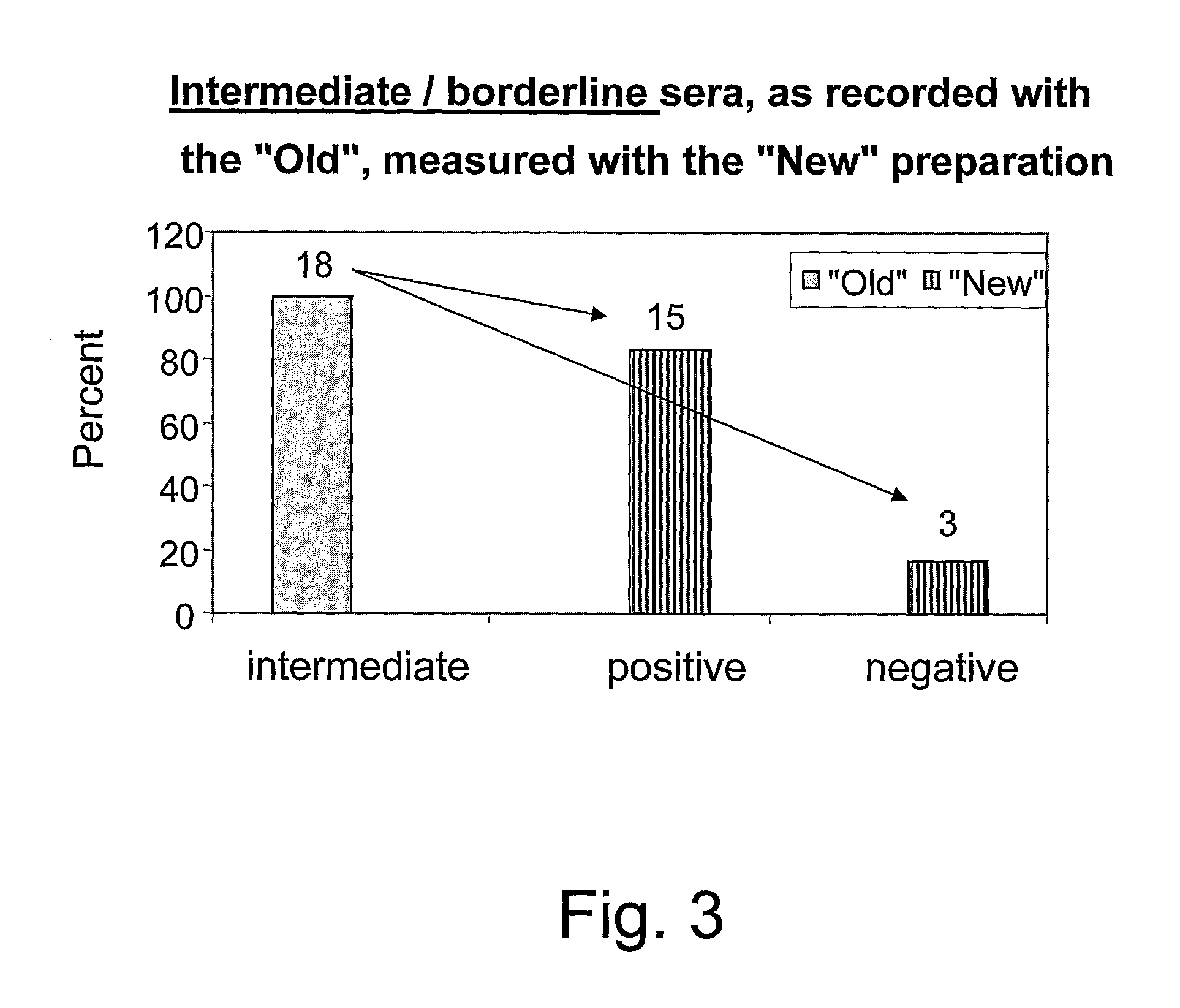
[0029]A mixture of ureaplasmal antigens from six different strains of the ureaplasmas species (three Ureaplasma parvum (Up) and three Ureaplasma urealyticum (Uu)) was prepared. The strains were isolated from patients as described in Example 1, and is designated “New”. The identity of the species was verified by the PCR. Sera samples from 113 patients with different diseases, which had been previously analyzed in our laboratory, were tested with an ELISA employing the “New” mixed antigen. In parallel, ELISA was performed, employing antigens prepared from Ureaplasma parvum ATCC # 27815 formerly designated Ureaplasma urealyticum serotype 3 (Up), and Ureaplasma urealyticum ATCC # 27816 formerly designated Ureaplasma urealyticum serotype 4 (Uu). Each of them was used separately, and both are designated here as “Old”. These antigens were used for testing said 113 sera samples. Thus, ELISA tests based on three different antigen systems were compared.

[0030]Several types of sera were used, r...


PUM

PropertyMeasurementUnit
Antigenicityaaaaaaaaaa

Abstract

A method and a kit are provided for detecting an infection by mycoplasma, particularly an urogenital infection in humans. The presence of specific anti-mycoplasma antibodies in a biological sample of a diagnosed subject is detected by reaction with an immobilized mixture of various antigenic determinants associated with a variety of pathological states.

Description

FIELD OF THE INVENTION[0001]The present invention relates to detecting an infection by mycoplasma, particularly an urogenital infection in humans. A kit for diagnosing urogenital infection caused by a broad range of mycoplasma strains is provided.BACKGROUND OF THE INVENTION[0002]Mycoplasmas are a group of unique bacteria that often cause silent infections in humans and animals and plants. In animals, they are notorious as causative agents of morbidity and mortality. In humans, a variety of disorders are associated with mycoplasma infections, an example being mycoplasmal pneumonia or common sexually transmitted infections whose precise diagnosing remains difficult [see, for example, Merck Manual, 17th Ed. (1999) p. 1326]. There is an increasing amount of evidence accumulated in the literature regarding mycoplasmal pathogenic mechanisms, but well-established tests, that could significantly promote the analysis of urogenital mycoplasmal diseases, are not generally available. WO 07 / 0331...

Claims


Application Information

Patent Timeline
IPC IPC(8): G01N33/53C07K14/195
CPCG01N2469/20G01N33/56933
Inventor HOROWITZ, SHULAMITH
Owner MOR RES APPL LTD
Learn More