Therapeutic modulation of autophagy
a technology of autophagy and therapeutic modulation, applied in the field of therapeutic modulation of autophagy, can solve the problems of severe liver injury, hepatocyte cell death, protein aggregate accumulation,
- Summary
- Abstract
- Description
- Claims
- Application Information
AI Technical Summary
Problems solved by technology
Method used
Image

Examples
example 1
Autophagy-Defective Tumor Cells Accumulate p62 in Response to Stress
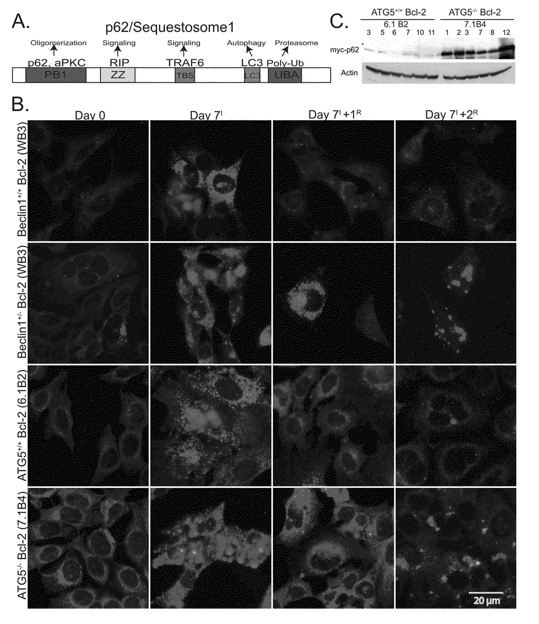
[0065]To address the potential role of autophagy-dependant protein quality control in tumor suppression, p62 modulation was assessed during metabolic stress and recovery in autophagy-competent (beclin1+ / + and atg5+ / +) and autophagy-defective (beclin1+ / − and atg5− / −) immortalized baby mouse kidney (iBMK) cells. Cells were engineered to express Bcl-2, as the assessment of autophagy is facilitated in an apoptosis-defective background (Degenhardt, K., et al. (2006), Autophagy promotes tumor cell survival and restricts necrosis, inflammation, and tumorigenesis, Cancer Cell 10, 51-64; Lum, J. J., et al.,. (2005), Growth factor regulation of autophagy and cell survival in the absence of apoptosis, Cell 120, 237-248). Under normal growth conditions, p62 levels were low in wild-type cells and slightly elevated in autophagy-defective iBMK cells (FIG. 1B). Following 7 days of metabolic stress there was dramatic p62 induction i...
example 2
Autophagy-Defective Tumor Cells Accumulate Damaged Mitochondria ER Chaperones and PDIs
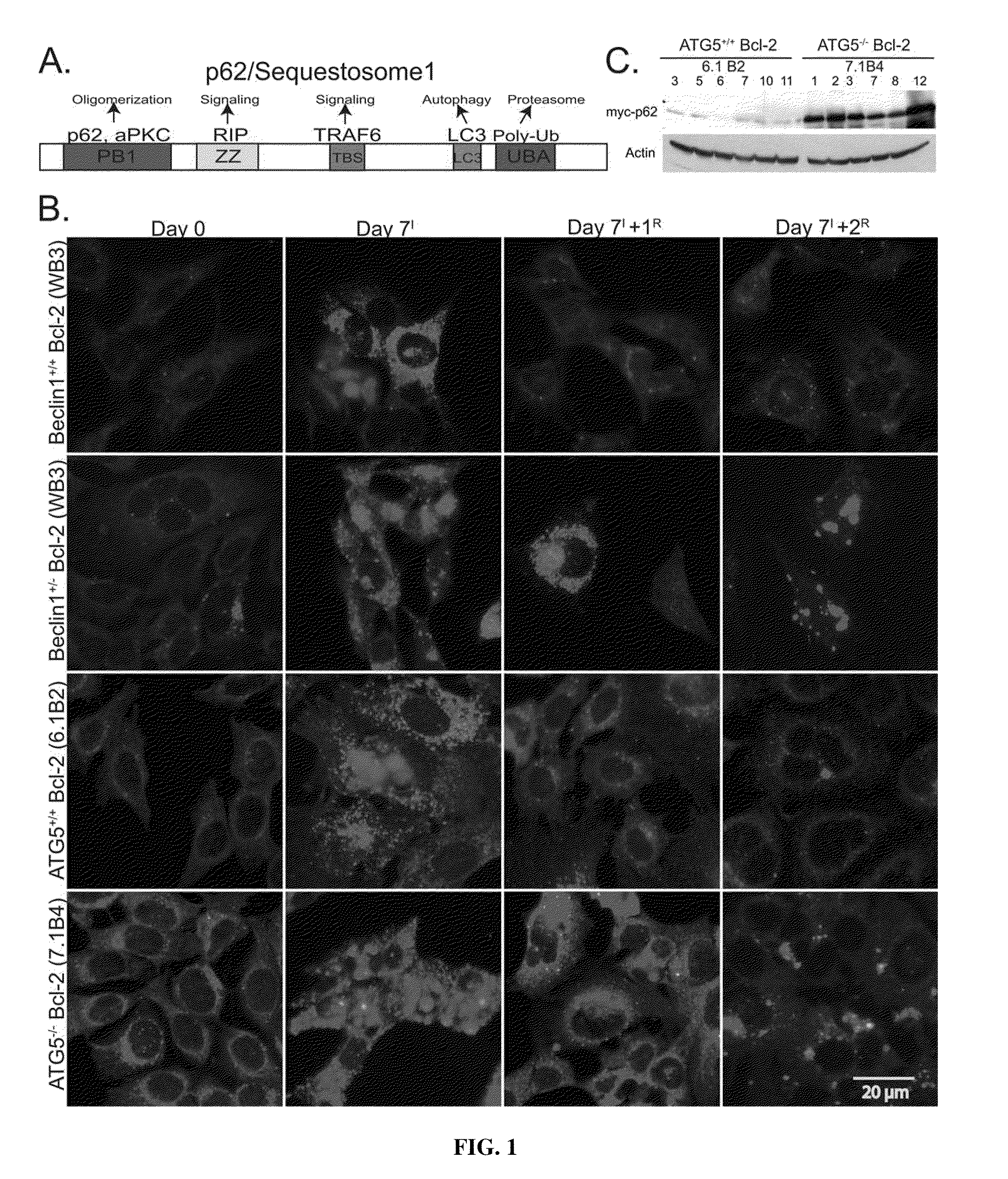
[0066]Apoptosis-deficient atg5+ / + iBMK cells responded to prolonged stress by undergoing progressive autophagy, yielding cells less than one-third their original size that retained numerous well-preserved mitochondria (M), and ER appeared slightly distended (E) indicative of an unfolded protein response (UPR) (FIGS. 2A and 2C). In contrast, Bcl-2-expressing atg5− / − (FIG. 2B) and beclin1+ / − (data not shown) iBMK cells showed disintegrating mitochondria with gross structural abnormalities (M) and large, abnormal cytoplasmic structures (A) resembling protein aggregates (FIGS. 2B and 2D), consistent with p62 aggregate accumulation (FIG. 1B). Thus, autophagy may function to prevent the accumulation of protein aggregates and damaged organelles during metabolic stress.
[0067]Since tumor cells with defective autophagy displayed failure of protein quality control, two-dimensional difference in gel electropho...
example 3
Autophagy Defects Cause Sensitivity to ER Stress
[0069]Since autophagy defects indicated an elevated demand for protein folding, the sensitivity of beclin1 cells to pharmacological induction of ER stress was investigated. Tunicamycin induces ER stress by inhibiting protein glycosylation and allelic loss of beclin1 caused increased sensitivity to this drug when compared to wild-type cells (FIG. 3B). Degradation of unfolded proteins via the proteasome pathway and ameliorates ER stress and to investigate if autophagy deficiency also elevated the burden on the ubiquitin-proteasome system, the sensitivity to the proteasome inhibitor epoxomycin was assessed. Autophagy-defective cells displayed increased sensitivity to proteasome inhibition, which was further exacerbated by metabolic stress (FIGS. 3C and 3D). This suggests an increased dependency of autophagy-defective cells on proteasome pathway particularly during metabolic stress. Thus, autophagy may function to maintain protein quality ...
PUM
 Abstract
Description
Claims
Application Information