Method for detection of adenoma or cancer by genetic analysis

a technology of genetic analysis and adenoma, applied in the direction of microbiological testing/measurement, biochemistry apparatus and processes, etc., can solve the problems of false positives and false positives, the validity of the process for measuring samples, and the reliability of the test is not considered at all, so as to reduce waste of time and cost for specifying the point, and the detection of sensitive and simpler cancers. , the effect of reliable test results

Inactive Publication Date: 2010-10-07
OLYMPUS CORP +1
View PDF3 Cites 9 Cited by
  • Summary
  • Abstract
  • Description
  • Claims
  • Application Information

AI Technical Summary

Benefits of technology

[0066]In the adenoma or cancer detection method of the present invention, the detection target is not a fragmented gene that involves various problems in the detection, but instead a housekeeping gene or its expression product which resides in both normal cells and adenoma or cancer-derived cells (more abundantly than a cancer specific gene) is extracted from a sample which is suspected to specifically include living adenoma or cancer-derived cells, and quantified. Therefore, as compared to prior art detections for a cancer specific gene and the like, more sensitive and simpler cancer detection can be achieved.
[0067]In addition, the adenoma or cancer testing method of the present invention can enable to check the quality and the quantity of a nucleic acid specimen (RNA, cDNA,

Problems solved by technology

In particular, the above-mentioned method (1) compares only the quantitative results as a comprehensive evaluation of quantitative values of a plurality of markers, and does not consider the validity of the process for measuring the samples.
For this reason, there is left a problem in that false negatives and false positives may occur if any error happens during the measurement process.
Moreover, the target of this m

Method used


Image

  • Method for detection of adenoma or cancer by genetic analysis
  • Method for detection of adenoma or cancer by genetic analysis
  • Method for detection of adenoma or cancer by genetic analysis

Examples


example 1

(Methods)

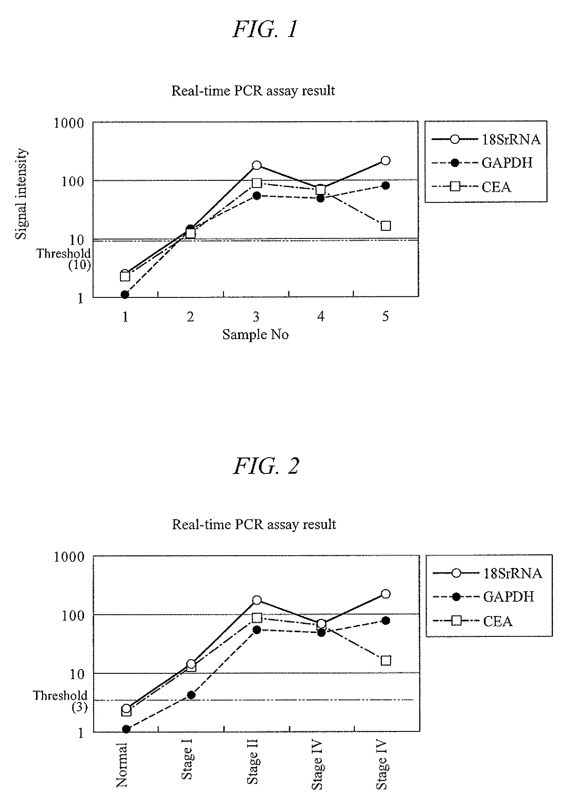
[0154]Feces was collected from a healthy subject, 6 g of which was placed in a 15 ml tube (manufactured by FALCON) and evenly mixed. Then, the mixture was divided into six samples of 1 g each. Of these, one sample was frozen at −20° C. for the analysis to come and preserved at −20° C. as it was. The remaining five samples were used right after the sampling, one of which was used as it was, and the remaining four were each added with 1 ml of the colon cancer cell line CCK-81. These samples were then respectively added with 5 ml of PBS, and mixed with a homogenizer to yield homogenized products. The homogenized samples were centrifuged at 4000×g for 10 minutes. Their supernatants were taken and RNA was extracted therefrom with the QIAGEN's RNeasy kit. A portion of each extracted RNA, 21.5 μl of RNase-free water, 25 μl of 2× TaqMan Universal PCR Master Mix, and 2.5 μl of a primer / probe set respectively for CEA, GAPDH, and 18S rRNA (manufactured by Applied Biosystems) were put ...

example 2

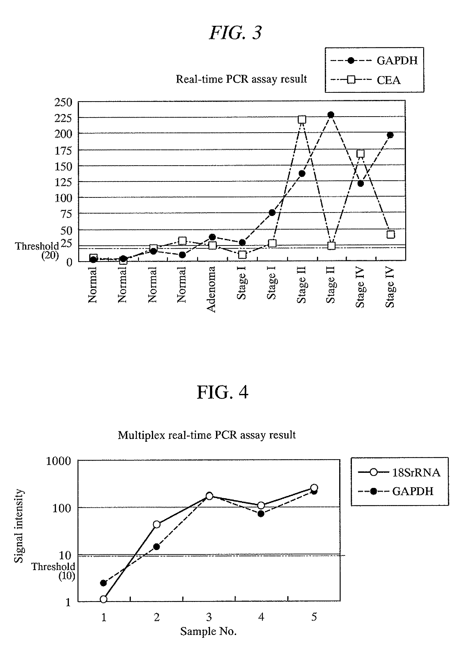
[0158]Feces were collected from one healthy subject and four colon cancer patients (stages I to IV), and 1 g of each sample was respectively placed in a 15 ml tube (manufactured by FALCON). Right after the sampling, the specimen samples were frozen at −80° C., and then added with an acid phenol-guanidine-chloroform solution. These samples were homogenized with a homogenizer. The homogenized samples were centrifuged at 4000×g for 10 minutes. Their supernatants were taken and RNA was extracted therefrom with the QIAGEN's RNeasy kit. A portion of each extracted RNA, 21.5 μl of RNase-free water, 25 μl of 2× TaqMan Universal PCR Master Mix, and 2.5 μl of a primer / probe set respectively for CEA, GAPDH, and 18S rRNA (manufactured by Applied Biosystems) were put in a 0.2 ml PCR tube and mixed therein. The probe used here was a reporter probe having a fluorophore at one end and a quencher at the other end. These mixtures were subjected to nucleic acid amplification with a real-time fluoresce...

example 3

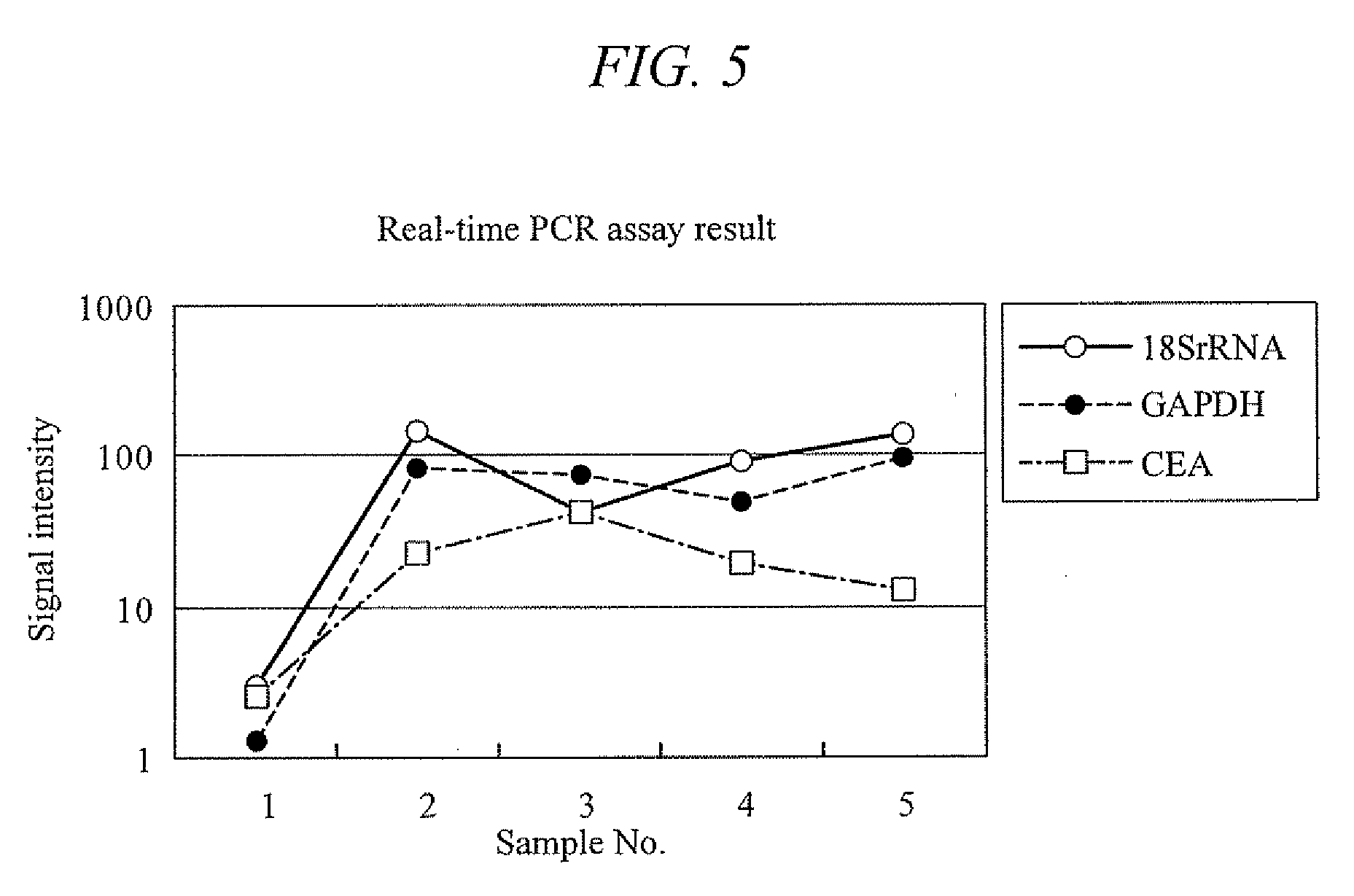
[0162]Feces were collected from four healthy subjects, one adenoma carrier, and seven colon cancer patients (stages I to IV), and 1 g of each sample was respectively placed in a 15 ml tube (manufactured by FALCON). Right after the sampling, the specimen samples were frozen at −80° C., and then added with an acid phenol-guanidine-chloroform solution. These samples were homogenized with a homogenizer. In the same manner as that of Example 2, RNA was extracted from the homogenized samples. A portion of each extracted RNA, 21.5 μl of RNase-free water, 25 μl of 2× TaqMan Universal PCR Master Mix, and 2.5 μl of a primer / probe set respectively for CEA and GAPDH (manufactured by Applied Biosystems) were put in a 0.2 ml PCR tube and mixed therein. The probe used here was a reporter probe having a fluorophore at one end and a quencher at the other end. These mixtures were subjected to nucleic acid amplification with a real-time fluorescence assay under a temperature condition consisting of on...


PUM

PropertyMeasurementUnit
Temperatureaaaaaaaaaa
Electrical conductanceaaaaaaaaaa
Nanoscale particle sizeaaaaaaaaaa

Abstract

Disclosed is a method which enables an early stage detection of an adenoma or cancer by gene expression analysis of a biomarker in a readily collectable sample. Specifically disclosed is a method for detecting an adenoma or cancer, which comprises the steps of: measuring the quantity of a sequence constituting at least one housekeeping gene or an expression product thereof contained in a body fluid sample or an excrement sample collected from an examinee; and calculating the concentration of the sequence in the sample.

Description

TECHNICAL FIELD[0001]The present invention relates to a method for detecting an adenoma or cancer by genetic analysis. More specifically, the present invention relates to a method which enables an early stage detection of an adenoma or cancer by genetic analysis of a biomarker in a readily collectable sample.BACKGROUND ART[0002]Colorectal cancer is the top leading cause of death in Japan and the second leading cause of cancer death in the United States. In the United States, about 1,300,000 cases of colorectal cancer are found each year, and of these about 50,000 people die, making it the third leading cause of death. Therefore, measures to counter cancer must be urgently adopted.[0003]In most cases, colorectal cancer is started from a small benign adenoma and is slowly developed into a malignant tumor over several tens of years. Thus, if it is found at an early stage, surgical treatments are effective and complete recovery is possible.[0004]In the case of a benign adenoma, a low in...

Claims


Application Information

Patent Timeline
IPC IPC(8): C12Q1/68
CPCC12Q1/6886C12Q2600/16C12Q2600/112C12Q2600/158
Inventor AKESAKA, TAMAONAGAOKA, TOMONORIKANAOKA, SHIGERUYOSHIDA, KENICHI
Owner OLYMPUS CORP
Why Eureka
Try Eureka