

Method of determining risk for cancer
a cancer risk and method technology, applied in the field of cancer, can solve the problem of risk in the increase of genomic cnvs in a mammal
- Summary
- Abstract
- Description
- Claims
- Application Information
AI Technical Summary
Benefits of technology
Problems solved by technology
Method used
Image

Examples
example 1
Methods
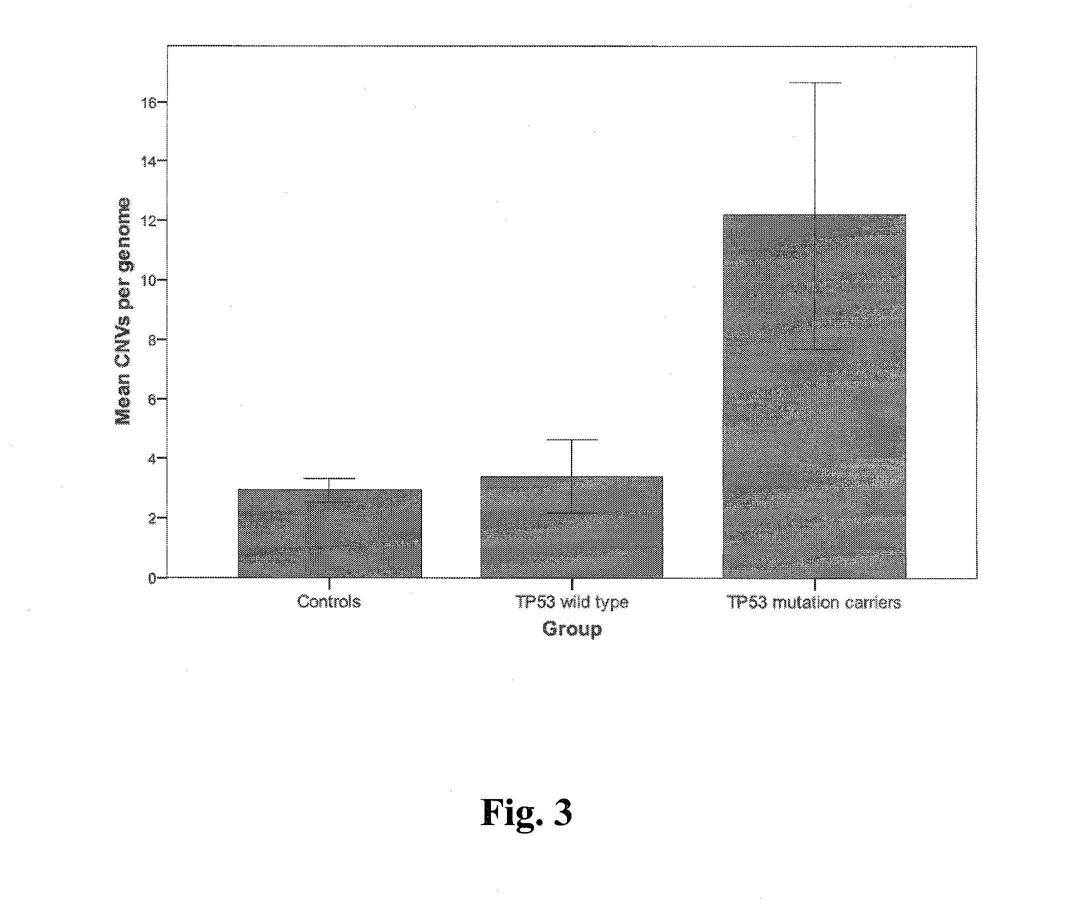
[0030]Subject recruitment. After obtaining written informed consent, DNA was extracted from peripheral blood leukocytes of 53 individuals from families with a germline TP53 mutation and from 70 unrelated controls. This included 20 TP53 wild type and 33 TP53 mutation carriers. Of these, one individual had been diagnosed as a TP53 mosaic and was grouped with the TP53 mutation carriers in the CNV analysis. In addition, genomic DNA from 5 frozen choroid plexus tumors was extracted. DNA was quantified using a NanoDrop Spectrophotometer (NanoDrop, Wilmington, Del.) and quality assessed by agarose gel electrophoresis. This study was approved by the Research Ethics Board at the Hospital for Sick Children in Toronto. Subject recruitment for the 500 individuals of European descent and the 270 individuals from the HapMap collection are described elsewhere (Nature 437, 1299-320 (2005); Matsuzaki, H. et al. Nat Methods 1, 109-11 (2004)).
[0031]DNA microarray analysis and CNV determination....
example 2
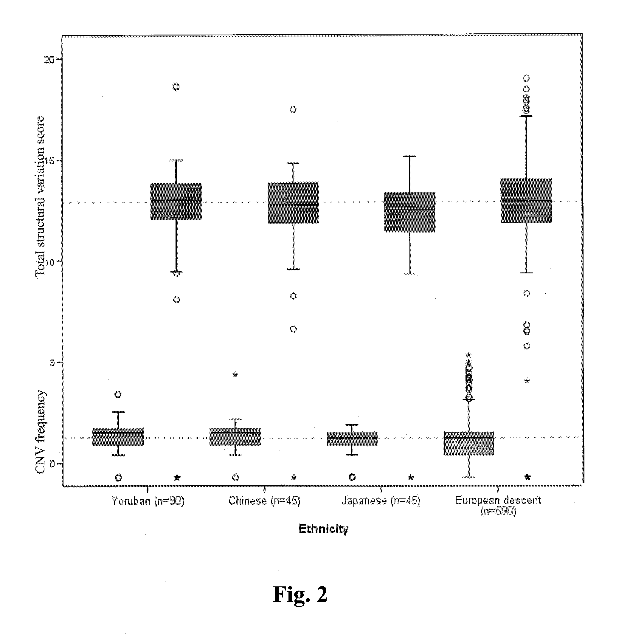
[0045]In a reference population, which included 500 persons of European descent and the multiethnic 270 person HapMap collection, 49 cancer-related genes encompassed or directly overlapped by a CNV were identified as set out in Tables 4A and 4B below.
TABLE 4AMost frequent cancer-related germline CNVsGeneGene nameRefSeq IDNLocationMLLT4Myeloid / lymphoid or mixed-lineage leukemia (trithoraxNM_005936136q27(trithorax homolog, Drosophila); translocated to, 4FHITFragile histidine triad geneNM_002012113p14.2TFGTRK-fused geneNM_00607073q12.2FANCFFanconi anemia, complementation group FNM_022725611p15MSH6mutS homolog 6 (E. coli)NM_00017962p16CENPKCentromere protein KNM_02214545q12.3MAML2Mastermind-like 2 (Drosophila)NM_032427411qPOT1POT1 protection of telomeres 1 homolog (S. pombe)NM_01545047q31.33RAD51L1RAD51-like 1 (S. cerevisiae)NM_133510414q23-q4.2RPS6KA2Ribosomal protein S6 kinase, 90 kDa, polypeptide 2NM_02113546q27
TABLE 4BAdditional cancer-related germline CNVsGeneSymbolGene NameRefSeq ...
example 3
[0051]Genomic DNA was extracted from patient blood samples using the standard phenol-chloroform method. Briefly, for each sample, 500 nanograms of genomic DNA was digested with Nsp I and Sty I restriction enzymes and ligated to adaptors. Fragments ranging from 200 to 1100 basepairs were amplified, purified, fragmented, labeled and hybridized on Affymetrix Human 6.0 GeneChip microarrays, a higher resolution platform than that utilized in Example 1. Microarrays were then washed, stained and scanned.
[0052]Array probe signal intensities were normalized and then CNVs were determined using a binary genomic segmentation informatics algorithm. CNVs (deletions or duplications) in regions with too few probes (<10) or with insufficient probe coverage (<1 probe per 5000 bp) were excluded. To avoid a high false positive rate, individuals with greater than 1000 CNVs were omitted.
[0053]FIG. 6 illustrates the CNV frequencies of TP53 wild type healthy controls (n=149) and TP53 mutation carriers affe...
PUM
| Property | Measurement | Unit |
|---|---|---|
| frequency | aaaaa | aaaaa |
| boxplots CNV frequency | aaaaa | aaaaa |
| frequencies | aaaaa | aaaaa |
Abstract
Description
Claims
Application Information