Nipah Virus Vaccines
a nipah virus and recombinant technology, applied in the field of recombinant vaccines against nipah virus, can solve the problems of disease, unreliable clinical efficacy, and inability to conduct controlled drug investigations, so as to facilitate infection and improve the preservation of the vector
- Summary
- Abstract
- Description
- Claims
- Application Information
AI Technical Summary
Benefits of technology
Problems solved by technology
Method used
Image
Examples
example 1
Constructs
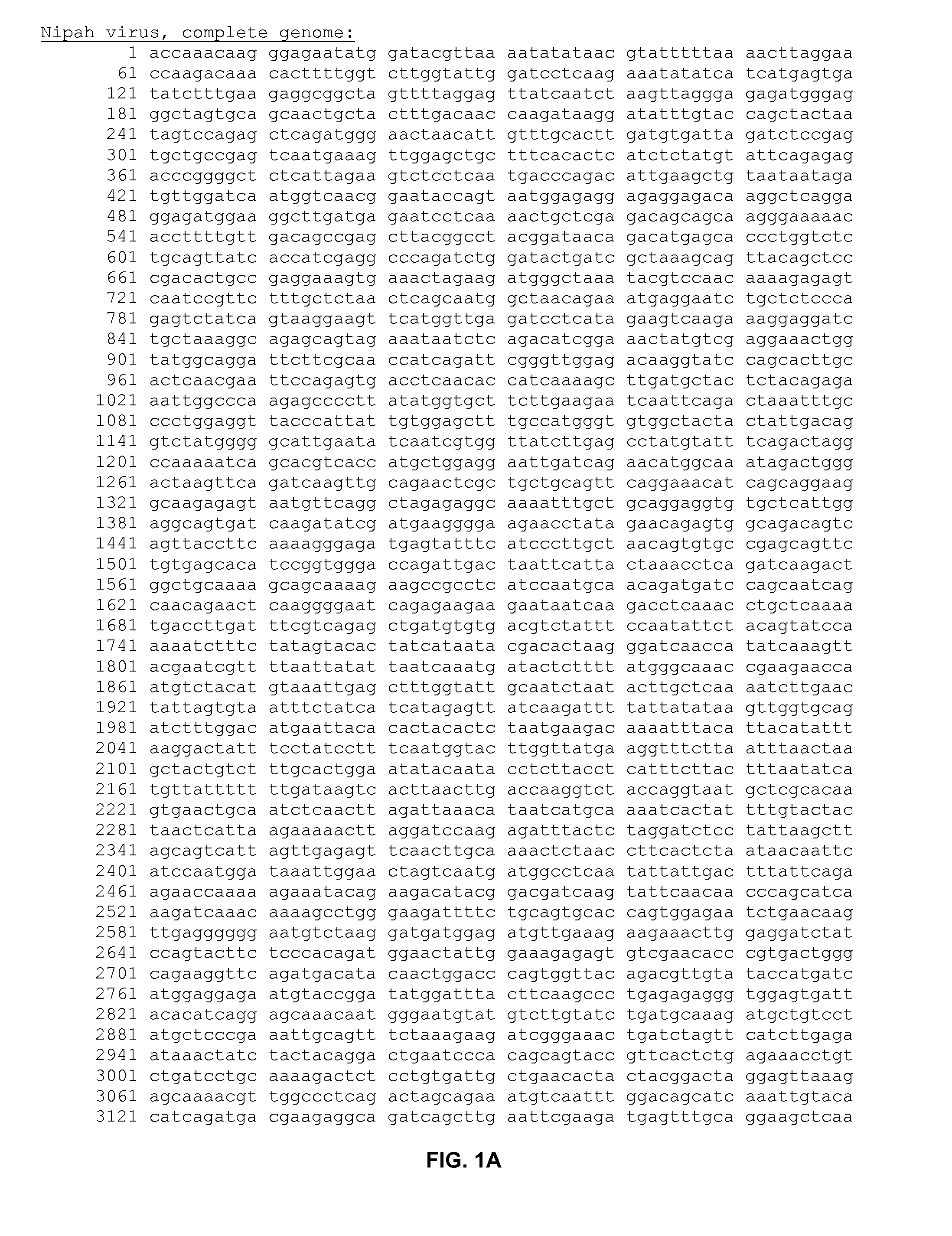
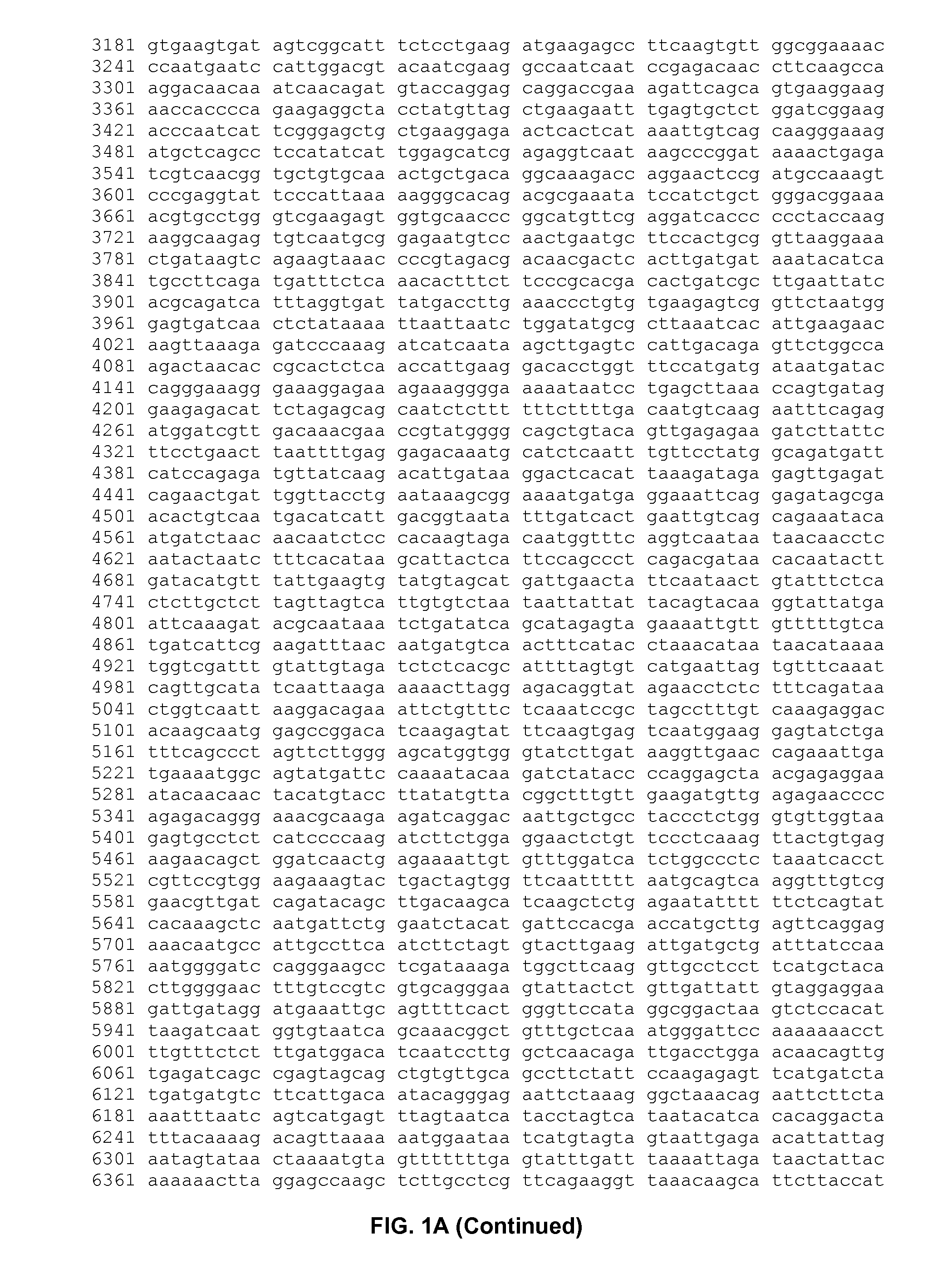
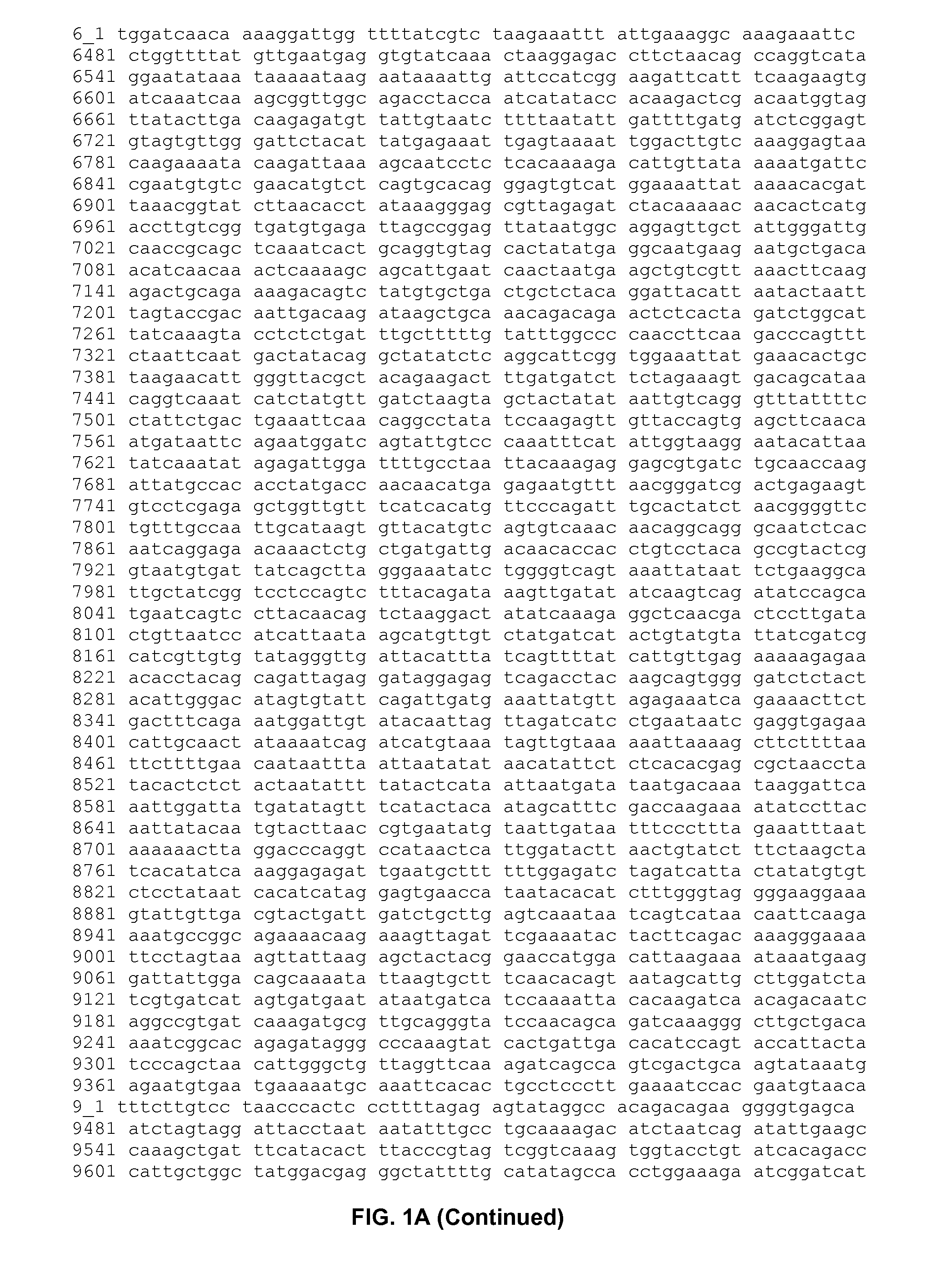
[0123]Construction of the plasmid pSL-6802-1-4. pSL-6802-1-4 comprises the flanking sequences of the C5 locus, H6 vaccinia promoter and G Nipah virus gene to generate VCP2199. The Nipah virus was isolated from human CSF. The Nipah G gene was PCR amplified and inserted into plasmid pTM1, generating pTM1 Nipah G. The purpose was to construct a pC5 H6p Nipah G donor plasmid for generation of an ALVAC canarypox virus recombinant expressing Nipah G. The plasmid name was pC5 H6p Nipah G, pSL-6802-1-4. The plasmid backbone was pCXL-148-2, pC5 H6p comprising the H6 vaccinia promoter, the left and the right arms corresponding to the C5 locus of insertion. The plasmid pCXL-148-2 is derived from the plasmid pNVQH6C5LSP-18 by a single base mutation from T to C in the C5 right arm. The plasmid pNVQH6C5LSP-18 is described in S. Loosmore et al US2005 / 0031641.
[0124]The Nipah G gene was PCR amplified using pTM1 Nipah G as template and primers 11470.SL and 11471.SL (FIG. 2B). The ˜1.8 kb PC...
example 2
Expression
[0139]Western blot of Fowlpox Nipah G, vFP2200 (FIG. 6). Primary CEF cells were infected with vCP2199 (ALVAC C5 H6p Nipah G) and vFP2200 (Fowlpox F8 H6p Nipah G) at MOI of 10 and incubated at 37° C. for 24 hrs. The cells and culture supernatant were then harvested. Sample proteins were separated on a 10% SDS-PAGE gel, transferred to Immobilon nylon membrane. The guinea pig antiserum and chemiluminescence system were used. Nipah G was expressed in cell pellets for vCP2199 and vFP2200. It did not show up in supernatant.
[0140]Western blot of ALVAC Nipah F, vCP2208 (FIGS. 7A and 7B). Primary CEF cells were infected with vCP2208. (ALVAC C5 H6p Nipah F) s at MOI of 10 and incubated for 24 hours. The supernatant was harvested and clarified. The cells were harvested and suspended in water to lyse. Lysate and supernatant were separated by 10% SDS-PAGE. The protein was transferred to nylon membrane and blocked with Western blocking buffer. Using guinea pig antiserum and chemilumines...
example 3
Serology and Protection
[0143]Sixteen pigs were allocated randomly into four groups. Group F animals were immunized with 108 pfu / dose of VCP2208 expressing Nipah virus F protein. Group G animals were immunized with 108 pfu / dose of VCP2199 expressing Nipah virus G protein. Group G+F animals were immunized with a mixture containing 108 pfu / dose of VCP2199 and 108 pfu / dose of VCP 2208 expressing respectively Nipah virus G and F proteins. Group challenge animals were unvaccinated control animals.
[0144]The pigs were injected by intramuscular route on Day 0 and Day 14. The pigs were challenged by intranasal inoculation of 2.5×10⁵ pfu of Nipah virus on Day 28. Seven days post challenge the presence of virus is identified by RT-PCR or virus isolation in various organs and in nasal swabs. Blood samples are collected on D0, D7, D14, D21, D28, D29, D30, D31, D32, D34, and D35 after the first injection and antibody titers are measured by IgG indirect ELISA or seroneutralisation assay. The neutra...
PUM
 Abstract
Description
Claims
Application Information
 - R&D
- Intellectual Property
- Life Sciences
- Materials
- Tech Scout
- Unparalleled Data Quality
- Higher Quality Content
- 60% Fewer Hallucinations
Browse by: Latest US Patents, China's latest patents, Technical Efficacy Thesaurus, Application Domain, Technology Topic, Popular Technical Reports.
© 2025 PatSnap. All rights reserved.Legal|Privacy policy|Modern Slavery Act Transparency Statement|Sitemap|About US| Contact US: help@patsnap.com


