
Enzyme activity assay using rolling circle amplification

a technology of enzyme activity and assay, applied in biochemistry apparatus and processes, pharmaceutical non-active ingredients, biocide, etc., can solve the problems of inability to establish satisfactory correlation between protein production and corresponding protein activity levels, and the determination of biological processes which take place in a single cell requires multiple levels of labor and time-consuming investigations

Inactive Publication Date: 2010-11-11
IN SITU RCP
View PDF0 Cites 52 Cited by
  • Summary
  • Abstract
  • Description
  • Claims
  • Application Information

AI Technical Summary

Benefits of technology

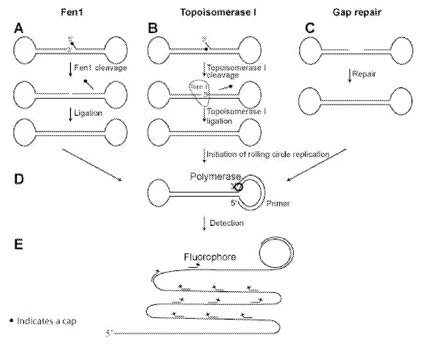
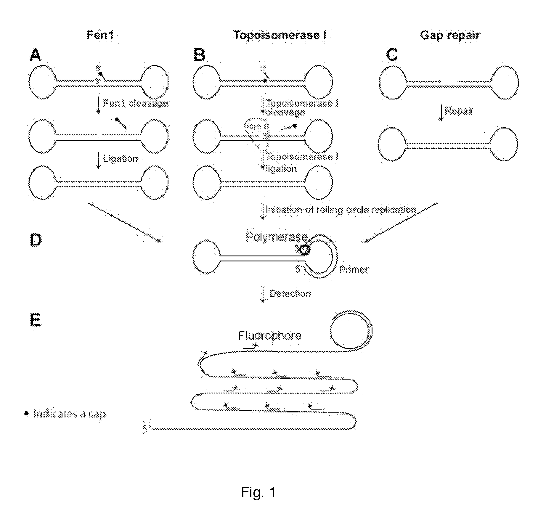
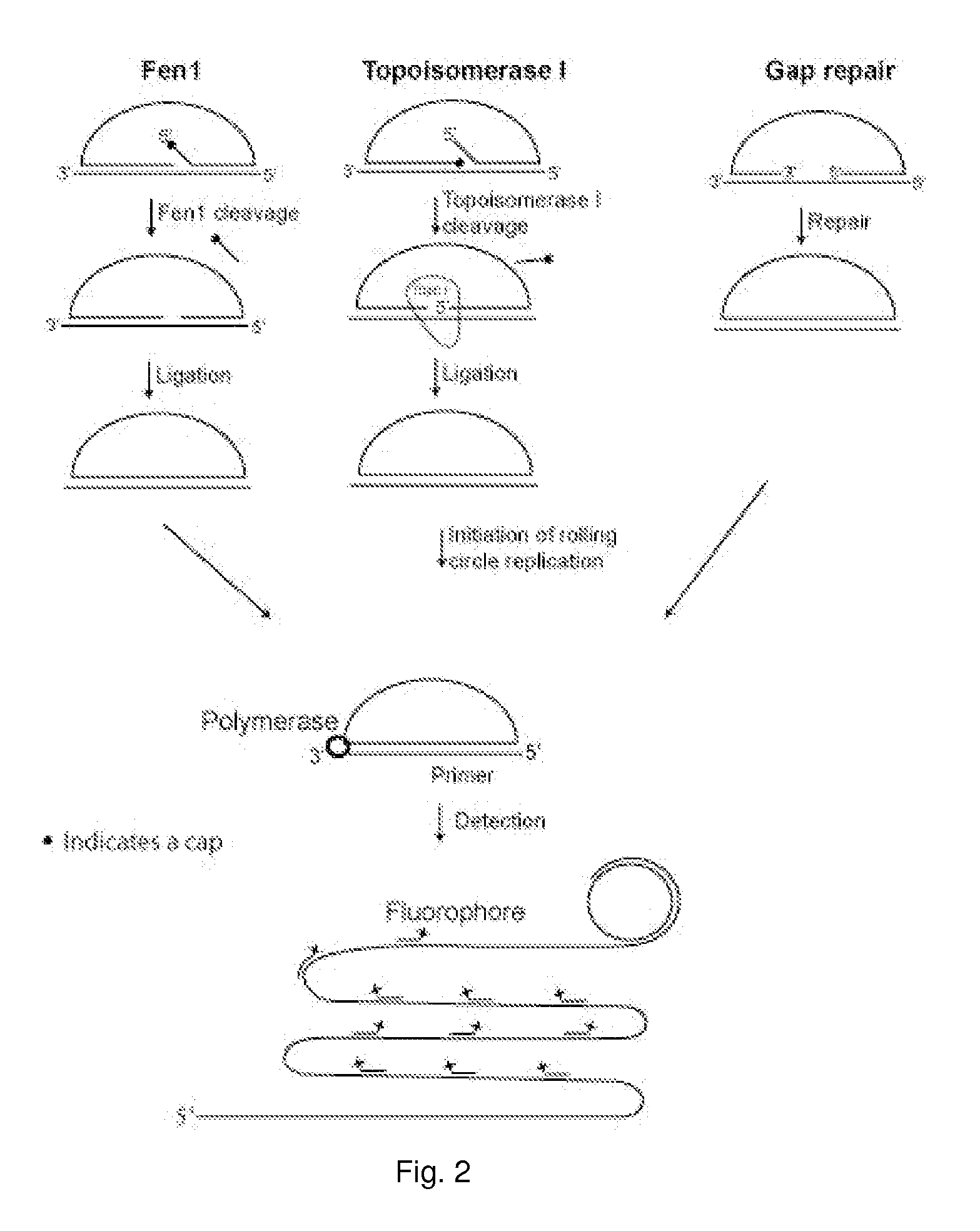
[0010]The present invention is directed to assays for the detection in a biological sample of enzyme activities, such as DNA modifying enzyme activities, such as nucleases and toposiomerases. The present invention makes it possible to design assays combining protein activity detection with RCR through the conversion of linear oligonucleotides into circular ones.
[0011]An oligonucleotide probe comprising a specific, unprocessed substrate moiety can be processed by a particular enzyme, or a particular class of enzymes, thereby generating a template for rolling circle amplification. Detection of the rolling circle amplification product serves as an indication for the presence in a biological sample of the enzyme activity in question. The assays of the present invention are both quantitative and qualitative, thereby enabling a more sensitive analysis of not only the presence of an enzyme, but also of the activity associated with the enzyme in question when the enzyme is present in a biological sample, such as a tissue sample and / or a body fluid sample.

Problems solved by technology

At present, a determination of the biological processes which take place in a single cell requires laborious and time consuming investigations at multiple levels.
In particular, detection of DNA modifying enzymes is dominated by techniques using radioactively labeled oligonucleotides, which are practical for monitoring different cleavage and ligation reactions in solution (Lisby et al., 2001; Friedrich-Heineken & Hubscher, 2004), but also inconvenient because of the radioactive labeling.
Furthermore, no satisfactory correlation can be established between protein production and corresponding protein activity levels.
A further complication is that many genes are alternatively spliced and that give rise to different proteins having different activities.

Method used


Image

  • Enzyme activity assay using rolling circle amplification
  • Enzyme activity assay using rolling circle amplification
  • Enzyme activity assay using rolling circle amplification

Examples


example 1

Methods

Oligonucleotides

[0322]Oligonucleotides (listed below) were purchased from DNA Technology A / S, Aarhus, Denmark.

NameSequenceFen15′-BIOTIN-GATATCGAAT TCCACTGTGA AGATCGCTTA TGGAATTCGATATCAAGCCC TCAATGCACA TGTTTGGCTC CGCTTGATAT-3′Fen15′-P-CGAATTCC ACTGTGAAGATCGCTTAT GGAATTCGAPOSTATCAAGC CCTCAATGCACATGTTTGGCTCC GCTTGATAT-3′Topo I5′-AGAAAAATTT TTAAAAAAAC TGTGAAGATC GCTTATTTTT TTAAAAATTTTTCTAAGTCT TTTAGATCCC TCAATGCTGC TGCTGTACTA CGATCTAAAAGACTTAGA-AMIN-3′Topo I5′-P-AGAAAAATTT TTAAAAAAAC TGTGAAGATC GCTTATTTTT TTAAAAATTTPOSTTCTAAGTCT TTTAGATCCC TCAATGCTGC TGCTGTACTA CGATCTAAAAA CTT-3′Primer15′-AMIN-CCAACCAACCAACCAA-ATAAGCGATCTTCACAGT-3′Primer25′-ATAAGCGATC TTCACAG-3′ID 165′-TAMRA-CCTCAATGCT GCTGCTGTAC TAC-3′ID 335′-FITC-CCTCAATGCA CATGTTTGGC TCC-3′

Fen1 Reaction

PAGE:

[0323]The Fen1 reaction was carried out in a mixture containing 40 mM Tris-HCl, pH 7.5, 10 mM MgCl2, 1 mM DTT, 0.2 μg / μl BSA, 0.1 μM probe, 30 pg / μl (1 fmol / μl) of Fen1 enzyme (Alexis Biochemicals), and 0.03 u / μl of T4 DNA ...

example 2

Gel-Based Detection Assays

Fen 1:

[0332]The experimental setup of the Fen1 activity assay was confirmed by PAGE (FIG. 2). The linear probe was degradable using a combination of exonuclease I and III (both exonucleases were used because the probe contained both single and double stranded regions) (compare FIG. 2, lane 1 and 2). The probe was also degradable when Fen1 and T4 DNA ligase were added separately (compare FIG. 2, lane 3 and 4 and lane 5 and 6). In contrast, when both Fen1 and T4 DNA ligase were included, a faster migrating product, resistant to exonucleases, appeared (compare FIG. 2 lane 7 and 8), indicating that the probe had been circularized. This circular product had the same migration speed as a circularized control probe with the same sequence as the Fen1 modified product (compare FIG. 2, lane 8 and 12). When only Fen1 was applied to the reaction, the linear product had a faster migration than without Fen1 (compare FIG. 2, lane 1 and 5), likely resulting from Fen1 remov...

example 3

Solid Support Amplification Assays

[0334]To verify that the resulting products were indeed circularized, solid support assays comprising RCR and fluorescent labeling were designed.

Fen1

[0335]Products corresponding to lane 1, 3, 5 and 7 from FIG. 2 were hybridized to a surface coated with 5′-amin-coupled oligonucleotides (primer 1). Following hybridization the covalently coupled oligonucleotide functioned as a primer for RCR. After amplification the products were detected by hybridizing a labeled oligonucleotide to the RCP. As expected, according to the gel-based assay, RCP's could only be detected when both Fen1 and ligase were present in the reaction mixture (FIG. 4). Since each Fen1 enzyme can prepare several probes for ligation, this assay does not correspond to single-molecule detection of Fen1 enzymes, but instead each green dot corresponds to a single circularization event (if two or more RCR reactions have not taken place in close proximity on the solid support, thereby giving ...


PUM

PropertyMeasurementUnit
temperatureaaaaaaaaaa
timeaaaaaaaaaa
timeaaaaaaaaaa

Abstract

The present invention relates to an enzyme activity assay using rolling circle amplification for verifying that a sample contains the enzyme activity in question. Thus, the present invention pertains to a method for determining the presence or absence of one or more enzyme activities involved in circularising a non-circular oligonucleotide probe in a biological sample. Furthermore, the present invention concerns liquid compositions comprising one or more oligonucleotide probes. Within the scope of the present invention is also a composition comprising a liquid composition and a tissue sample, and solid support of one or more oligonucleotides of the present invention. Disclosed is also a microfluidic device with one or more compartments for performing rolling circle amplification events, and a method for correlating one or more rolling circle amplification events. Methods for testing the efficacy of a drug, for diagnosing or prognosing a disease, for treating a disease, or for treating prophylactically a disease is furthermore disclosed.

Description

[0001]All patent and non-patent references cited in this application are hereby incorporated by reference in their entirety.FIELD OF INVENTION[0002]The present invention relates to an enzyme activity assay using rolling circle amplification for verifying that a sample contains the enzyme activity in question.BACKGROUND OF INVENTION[0003]At present, a determination of the biological processes which take place in a single cell requires laborious and time consuming investigations at multiple levels.[0004]DNA can be evaluated using different in situ hybridization techniques, such as FISH (Levsky & Singer, 2003), PRINS (Koch et al., 1989) or target primed amplification of padlock probes (Larsson et al., 2004). RNA detection is mainly performed using FISH techniques.[0005]Detection of proteins are routinely performed using antibodies, but new techniques are emerging, e.g. proximity ligation, where oligonucleotide tagged antibodies are used for the detection interacting proteins by either ...

Claims


Application Information

Patent Timeline
Patent Type & Authority Applications(United States)
IPC IPC(8): A61K47/00C12Q1/68A61P35/00
CPCC12Q1/6844G01N33/57484G01N33/6896G01N2333/91245G01N2500/00C12Q2521/307C12Q2531/125C12Q2521/507C12Q2521/519A61P35/00
Inventor LOHMANN, JAKOB SCHWALBESTOUGAARD, MAGNUSKOCH, JORN ERLAND
Owner IN SITU RCP
Learn More