Method of using an extracellular matrix to enhance cell transplant survival and differentiation

a cell transplant and extracellular matrix technology, applied in the direction of biocide, embryonic cells, drug compositions, etc., can solve the problems of inability to differentiate, affect the survival and differentiation of cells, and the limitations of existing therapies, so as to and increase the survival and/or differentiation of target cells

Inactive Publication Date: 2010-11-25
UNIV OF MEDICINE & DENTISTRY OF NEW JERSEY
View PDF2 Cites 16 Cited by
  • Summary
  • Abstract
  • Description
  • Claims
  • Application Information

AI Technical Summary

Benefits of technology

[0012]In another aspect, the invention provides a method of increasing survival and / or differentiation of target cells on a base matrix, the method comprising: creating a cell-made extracellular matrix on the base matrix to produce a modified base matrix and administering the target cells to the modified base matrix. In different embodiments of the invention, the matrices include, without limitations, those described above.
[0013]In another aspect, the invention provides a method of increasing survival and / or differentiation of target cells on a base matrix through providing a soluble formulation of the extracellular matrix or conditioned media to the apical surface of the cells to stimulate self-assembly and deposition of extracellular matrix and / or stimulation of mechanisms for cell survival and differentiation.

Problems solved by technology

Disease-related changes may mask extracellular matrix ligand availability to transplanted cells, impairing post-attachment events and leading, in turn, to cell death or inability of the cells to differentiate.
In addition, disease-related changes in the extracellular matrix can promote cell death, leading to the clinical situation in which cell transplantation is contemplated.
Existing therapy has significant limitations.
Antioxidants, for example, do not seem to be effective in the prevention of early AMD (i.e., drusen, retinal pigmentary changes).
Also, these medications currently are administered via repeated intravitreal injection, which entails some risk and inconvenience for the patient.

Method used


Image

  • Method of using an extracellular matrix to enhance cell transplant survival and differentiation
  • Method of using an extracellular matrix to enhance cell transplant survival and differentiation
  • Method of using an extracellular matrix to enhance cell transplant survival and differentiation

Examples


example 1

Long-Term Survival of Fetal RPE on Aged Submacular Human Bruch's Membrane is Impaired

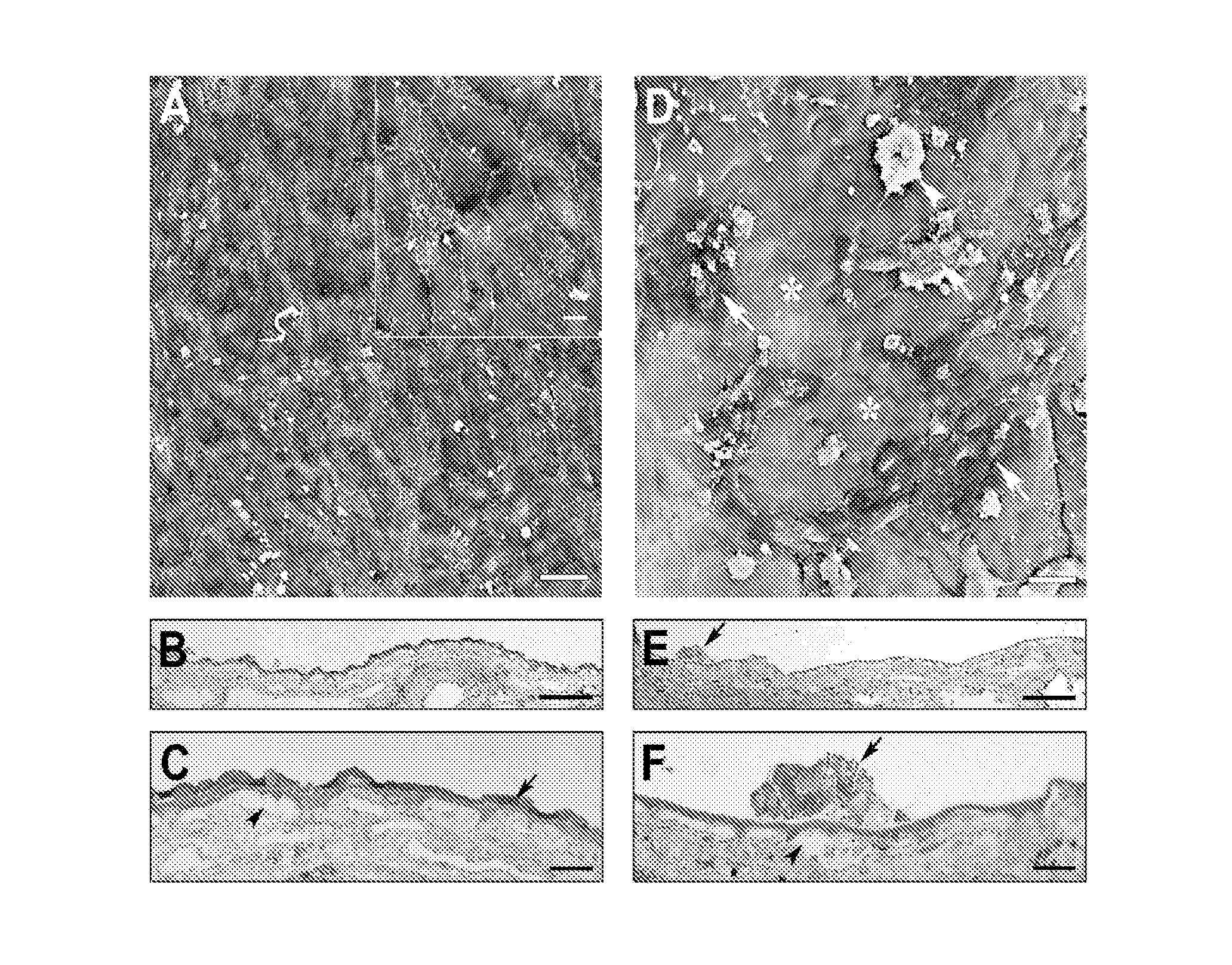
[0086]Fetal RPE (3164 cells / mm2) were seeded on aged human submacular Bruch's membrane debrided to expose the superficial surface of the inner collagenous layer. To create surfaces exposing the RPE basement membrane, RPE were gently wiped off the RPE / choroid / sclera explant using a wet surgical sponge. To create surfaces exposing the surface of the inner collagenous layer beneath the RPE basement membrane (i.e., superficial ICL), following RPE removal as indicated previously, a moistened surgical sponge was use to abrade the RPE basement membrane. In general, the area of RPE basement membrane debridement was created by approximately 5 wipes of the moistened sponge in each of 4 directions (rotating the explant 90 degrees after each series of 5 wipes). (V. K. Gullapalli, et al., Exp Eye Res 2005; 80(2):235-248). Cells were seeded onto the sclera / choroid explant and cultured for 21 days and evaluated fo...

example 2

Fetal RPE Resurfacing on Aged Bruch's Membrane Resurfaced with Bovine Corneal Endothelial Matrix (BCE-ECM)

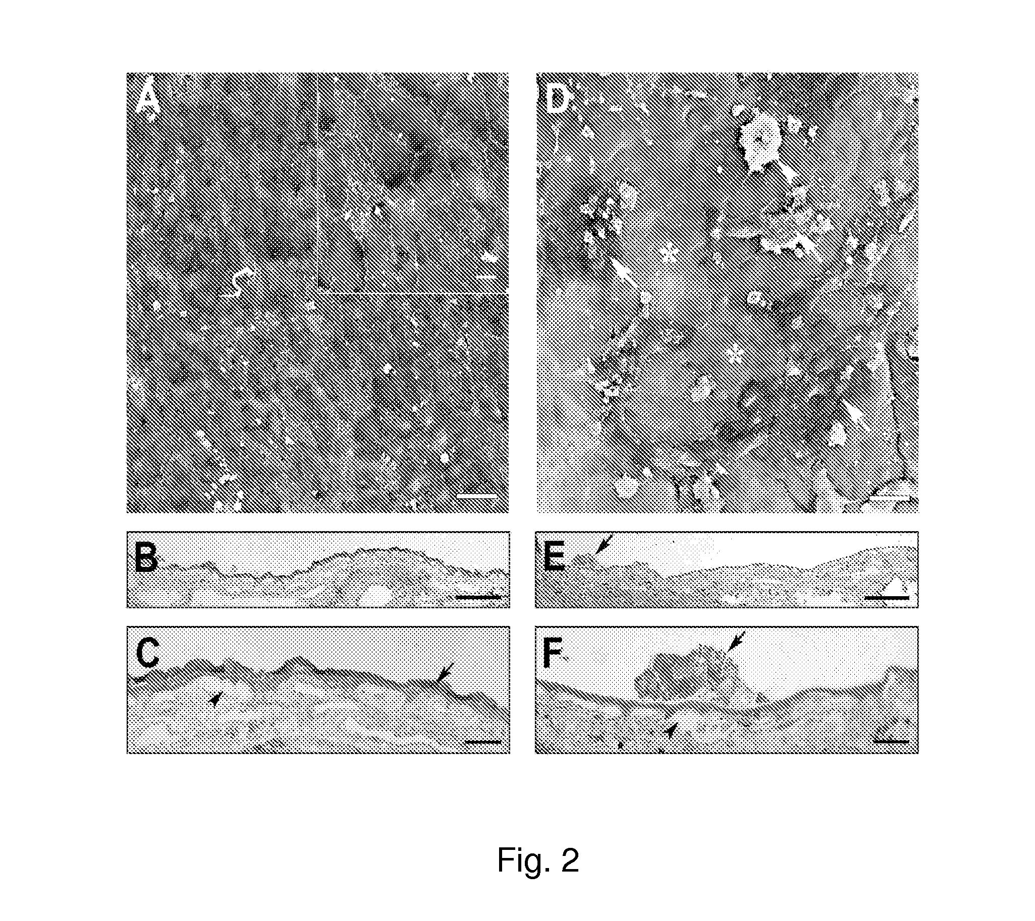
[0088]BCE (3164 cells / mm2) were cultured on the inner collagenous layer of aged human submacular Bruch's membrane (65 yr. old donor) for 14 days to allow ECM deposition. Cells were culture in the same way as cells cultured for ECM deposition on culture dishes (see paragraph 0056). Following BCE removal with NH4OH to expose the newly deposited ECM and extensive washing with PBS, explants were seeded with fetal RPE (3164 cells / mm2) and cultured for 21 days. The results of these experiments are illustrated in FIG. 2.

[0089]FIG. 2A is a scanning electron micrograph (SEM), showing that fetal RPE fully resurfaced the treated explant with large, flat polymorphic cells. Cells showed varying amounts of short apical processes on their surfaces (insert). Mag. bar 50 μm; insert mag bar 10 μm.

[0090]FIGS. 2B and 2C are light micrographs (LMs). As shown in FIG. 2B, cells fully resurfaced the tr...

example 3

Resurfacing Bruch's Membrane with a Biologically Deposited Extracellular Matrix (ECM) Improves Cell Survival

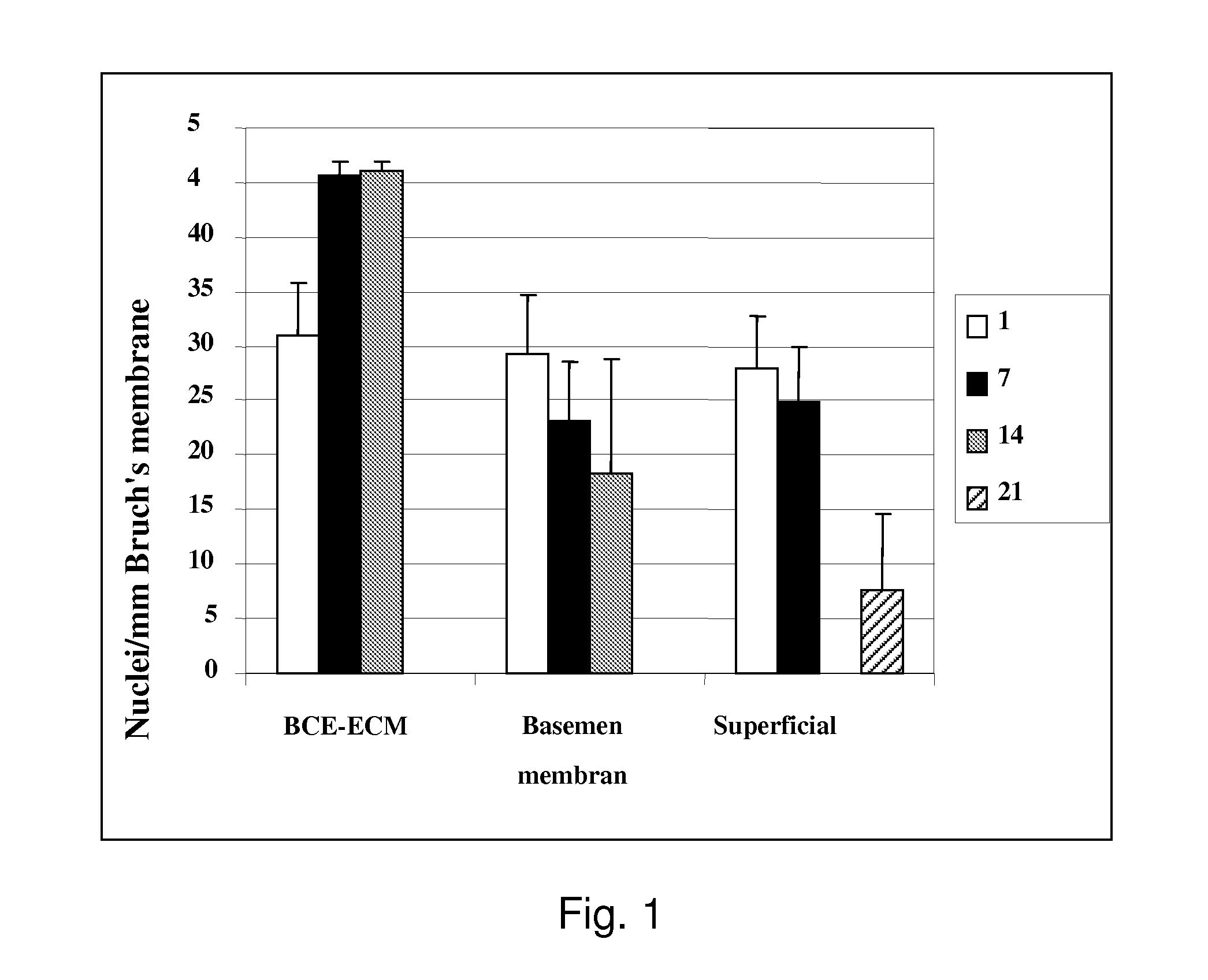
[0094]Bovine corneal endothelial cells (BCE, passage-2) were seeded onto human submacular superficial ICL of Caucasian donors over 55 years old at a density of 3164 cells / mm2 and cultured for 14 days to allow ECM deposition or treated for days with serum-free media only. Following BCE removal with NH4OH and extensive rinsing, fetal RPE (passage-2-5) were seeded at the same density onto the treated Bruch's membrane surface and cultured for 21 days. The fellow eye was treated similarly except no BCE were seeded.

[0095]RPE seeding density was 3164 cells / mm2 for 21 day incubations to determine long-term survival and morphology. FIG. 3 shows the cumulative data from 9 explant pairs. Counts are mean fetal RPE nuclei / mm Bruch's membrane (±SEM).

[0096]A statistically significant 230% (p=0.006) increase in cell density is seen at day-21 on treated explants compared to explants treated wi...


PUM

PropertyMeasurementUnit
diameteraaaaaaaaaa
diameteraaaaaaaaaa
timeaaaaaaaaaa

Abstract

Provided is a matrix for promoting survival and differentiation of cells transplanted thereon, comprising a base matrix and a cell-made matrix thereon. Methods and means for making and using same are also provided.

Description

RELATIONSHIP TO PRIOR APPLICATIONS[0001]This application claims priority under 35 U.S.C. §119(e) from U.S. Provisional application No. 60 / 991,601 filed on Oct. 19, 2007.GOVERNMENTAL SUPPORT[0002]The Research leading to the present invention was supported in part, by National Institutes of Health Grant No. NIH RO3 EY013690. Accordingly, the U.S. Government has certain rights in this invention.FIELD OF INVENTION[0003]This invention relates to the production of an extracellular matrix and methods of use for clinical treatment of disease in the central nervous system.BACKGROUND[0004]Disease-related changes may mask extracellular matrix ligand availability to transplanted cells, impairing post-attachment events and leading, in turn, to cell death or inability of the cells to differentiate. In addition, disease-related changes in the extracellular matrix can promote cell death, leading to the clinical situation in which cell transplantation is contemplated.[0005]One of the conditions in w...

Claims


Application Information

Patent Timeline
Patent Type & Authority Applications(United States)
IPC IPC(8): A61K9/00C12N5/07A61K35/12C12N5/074C12N5/0735C12N5/079A61P27/02
CPCC12N5/0068C12N5/0621C12N2500/99C12N2533/90C12N2533/30C12N2533/40C12N2502/28A61P27/02C12N2500/90
Inventor SUGINO, ILENEGULLAPALLI, VAMSIZARBIN, MARCO
Owner UNIV OF MEDICINE & DENTISTRY OF NEW JERSEY
Why Eureka
Try Eureka