
Scleral buckles for sutureless retinal detachment surgery

a retinal detachment and buckle technology, applied in the field of eye surgery, can solve the problems of surgical failure and permanent damage, high surgical procedure with potential major complications, and accidental perforation of the sclera,

Active Publication Date: 2010-12-02
THE CHINESE UNIVERSITY OF HONG KONG
View PDF9 Cites 12 Cited by
  • Summary
  • Abstract
  • Description
  • Claims
  • Application Information

AI Technical Summary

Benefits of technology

[0016]The device and associated procedure reduce intraoperative risk of globe perforation during the procedure, thus reducing the chance of disastrous complications such bleeding, retinal hole formation and softening of the globe. In addition, the device simplifies the procedure, so the length of time of surgery is reduced and patient comfort is increased.
[0017]A device according to the invention could be easily combined with other options of retinal detachment treatment including pars plana vitrectomy, increasing surgical effects synergistically.
[0018]The buckle and encircling band according to the invention can be more securely implanted onto the globe. Therefore the postoperative risk of a loosened buckle can be reduced, enhancing the surgical success rate and minimizing the need for repeated surgery.

Problems solved by technology

Although scleral buckling surgery is effective, it is also a highly demanding surgical procedure with potential major complications.
If the placement of needle is too deep, the sclera will be accidentally perforated.
Perforation of sclera may damage the underlying tissue, leading to bleeding or retinal hole formation, surgical failure and permanent damage.
However, if the placement of the needle is too shallow, the buckle or encircling band could be loosened or even exposed out from the eye after the surgery.
A loosened or exposed buckle also leads to surgical failure, infection and unwanted pain.
Apart from perforation of the globe, suturing poses at least one more disadvantage.
However, the posterior anchor is usually located posterior to the equator of the globe, and this location is relatively difficult to reach.
The working space for making the posterior anchor is usually very limited because of the funnel shape of the bony orbit and the posterior location of the anchor site.
If the vortex vein is accidentally perforated, profound bleeding can lead to failure of the whole surgery.
However, it can swell unexpectedly up to four fold of its original size years after the surgery because of hydrolytic degradation, thus potentially causing serious complications such as buckle extrusion, intrusion, squint and subcutaneous mass, etc.
The scleral buckle needs to be fixed in place by sutures or adhesives, which inevitably pose the same hazards as conventional scleral buckling surgery.
The scleral band is inserted into the Tenon's space, but there is no mechanism described to stabilize the scleral band onto the eyeball.

Method used


Image

  • Scleral buckles for sutureless retinal detachment surgery
  • Scleral buckles for sutureless retinal detachment surgery
  • Scleral buckles for sutureless retinal detachment surgery

Examples


embodiment i

[0046]Main Scleral Buckle

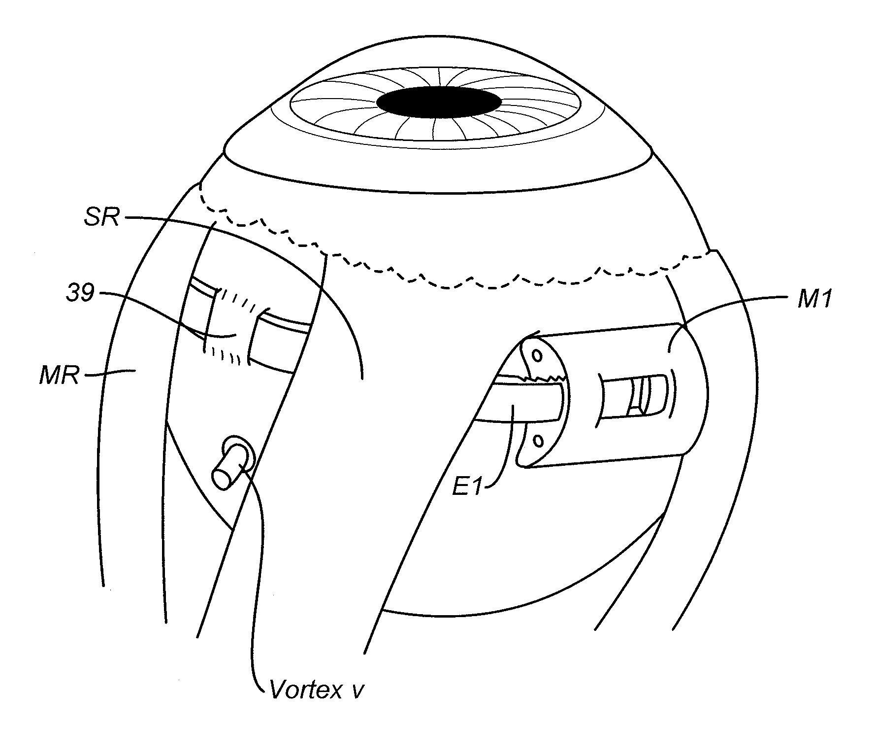
[0047]FIG. 12 depicts a perspective view of a main circumferential scleral buckle in a first embodiment of the invention in use, and FIG. 13 depicts an additional perspective view of a main radial scleral buckle in the first embodiment in use. In the first embodiment, there are two types of scleral buckles: the main scleral buckle and the optional supplementary scleral buckle. All use an encircling band E1. All are made with medical grade silicone rubber. Other embodiments of the scleral buckles are mounted in a similar fashion and are therefore not shown as mounted.

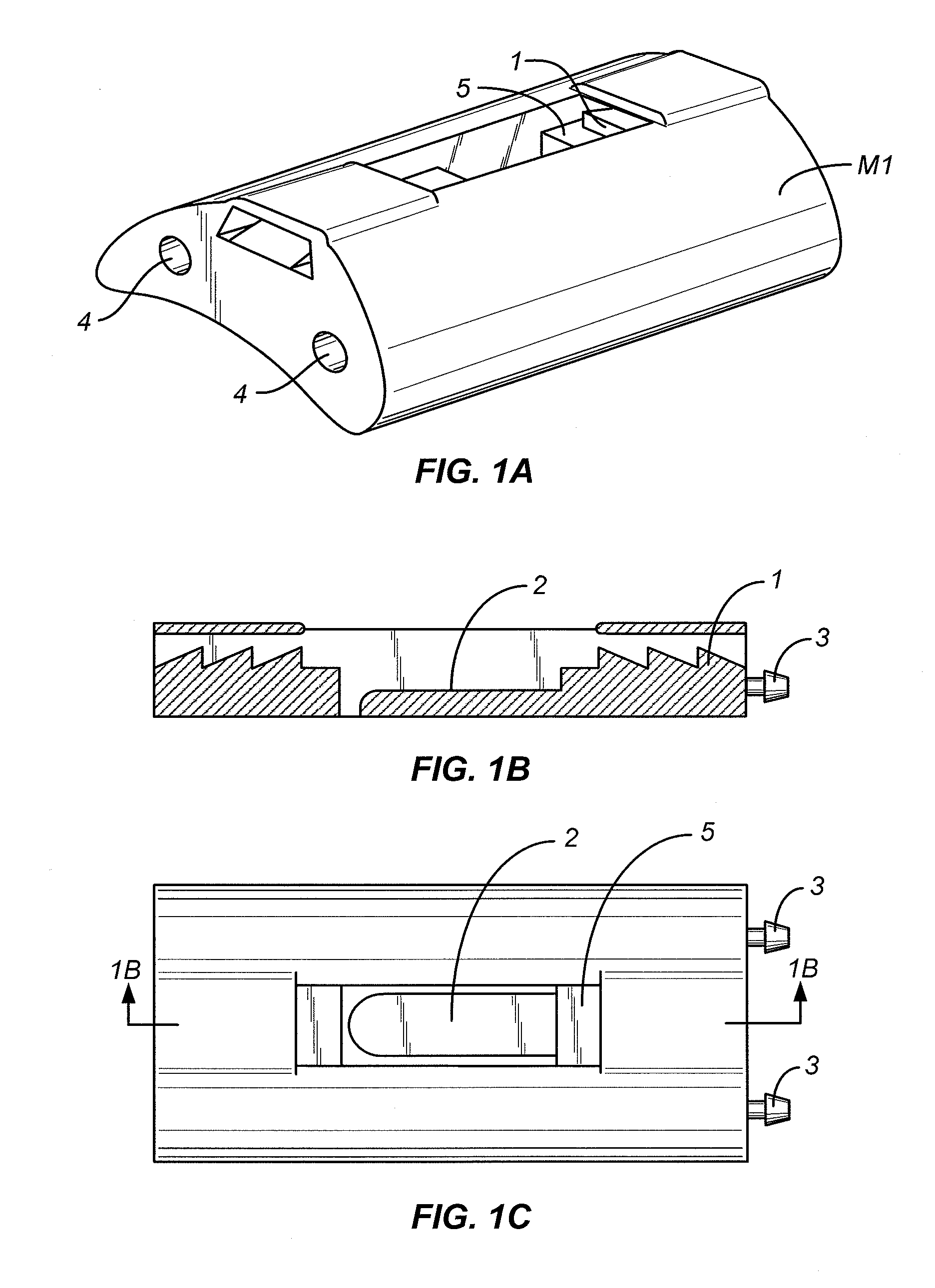
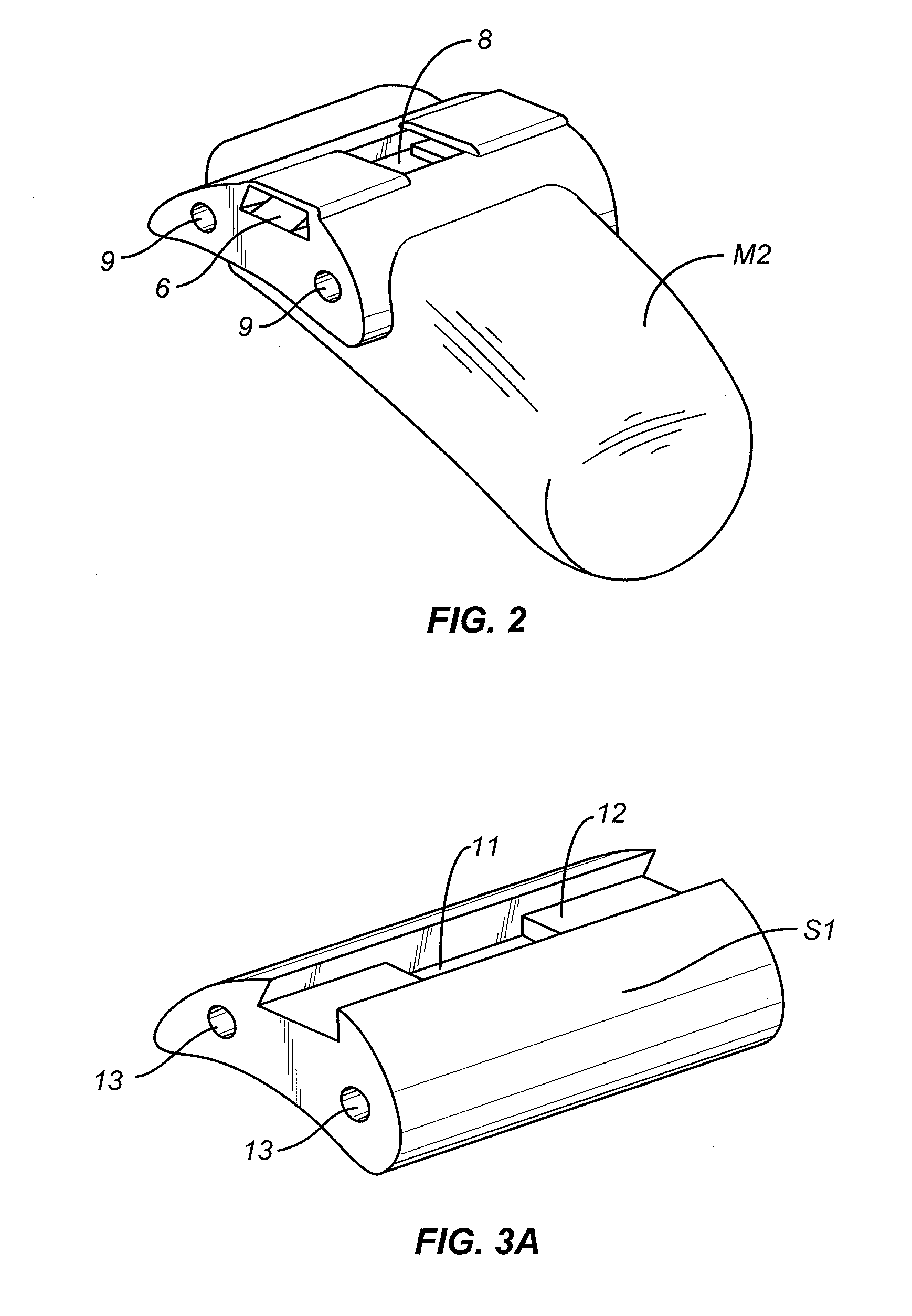
[0048]The structure features a main or major scleral buckle and an optional supplementary scleral buckle and an encircling band. The main scleral buckle has two configurations, circumferential M1 (FIGS. 1A-1C and FIGS. 12) and radial M2 (FIG. 2 and FIG. 13). The main scleral buckle M1 or M2 directly locks and tightens the two ends of the encircling band E1 (FIGS. 12, 13) with locks 1, 6 and forms...

embodiment ii

[0064]In a second embodiment, there is only one type of scleral buckle (with 2 configurations: radial (FIG. 6A-6C) and circumferential (FIG. 7) to assemble with an encircling band E2. The basic principle, material and rationale for choice of configuration of scleral buckle are the same as that of embodiment I. The difference between embodiment I & II lies in the method of locking between the scleral buckle and encircling band. In this embodiment, both ends of the encircling band are locked together directly by a silicone sleeve (not shown) after the encircling band E2 has passed through the four scleral tunnels. The scleral buckle does not function as the main lock to hold and tighten the two ends of the encircling band. The buckle is assembled with the encircling band to exert indentation effect onto the eyeball. (FIG. 8).

[0065]The Scleral Buckle

[0066]The scleral buckle of a second embodiment has two configurations: circumferential II1 (FIG. 6A-6C) and radial II2 (FIG. 7). The scle...

embodiment iii

[0071]Embodiment III is similar to embodiment II where there is only one type of scleral buckle (with 2 configurations: circumferential (FIG. 9) and radial (FIG. 10) to assemble with an encircling band E3. The basic principles, materials and rationale for choice of configuration of the scleral buckle are the same as that of embodiments I and II. The difference between embodiment II and embodiment III lies in the method of locking between the scleral buckle and the encircling band. In this embodiment, like embodiment II, a sleeve is used to lock and tighten the two ends of the encircling band together after it has passed through the four scleral tunnels 39. The buckles III1, III2 are assembled with the encircling band E3 to exert an indentation effect onto the eyeball by a different mechanism from embodiment II.

[0072]The Scleral Buckle

[0073]The scleral buckle has two configurations: circumferential III1 and radial III2. The scleral buckles III1, 1112 are assembled with the encircling...


PUM


Abstract

A set formed of a scleral buckle and an encircling band is provided for use in connection with retinal detachment surgery to enable the implantation of both the scleral buckle and encircling band free of any suture. A self-assembling scleral buckle-encircling band combination is secured in place by surface scleral tunnels operative as belt loops to enable the securing of a scleral buckle and encircling band on the eyeball to exert an intended indentation effect for treatment of retinal detachment.

Description

CROSS-REFERENCES TO RELATED APPLICATIONS [0001]NOT APPLICABLESTATEMENT AS TO RIGHTS TO INVENTIONS MADE UNDER FEDERALLY SPONSORED RESEARCH AND DEVELOPMENT[0002]NOT APPLICABLEREFERENCE TO A “SEQUENCE LISTING,” A TABLE, OR A COMPUTER PROGRAM LISTING APPENDIX SUBMITTED ON A COMPACT DISK[0003]NOT APPLICABLEBACKGROUND OF THE INVENTION[0004]This invention relates to eye surgery and more particularly to aids for retinal detachment surgery.[0005]The retina is a thin sheet of light sensitive nerve tissue lining the inner posterior of the eye and which functions to capture focused images like the film of a camera. Retinal detachment (RD) occurs when the retina separates from the posterior wall of the eye. It is usually caused by a break in the retina. Retinal detachment is one of the major human eye diseases which can lead to severe visual impairment or even blindness unless it is treated surgically.[0006]Scleral buckling surgery is one of the most commonly performed surgical operations to tre...

Claims


Application Information

Patent Timeline
Patent Type & Authority Applications(United States)
IPC IPC(8): A61F2/14A61B17/00
CPCA61F9/00727
Inventor LEE, YAU WING VINCENTLAM, SHUN-CHUI DENNISLIU, TA LI DAVIDLI, CHI LAI
Owner THE CHINESE UNIVERSITY OF HONG KONG
Learn More