Ordered Assembly of Membrane Proteins During Differentiation of Erythroblasts
a technology of membrane proteins and erythroblasts, which is applied in the field of identification and isolation of red blood cell precursors, can solve the problems of loss of mechanical integrity and hemolytic anemia, lack of clear differentiation between stages, and overlap of proerythroblast to mature functional red blood cells (rbc) maturation studies, etc., to achieve the effect of increasing the level of proteins important, reducing the expression of cd44 surface, and increasing the protein level
- Summary
- Abstract
- Description
- Claims
- Application Information
AI Technical Summary
Benefits of technology
Problems solved by technology
Method used
Image
Examples
example 1
Ordered Assembly of Membrane Proteins during Differentiation of Erythroblasts
[0038]Expression of Transmembrane Proteins during Erythropoiesis.
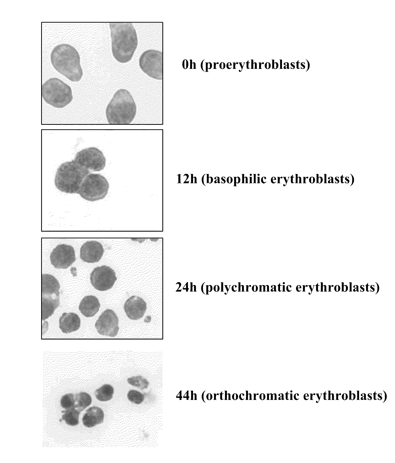
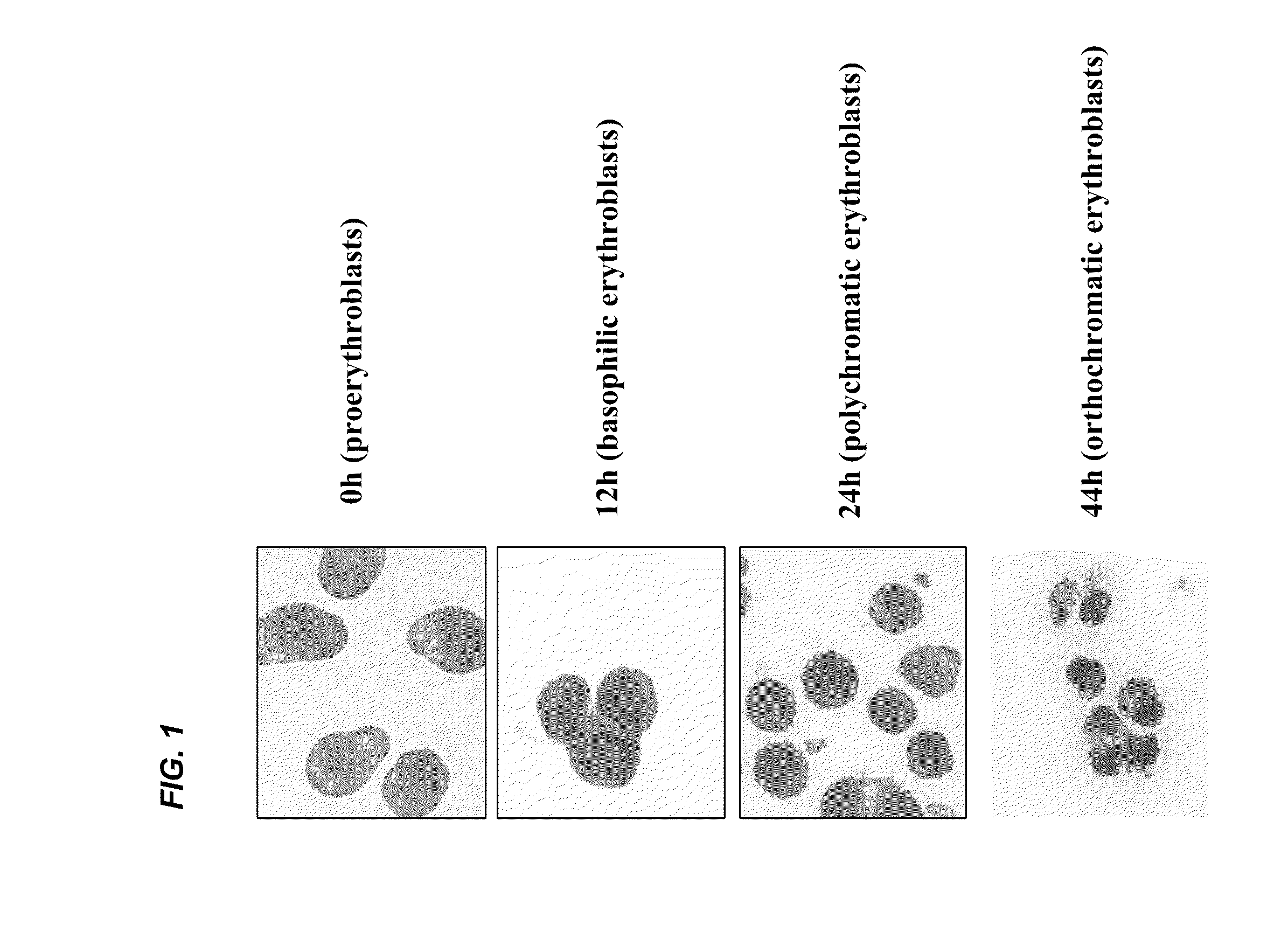
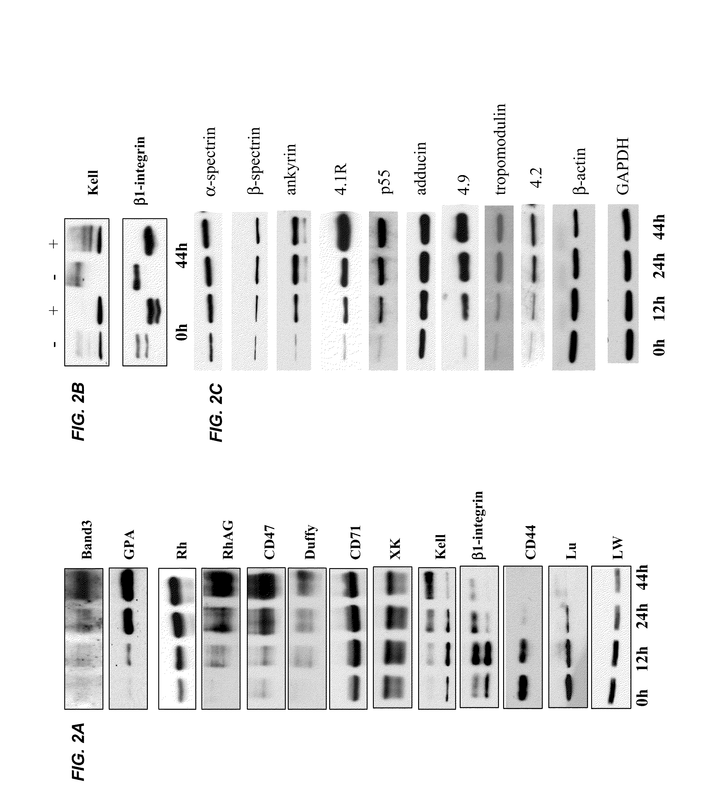
[0039]It is expected that the differentiation of erythroblasts would be accompanied by the changes in protein composition and properties of the plasma membrane, however a comprehensive study has not been reported. Using FVA system (Koury et al. J. Cell Physiol. 121:526-63, 1984) near homogenous populations of erythroblasts were obtained at proerythroblast, basophilic erythroblast, polychromatic erythroblast, orthochromatic erythroblast stages (depicted in FIG. 1) and examined the expression of 13 transmembrane proteins by Western blotting. During 44 hours of culture in this system, proerythroblasts (0 hr) progressively differentiated into basophilic erythroblasts (12 hr), polychromatic erythroblasts (24 hr) and orthochromatic erythroblasts and reticulocytes (44 hr). FIG. 2A depicts the relative concentrations of these proteins as assessed by W...
example 2
Quantitative Analysis of Erythropoiesis in Mouse Bone Marrow
[0062]Two distinct erythroid progenitors have been functionally defined in colony assays, namely, the early-stage burst forming unit-erythroid (BFU-E), and the later stage colony forming unit-erythroid (CFU-E) progenitor. The earliest morphologically recognizable erythroblast in hematopoietic tissues is the proerythroblast, which undergoes 3 to 4 mitoses to produce reticulocytes. Morphologically distinct populations of erythroblasts are produced by each successive mitosis, beginning with proerythroblasts and followed by basophilic, polychromatic and orthochromatic erythroblasts. Based on the changes in expression levels of CD44, GPA and cell size, a method was developed to isolate populations of erythroblasts at each stage of development, in a more homogenous state than previously achieved, dependent on the expression levels of the transferrin receptor, CD71. In this study, the ratio of proerythroblast:basophilic:polychroma...
example 3
Analysis of In Vitro Human Erythropoiesis
[0068]Erythropoiesis is the process by which nucleated erythroid progenitors proliferate and differentiate to generate, every second, millions of non-nucleated red cells with their unique discoid shape and membrane material properties. The time-course appearance of individual membrane protein components during murine erythropoiesis was studied and distinct changes of individual proteins were found during terminal erythroid differentiation, particularly a progressive and dramatic decrease of CD44 from proerythroblast to reticulocyte. These findings have allowed the development of a new strategy for quantifying the in vivo erythropoiesis and defining stage-specific defects in erythroid maturation in mouse. To examine whether the similar strategy can be applied to study human erythropoiesis, the expression of various red cell membrane proteins were examined during terminal differentiation of human erythroid progenitors using a human unilineage e...
PUM
| Property | Measurement | Unit |
|---|---|---|
| pH | aaaaa | aaaaa |
| size | aaaaa | aaaaa |
| cell size | aaaaa | aaaaa |
Abstract
Description
Claims
Application Information
 - R&D
- Intellectual Property
- Life Sciences
- Materials
- Tech Scout
- Unparalleled Data Quality
- Higher Quality Content
- 60% Fewer Hallucinations
Browse by: Latest US Patents, China's latest patents, Technical Efficacy Thesaurus, Application Domain, Technology Topic, Popular Technical Reports.
© 2025 PatSnap. All rights reserved.Legal|Privacy policy|Modern Slavery Act Transparency Statement|Sitemap|About US| Contact US: help@patsnap.com


